Nanoscale brain imaging with electron microscopy
Communication between the billions of neurons in our brains underlies the thoughts, feelings, and behaviors that make us who we are. The basic unit of that communication is the synapse: a physical structure essential for the interaction of neurons. Using electron microscopy, cellular and molecular facets of these three-dimensional synapses can be directly visualized, allowing scientists to explore the brain at the nanometer scale. This offers an unparallelled view of the structures and connections that define neuronal function and can aid in our understanding of diseases that alter these networks.
In a recent publication, research led by Dr. Jill Glausier and Dr. Zachary Freyberg from the University of Pittsburgh investigated whether the ultrastructure of postmortem human tissue can reveal information about the functional dynamics that existed during life.
“Our application of three-dimensional FIB-SEM imaging to brain tissue has provided insights into the nano-architecture of the connections that foster communications between neurons and local support cells. And all at resolutions that also enable us to generate spatial maps of the molecules that comprise these connections.
– Dr. Zachary Freyberg, University of Pittsburgh
FIB-SEM generates 3D images of prefrontal cortex
The study focused on the human dorsolateral prefrontal cortex (DLPFC), a region tied to some of the most advanced cognitive processes, including reasoning, memory, and decision-making. It is also a region profoundly affected by disorders like schizophrenia. Using a Thermo Scientific Helios 5CX DualBeam (a focused ion beam scanning electron microscope, or FIB-SEM), the researchers were able to generate high-resolution 3D datasets from human brain specimens. This acquisition method is one of the ways to generate volume electron microscopy (volume EM) data, which provides detailed ultrastructural 3D information about biological specimens. In this case, volume EM enabled the detailed exploration of structures and connections in the DLPFC.


This approach enabled the visualization of synaptic complexes, mitochondria, and sub-synaptic structures down to nanometer resolution. Such a level of tissue preservation is critical, as it validates the reliability of postmortem data for the inference of functional relationships.
The team reconstructed 50 glutamate axo-spinous synapses in 3D, a feat that was previously unimaginable with traditional microscopy. These reconstructions revealed preserved relationships between pre- and post-synaptic structures, including correlations between pre-synaptic bouton volumes and post-synaptic density sizes, which are key indicators of synaptic strength and activity. This aligns with known models of synaptic function in living systems and reinforces the potential of postmortem studies to support the investigation of cognition and cognitive impairment.

Brain tissue segmentation from 3D imaging
A significant finding in the 3D data was a complex dendritic shaft that exhibited a large number of mitochondria and synaptic connections. This suggests that it may serve as a hub for heightened synaptic communication and plasticity. Such a discovery is not only novel but also opens up new avenues of exploration into how specific neuronal features may contribute to higher-order functions or vulnerabilities in psychiatric and neurological conditions.
“In the near future, a major goal is integrating the volume EM approaches we have established in this paper with molecular approaches, such as transcriptomics or proteomics, to further enhance the interpretative power of microscopy data.”
– Dr. Jill Glausier, University of Pittsburgh
Spotlight on the researchers

Jill Glausier, PhD
Dr. Glausier’s research focuses on cortical and cognitive dysfunction in schizophrenia. Using postmortem human brain tissue, her work leverages molecular and imaging techniques, including FIB-SEM, to unravel the synaptic alterations underpinning cognitive deficits. Her findings, such as the role of parvalbumin basket cell synapses and mitochondrial dynamics in the DLPFC, pave the way for novel therapeutic targets.

Zachary Freyberg, MD, PhD
Dr. Freyberg’s interdisciplinary research bridges cell biology and psychiatry. His lab investigates dopamine’s roles in neurodegeneration and metabolism. Collaborating with Dr. Glausier, he employs advanced imaging methods, including cryo-electron tomography and FIB-SEM, to study synaptic nanoarchitecture. His discovery of ribosome-associated vesicles has helped shed light on the interaction of protein synthesis, neuronal plasticity, and cognitive function.
Powerful insights into cognitive functions enabled by FIB-SEM imaging
The work of Glausier et al. is a testament to the transformative power of advanced imaging technologies like FIB-SEM. By visualizing the 3D ultrastructure of postmortem human brain tissue with the Helios 5CX DualBeam, vital insights into synaptic function were obtained, enhancing our structural understanding of cognitive disorders like schizophrenia. This exciting research not only advances our knowledge of the brain but also paves the way for future innovations in microscopy that support the neurosciences.
To learn more about plasma FIB-SEM for the life sciences, visit thermofisher.com/hydrabio

Researchers at Guangzhou Medical University and Nantong University discover intercellular organelle reservoir in the epididymal epithelium using FIB-SEM imaging
The critical need to study male infertility: revisiting the ...
Read More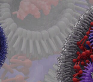
Cryo-TEM unlocks the potential of lipid nanoparticles for drug delivery
Lipid nanoparticles in drug development Lipid nanoparticles ...
Read More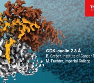
Glacios 2 Cryo-TEM and the Evolution of 200 kV Cryo-EM
Glacios 2 Cryo-TEM and the Evolution of 200 kV Cryo-EM Cryo-...
Read More
Unraveling Brain Evolution Through the Study of Teleost Fish Intelligence
Higher order brain function in different animals Once, highe...
Read More
Leave a Reply