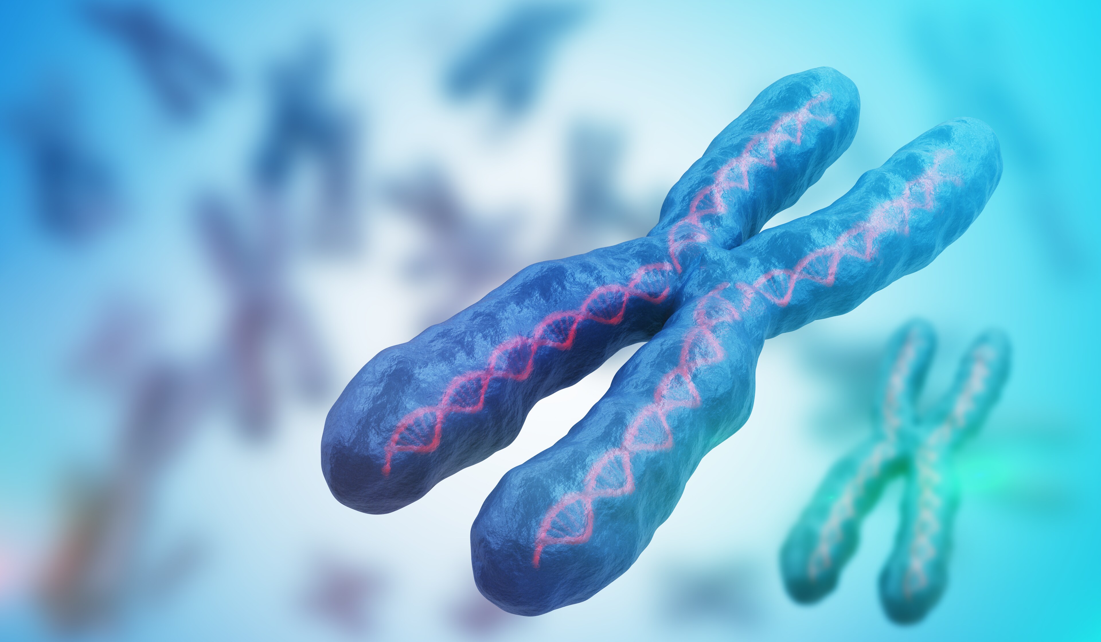
 Chromosomal abnormalities are at the heart of numerous cancers, effecting enormous changes in genetic activity. These alterations come in several varieties, each of which poses a detection challenge. Chromosomal abnormalities are different enough from smaller-scale deviations from a healthy state that ordinary genetics tools are often not able to accurately identify them, so properly understanding them requires a continually improving array of next-generation tools. One form of chromosomal abnormality that is attracting increasing attention is chromothripsis, and Thermo Fisher Scientific tools are up to the task.
For research use only. Not for use in diagnostic procedures.
Chromothripsis, from the Greek thripsis or “shattering,” refers to a process in which a single catastrophic event shatters a chromosomal region into numerous pieces. Repair mechanisms then try to reassemble the chromosome, but the extent of the damage means that hundreds of distinct mutations are incorporated into the “repaired” chromosome at once. These mutations span the whole range of duplications, deletions, inversions and more, and the mix of copy number variations (CNVs) and other changes makes them both uniquely dangerous and particularly difficult to fully explore with most genomics tools. PJ Stephens and his team first discovered chromothripsis in 2011,1 reporting that its apparent signatures can be found in up to 3% of all cancers and up to 25% of bone cancers. Since Stephens’s discovery, thousands of scientific publications about chromothripsis have documented, defined, and explored this new frontier in cancer research.2 Chromothripsis has been discovered in neuroblastoma,3 medulloblastoma,4,5 multiple myeloma,6 and lung, renal and thyroid cancers,7 among others, indicating that this phenomenon is widespread.
Finding chromothripsis everywhere is not the same as understanding it. When repair mechanisms fail, not all of the results are cancerous. Many cancers accumulate “passenger” mutations that do not contribute to their malignancy, but which are still useful for detecting and characterizing them. Chromothripsis could induce malignancy in healthy cells, occur within tumors as other repair mechanisms fail and reproduction becomes more chaotic, or both, likely differing between tumors. It could even be a tool to destabilize cancer growth, damaging cells to the point of apoptosis. Whether and when chromothripsis induces, tags along with or fights cancer is being explored in ongoing research, which requires the most up-to-date tools. Conventional karyotyping does not have the resolution necessary to completely characterize such complex events, though it can detect some of them. Next-generation sequencing (NGS) can generate complete genome reads at high levels of resolution, but is expensive and often challenging. Fluorescence in situ hybridization (FISH) is useful for recognizing particular kinds of chromosome damage, but many versions of FISH must be combined with one another to study chromothripsis, dramatically increasing the complexity of observations. Chromosomal microarrays are ideal as they provide a compromise between the expansive reads of NGS and its cost, but can be limited in their coverage. Chromothripsis combines additions, deletions, inversions, single-nucleotide polymorphisms (SNPs) and CNVs, which different types of assays can detect with different efficacies.
The solution is research using high-resolution, whole-genome chromosomal microarrays that can accurately probe both SNPs and CNVs, offering a complete picture of the kinds of changes that can accumulate in a chromothripsis event. The Applied Biosystems™ OncoScan™ CNV and CNV Plus Assays are whole-genome arrays that can efficiently detect chromothripsis as well as breakpoint determination, ploidy level, genomic low-level mosaicism, and other copy number gains and losses in solid tumors. The Applied Biosystems™ CytoScan™ HD Suite, in turn, offers similar possibilities for hematological malignancies.
For more information about the OncoScan CNV Assay, OncoScan CNV Plus Assay, and CytoScan HD Suite, follow these links.
For research use only. Not for use in diagnostic procedures.
- Stephens, PJ., et al. (2011) “Massive genomic rearrangement acquired in a single catastrophic event during cancer development,” Cell, 144, pp. 27–40.
- Rode, A., et al. (2016) “Chromothripsis in cancer cells: An update,” Int J Cancer, 138, pp. 2322–2333.
- Molenaar, J.J., et al. (2012) “Sequencing of neuroblastoma identifies chromothripsis and defects in neuritogenesis genes,” Nature, 483, pp. 589–593.
- Northcott, P.A., et al. (2012) “The clinical implications of medulloblastoma subgroups,” Nat Rev Neurol, 8, pp. 340–351.
- Rausch, T., et al. (2012) “Genome sequencing of pediatric medulloblastoma links catastrophic DNA rearrangements with TP53 mutations,” Cell, 148, pp. 59–71.
- Magrangeas, F., et al. (2011) “Chromothripsis identifies a rare and aggressive entity among newly diagnosed multiple myeloma patients,” Blood, 118, pp. 675–678.
- Forment, J.V., et al. (2012) “Chromothripsis and cancer: Causes and consequences of chromosome shattering,” Nat Rev Cancer, 12, pp. 663–670.
Leave a Reply