Need for Standardization
Applications of flow cytometry in multi-parametric analysis of single cells or cell populations are widely known. But standardization of flow cytometric assays, not as much. It is extremely important for comparison of data across studies, instruments, and locations. There are many variables in flow cytometry such as instrument setup, reagents, controls, sample handling, data analysis, etc. It’s like making a great burger (only flow cytometric assay standardization is numerous times more difficult) is nice, but for universal acceptance, standardization is necessary in the cooking process, i.e. quality of ingredients, etc., across all locations.
Why is it Difficult to Standardize Assays?
Standardization is critical for comparing data among samples within an experiment or across experiments, to ascertain the differences observed are biological and not due to variables. Standardization is also important for comparing data between different instruments, scientists, laboratories, and sites. It is relatively less difficult to standardize steps for flow cytometry analysis in a single laboratory (yes, “relatively” as scientists are aware of the challenges with standardization processes). It is much more challenging and daunting to implement standardization across sites and laboratories. Process of standardization is time consuming and takes technical expertise in error management and variability. Bead standards are useful for instruments controls, as standards in studies and compensation. Consistent data from instruments with minimal variation for alignment, size calibration and sort set-up; sample conservation; and comparable data between experiments and instruments are some of the many advantages offered by bead standards.
Bead Standards to The Rescue!
We now know the challenges involved with standardization of flow cytometry assays, but where to start with standardization? Well, the instrument is the first place to look. Standardize optical and fluidics systems, to ensure that cells pass the lasers at a specific interrogation point. At this point, light scatter and fluorescent light are generated, ensuring no loss of sensitivity or errors in measurements.
The InvitrogenTM AccuCheckTM ERF Reference Particles are used for standardization and calibration of an intensity unit of fluorescence for flow cytometry analysis. For a given fluorophore, determine instrument sensitivity and accuracy through the application of the assigned NIST-traceable intensity standard. This permits data comparison between flow cytometry instruments. In combination with a biological standard, you can use these particles to quantify the level of biomarker expression. This solution offers confidence, reliability, and compatibility especially where comparison of results over time is important; because ERF values of AccuCheckTM ERF Reference Particles are NIST-traceable and comparable between manufacturers unlike MESF beads available through other manufacturers.
Now going deep into the assays, fluorochrome-conjugated antibodies are important components of flow cytometry studies; their performance is an area of focus in the context of large, multi-parameter panels, in terms of standardization. Antibodies tend to underperform in large panels due to compensation, unmixing in the detectors or unexpected interactions between dyes, cells, or buffers. Using appropriate controls can be helpful, such as The InvitrogenTM Flow Cytometry Compensation Beads. These beads help set voltages and gating parameters for obtaining accurate fluorescence signals. Compensation beads are particularly effective in cases where multiple fluorophore emissions overlap, limited sample is available (which is often the case with immunology studies), large multicolor immunophenotyping panels, or large experiments.
Count the Benefits of Counting Beads
Flow Cytometry offers a rapid method to quantify cell characteristics, but most flow cytometers cannot provide cell concentration or absolute cell counts in a sample. There are two methods to obtaining an absolute cell count. One method is to use a hematology analyzer by combining separate cell concentration numbers. Another method involves adding an internal microsphere counting standard to the samples, also known as single platform testing. The single platform testing method is preferential due to lack of complications and it avoids interlaboratory variability and underestimations. Various Invitrogen Flow Cytometry Cell Counting Beads are available for cell as well as bacterial counting.
Standardization in flow cytometry assays is a complex process with multiple variables; however, a suite of bead standards can make your studies more manageable while providing high quality data.
Learn more about the product in this blog: InvitrogenTM AccuCheckTM ERF Reference Particles, InvitrogenTM Flow Cytometry Compensation Beads, Invitrogen Flow Cytometry Cell Counting Beads

Flow Cytometry Resources and Solutions
- Flow Cytometry Panel Builder – this online tool helps you design an effective reagent panel for your specific application on your conventional or spectral flow cytometer
- Flow Cytometry Protocols Handbook – protocols that fit your needs in flow cytometry ranging from sample preparation to numerous cell stimulation conditions, staining, immunophenotyping and data analysis strategies
- Immunology at Work Resource Center – practical guidance and protocols to help you get started quickly and confidently
Related Blogs
- https://www.thermofisher.cn/blog/behindthebench/clog-resistance-of-non-pressure-based-flow-cytometers/
- https://www.thermofisher.cn/blog/behindthebench/is-it-a-t-cell-or-b-cell-antibodies-for-immunophenotyping/

How to Achieve More Sustainable mRNA Synthesis and Purification
mRNA therapeutics have delivered on the promise of simple an...
Read More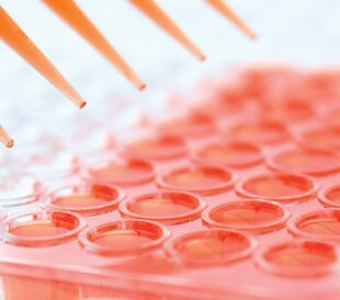
Narrow down your ELISA reagent choices
Faster Decisions ELISA Design Considerations The enzyme link...
Read More
Recipe for Success
Why are Protocols Hard to Find? Immunology research is advan...
Read More
Two Applications One Instrument
What are the Benefits of Performing Proteomics and Genomics ...
Read More
Leave a Reply