Isoaspartyl formation is a nonenzymatic reaction in which the carboxylic acid of an aspartate or a deamidated asparagine residue undergoes a nucleophilic attack on the amino group on the polypeptide backbone. The resulting structure is an unstable cyclical succinimide intermediate. This intermediate can resolve by hydration to either the original aspartyl residue or into an isoaspartyl residue.1 An isoaspartyl residue is a unique branching of the polypeptide backbone where the carbonyl of the original aspartate residue acts as the carbonyl, adding an additional methylene group and causing a kink in the backbone of the protein. Accumulation of isoaspartate residues results in decreases in enzymatic activity and potentially increased proteolysis. Because this is a nonenzymatic process, this damage can accumulate even in a purified protein, such as in a protein pharmaceutical during transport or storage.2
Cells have adapted a way to potentially repair these isomerization events through the methylation of the carboxylic acid of the isoaspartyl residue. The addition of the methyl group from S-adenosylmethionine to the isoaspartate residue results in recyclization back to the unstable succinimide intermediate and potentially back into the native aspartate.1,3 The methylation is carried out by protein isoaspartyl methyltransferase (PIMT) and is nearly universally conserved in all organisms. PIMT is not an essential protein, but deletion of PIMT from cells is known to affect protein quality control and growth rates.3 Conversely, overexpression of PIMT improves growth for at least some organisms.4
Although much is known about the formation of these aberrant residues and the biological mechanism for the repair of this damage, the fate of the proteins that can no longer be repaired is not currently well understood. The prevailing theories involve proteolysis and/or excretion of the proteins out of the cell.3 In Dai et al.,1 a systematic analysis of the isoaspartyl-containing proteins found in mouse urine from PIMT-deficient mice was performed. Compared with wild-type mice, PIMT-deficient mice have elevated levels of isoaspartyl proteins in their urine. Subsequent proteomic analysis revealed the modified proteins to be major urine-associated proteins, or MUPs. Using an LTQ Orbitrap XL mass spectrometer (Thermo Scientific) with an electron transfer dissociation (ETD) ion source, 38 isoaspartyl modification sites were identified in the protein set by selected reaction monitoring (SRM) for specific reporter ions from isoaspartyl residues. Five of those sites were asparagine residues that had undergone deamidation, and the rest were native aspartate residues.1
The significance of finding so many isoaspartyl modification sites in major urine-associated proteins is twofold. First, the accumulation of protein fragments with isoaspartyl residues provides evidence that some of these proteins may be degraded. Also, the high levels of these modified proteins in urine demonstrate that excretion of these proteins is a pathway for their removal from a cell or organism.1 In addition to finding a large number of damaged proteins, the use of ETD-SRM-targeted proteomics to locate the sites is a significant breakthrough for the study of this modification. The odd nature of the isomerization makes identification of this modification difficult for classic mass spectrometry search engines. The use of SRM to identify reporter ions of the kinked branching caused by isoaspartyl residues allows for quick identification from a complex mixture of proteins.
Proteomic mapping of isoaspartyl sites in urine proteins may provide information about this modification and provide additional evidence for the excretion process by which these proteins may be removed from cells. ETD-SRM mass spectrometry provides a potentially useful tool for the identification and quantification of isoaspartyl damage for proteins from a complex mixture. This advance may aid in the evaluation of protein pharmaceuticals that can accumulate this sort of damage in the process of normal handling. While the fate of these damaged proteins remains unclear from an in vivo point of view, the ability to selectively target damaged peptides from proteins within a complex mixture is a significant step forward in understanding this protein modification pathway and may have implications in pharmaceutical quality control.
REFERENCES
1. Dai, S., et al. (2013) “Integrated proteomic analysis of major isoaspartyl-containing proteins in the urine of wild type and protein l-isoaspartate O-methyltransferase-deficient mice,” Analytical Chemistry, 85(4) (pp. 2423–30).
2. Doyle, H.A., Gee, R.J., and Mamula, M.J. (2007) “Altered immunogenicity of isoaspartate containing proteins,” Autoimmunity, 40(2) (pp. 131–7).
3. Shimizu, T., Matsuoka, Y., and Shirasawa, T. (2005) “Biological significance of isoaspartate and its repair system,” Biological and Pharmaceutical Bulletin, 28(9) (pp. 1590–96).
4. Kindrachuk, J., et al. (2003) “Overexpression of L-isoaspartate O-methyltransferase in Escherichia coli increases heat shock survival by a mechanism independent of methyltransferase activity,” Journal of Biological Chemistry, 278(51) (pp. 50880–86).
Analysis of Ketosteroids by Girard P Derivatization
Dehydroepiandrosterone (DHEA); 4-androstene-3,17-dione (AD);...
Read More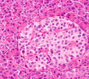
Profiling Human Islet Cells with Laser Capture Microdissection
Composed mainly of β-cells, human islet cells secrete h...
Read More
Intact Proteins in Native Conditions with Quadrupole-Orbitrap Mass Spectrometry
Successful research focusing on antibody and drug interactio...
Read More
Autophagy Helps Control Endothelial Permeability
A research team in Glasgow has identified autophagy as a str...
Read More
Leave a Reply