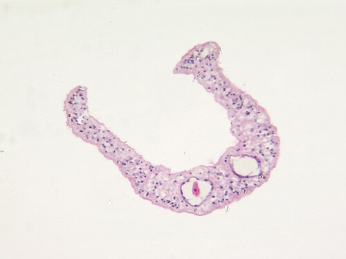
 The current tools for diagnosing and monitoring the human helminth infection schistosomiasis include microscopy to detect Schistosoma mansoni eggs in stool, assay for circulating antigens or antibodies, ultrasound for liver assessment and polymerase chain reaction using fecal samples. Unfortunately, these options bring challenges related to expense, complexity, sensitivity issues and cross-reactivity with other helminth infections. The majority also can’t discriminate between past infection and active infection.
Mass spectrometry (MS) is a promising option for modernizing schistosomiasis diagnosis and monitoring. Toward this end, Kardoush, Ward and Ndao (2016) recently searched for specific biomarkers and proteome profiles capable of identifying schistosomal infection and differentiating between pathological stages of the disease (early: before egg production, acute: with egg deposition in liver, chronic: with granulomatous reaction in liver).1 To do this, they performed sample fractionation and differential gel electrophoresis on mouse serum samples before MS analysis using multiple platforms, including an LTQ Orbitrap Velos mass spectrometer (Thermo Scientific).
First, the research team applied surface-enhanced laser desorption/ionization time-of-flight (SELDI-TOF) MS for proof of principle, identifying 66 candidate biomarkers. Then, they used matrix-assisted laser desorption/ionization time-of-flight (MALDI-TOF) MS on sodium dodecyl sulfate polyacrylamide gel electrophoresis (SDS-PAGE) slices containing pooled sera collected from groups of mice at three time points corresponding with the three disease stages (three, six and 12 weeks post-infection). They identified eight potential biomarkers, including two candidates they highlighted as particularly convincing: serotransferrin and alpha-1 antitrypsin.
Turning to the Orbitrap mass spectrometer, the researchers analyzed mouse sera from both acute and chronic infection, identifying 200 and 105 proteins, respectively, that met their criteria. They applied filtering software and whittled this number to 28 candidate biomarkers from both stages. Notable candidates included actin (the most frequently identified protein), ryanodine and protocatechuate dioxygenase. They note that because of low parasite antigen concentrations in infected serum, many of the identifications relied on single peptides, which is not ideal. However, they used Western blotting to confirm some of these single peptide MS identifications, thereby increasing confidence in the results.
Of course, another means of validating results is instrument overlap. All eight potential biomarkers identified by MALDI-TOF also appeared in the more comprehensive list of upregulated proteins detected by orbitrap. All but one also populated in the corresponding SELDI peaks. Of the 66 peaks detected by SELDI, approximately 70% (46 peaks) appeared in the database generated from Orbitrap mass spectrometer data.
Table 1. Overlapping candidate biomarkers
| Hemoglobin subunit beta-1 | Apolipoprotein A-IV |
| Apolipoprotein A-I | Alpha-1 antitrypsin |
| Serotransferrin | Beta-2-globin |
| Albumin 1 | Hemopexin (not detected by SELDI) |
Kardoush et al. highlight the remarkably high number of schistosome protein identifications produced by the Orbitrap instrument, likely due to their inclusion of later-time-point serum samples as well as platform sensitivity. They indicate that even the low-abundance proteins could provide insight into schistosomiasis biology. They call for further studies to validate the candidate biomarkers they identified here, including human serum studies, with the goal of producing a rapid, accurate, inexpensive diagnostic field test.
Reference
1. Kardoush, M.I., Ward, B.J., and Ndao, M. (2016) “Identification of candidate serum biomarkers for Schistosoma mansoni infected mice using multiple proteomic platforms,” PLOS One, 11(5) (e0154465), doi: 10.1371/journal.pone.0154465.
Leave a Reply