Search

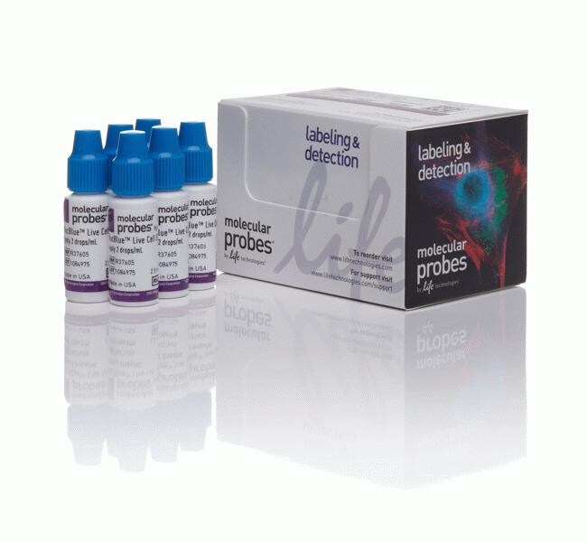
Reactivo NucBlue™ Live ReadyProbes™ (Hoechst 33342)
| Número de catálogo | Cantidad |
|---|---|
| R37605 | 6 vial(es) |
Hoechst 33342 es una contratinción nuclear popular que penetra en la célula y emite una fluorescencia azul cuando se une al ADN. Con el reactivo de células vivas NucBlue™ ReadyProbes™, hemos formulado esta tinción clásica para convertirla en una solución estable a temperatura ambiente que se suministra en un cómodo frasco cuentagotas. Para teñir las células, solo tiene que añadir dos gotas por ml.
• No es necesario diluir, pesar ni pipetear
• Frasco cuentagotas cómodo: usar solo dos gotas por ml
• Estable a temperatura ambiente: mantener a mano en su zona de trabajo o área de cultivo celular
• Se excita con luz UV y emite fluorescencia azul a 460 nm cuando se une al ADN
Ver otros reactivos ReadyProbes™ para la tinción celular
Consulte otras tinciones nucleares para adquisición de imágenes
Aplicaciones de adquisición de imágenes celulares
as propiedades espectrales de Hoechst 33342 (2'-[4'-etoxifenil]-5-[4-metilpiperazin-1-il]-2,5'-bis-1h-benzimidazol), incluido un gran desplazamiento de stokes, hacen que sea perfecto para el uso con fluorósforos verdes (Alexa Fluor™ 488, fluoresceína [FITC], proteína verde fluorescente [GFP]) y rojos (Alexa Fluor™ 594, Texas Red™, rodamina, mCherry, mKate-2) en experimentos multicolor. Debido a su alta afinidad con el ADN, Hoechst 33342 también se utiliza con frecuencia en estudios de recuento, ciclo y replicación celular para distinguir núcleos condensados en células apoptóticas, para los estudios de ciclos celulares en combinación con la tinción de BrdU o EdU Click-iT™, como herramienta de segmentación nuclear en el análisis de imágenes de alto contenido y para clasificar las células en función de su contenido de ADN.
Sugerencias de uso
La tinción de células vivas NucBlue™ se puede añadir directamente a las células en medios completos o soluciones de tampón.
• En la mayoría de los casos, 2 gotas/ml y una incubación de 15 a 30 minutos producirán una tinción nuclear brillante. Sin embargo, puede ser necesario realizar ajustes para algunos tipos de células, condiciones y aplicaciones. En estos casos, basta con añadir más o menos gotas hasta que se obtiene la intensidad de tinción óptima. En la mayoría de los casos, la intensidad de tinción aumenta con el tiempo si las células no se lavan antes de la adquisición de imágenes.
• La tinción de células vivas NucBlue™ se excita con luz UV a 360 nm cuando se une al ADN, con un nivel máximo de emisión a 460 nm. Se detecta mediante un filtro azul/cián, como un filtro DAPI, filtros GFP azules o el conjunto de filtros del colorante Semrock BrightLine™ Alexa Fluor™ 350.
Almacenar a ≤ 25 °C