Search


Zitierungen und Referenzen (13)
Invitrogen™
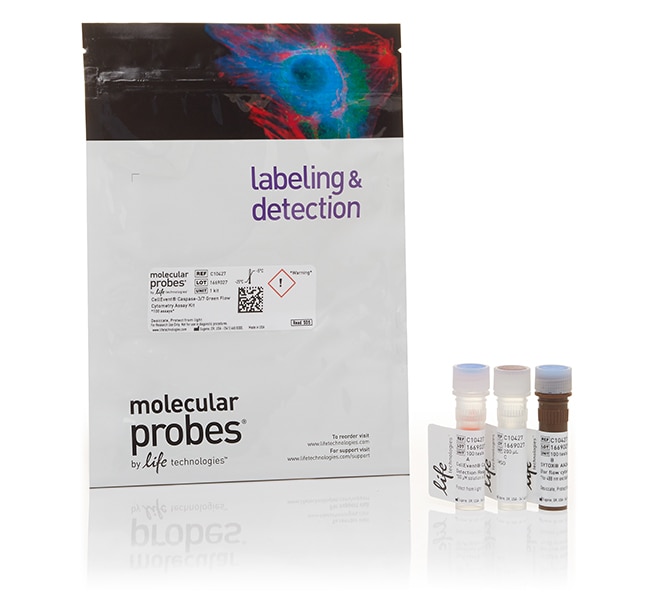
CellEvent™ Caspase-3/7 Green Flow Cytometry Assay Kit
Das CellEvent™ Caspase-3/7 Durchflusszytometrie-Assay-Kit Grün ermöglicht die durchflusszytometrische Detektion aktivierter Caspase-3 und Caspase-7 in apoptotischen Zellen. Das Kit enthält dasWeitere Informationen
Have Questions?
Ansicht ändern

| Katalognummer | Menge |
|---|---|
| C10427 | 100 Assays |
| C10740 | 20 Assays |
Katalognummer C10427
Preis (EUR)
528,00
Each
Menge:
100 Assays
Preis (EUR)
528,00
Each
Das CellEvent™ Caspase-3/7 Durchflusszytometrie-Assay-Kit Grün ermöglicht die durchflusszytometrische Detektion aktivierter Caspase-3 und Caspase-7 in apoptotischen Zellen. Das Kit enthält das neuartige fluorogene Substrat CellEvent™ Caspase-3/7 Nachweisreagenz Grün sowie den SYTOX™ AADvanced™ Totzellfarbstoff.
• Caspase-3/7-spezifisch—Enthält die Erkennungssequenz für aktivierte Caspase-3 und Caspase-7
• Einfache Identifizierung—Klare Identifizierung von lebenden, toten und apoptotischen Zellpopulationen
• Schnelle Analyse—Kein Waschen und Fixieren erforderlich
•Mehrfarben-Kompatibilität—Kombinierbar mit anderen Farbstoffen, die durch den 488 nm-Laser oder andere Laser anregbar sind
CellEvent™ Caspase-3/7 Nachweisreagenz Grün ist ein zellpermeantes Reagenz, das aus einem Vier-Aminosäure-Peptid (DEVD) besteht, welches mit einem Nukleinsäure-bindenden Farbstoff konjugiert ist. Bei der Apoptose werden die Proteine Caspase-3 und Caspase-7 aktiviert und können die im DEVD-Peptid kodierte Erkennungssequenz für Caspase 3/7 spalten. Die Spaltung der Erkennungssequenz und die Bindung der DNA durch das Reagenz markiert die apoptotischen Zellen mit einem hellen, fluorogenen Signal, das ein Absorptions-/Emissionsmaximum von ca. 511/533 nm besitzt. In Verbindung mit dem SYTOX™ AADvanced™ Totzellfarbstoff können apoptotische Zellen leicht von lebenden und nekrotischen Zellen differenziert werden.
Da kein einzelner Parameter die Apoptose in allen Systemen definiert, empfehlen wir dringend, für den zuverlässigen Nachweis der Apoptose eine Kombination verschiedener Messungen zu verwenden.
Nur für Forschungszwecke. Nicht zur Verwendung bei diagnostischen Verfahren.
Specifications
Anregung/EmissionSYTOX AADvanced: 546/647
CellEvent Caspase 3/7 Nachweisreagenz Grün: 511/533
CellEvent Caspase 3/7 Nachweisreagenz Grün: 511/533
Durchflusszytometer-Laserlinien488
Zur Verwendung mit (Anwendung)Durchflusszytometrie
Zur Verwendung mit (Geräte)Attune™ Akustisches Fokussierzytometer, Durchflusszytometer
MarkertypAndere Labels oder Farbstoffe
Marker oder FarbstoffCellEvent™ Caspase 3/7 Nachweisreagenz Grün, SYTOX AADvanced Totzellfarbstoff
ProduktlinieCellEvent
ProdukttypCaspase-Assay-Kit
Menge100 Assays
VersandbedingungNasseis
LöslichkeitDMSO (Dimethylsulfoxid)
NachweisverfahrenFluoreszenz
FormatKit
Unit SizeEach
Inhalt und Lagerung
Enthält 1 Fläschchen CellEvent™ Caspase 3/7 Nachweisreagenz Grün, 1 Fläschchen SYTOX AADvanced Totzellfarbstoff und 1 Fläschchen DMSO.
Alle Fläschchen aufrecht bei oder unter -20 °C lagern, mit Trockenmittel und lichtgeschützt.
Alle Fläschchen aufrecht bei oder unter -20 °C lagern, mit Trockenmittel und lichtgeschützt.
Häufig gestellte Fragen (FAQ)
I want to study apoptosis using an Annexin V conjugate, but with adherent cells via microscopy instead of flow cytometry. Can this be done?
Can I use CellEvent Caspase-3/7 Green Detection Reagent with fixed samples?
With the CellEvent Caspase-3/7 Green Detection Reagent, may I stain cells with nucleic acid stains?
With the CellEvent Caspase-3/7 Green Detection Reagent, may I multiplex with fluorescent immunolabeling?
Is the CellEvent Caspase-3/7 Green Detection Reagent fixable?
Zitierungen und Referenzen (13)
Zitierungen und Referenzen
Abstract
Counteracting autophagy overcomes resistance to everolimus in mantle cell lymphoma.
Journal:Clin Cancer Res
PubMed ID:22879389
'Mantle cell lymphoma (MCL) is an aggressive B-lymphoid neoplasm with poor response to conventional chemotherapy and short survival. The phosphatidylinositol 3-kinase/Akt/mTOR survival pathway is constitutively activated in MCL cells, thereby making the mTOR inhibition an attractive therapeutic strategy. The first clinical studies of everolimus (RAD001), an mTOR inhibitor, in relapsed
The cytotoxicity of bupivacaine, ropivacaine, and mepivacaine on human chondrocytes and cartilage.
Journal:
PubMed ID:23749443
'Intraarticular injections of local anesthetics are frequently used as part of multimodal pain regimens. However, recent data suggest that local anesthetics affect chondrocyte viability. In this study, we assessed the chondrotoxic effects of mepivacaine, ropivacaine, and bupivacaine. We hypothesized that specific cytotoxic potencies directly correlate with analgesic potencies, and that
Proteomic analysis reveals that pardaxin triggers apoptotic signaling pathways in human cervical carcinoma HeLa cells: cross talk among the UPR, c-Jun and ROS.
Journal:Carcinogenesis
PubMed ID:23615400
Pardaxin, an antimicrobial peptide secreted by the Red Sea flatfish Pardachirus marmoratus, inhibits proliferation and induces apoptosis of human cancer cell lines. However, the underlying molecular mechanisms are only partially understood at present. In this study, we used proteomic approaches and network reconstruction to clarify the mechanism of pardaxin-induced apoptosis
Salinomycin inhibits breast cancer progression via targeting HIF-1a/VEGF mediated tumor angiogenesis in vitro and in vivo.
Journal:Biochem Pharmacol
PubMed ID:31028743
'Cancer is a complex disease wherein cells begin to divideabnormally and spread into surrounding tissues. Angiogenesis plays a crucial role in tumor progression as it is required for sustained growth and metastasis, therefore targeting angiogenesis is a promising therapeutic approach for breast cancer management. Salinomycin (SAL) has been reported to
Gel-based cell manipulation method for isolation and genotyping of single-adherent cells.
Journal:Analyst
PubMed ID:30302469
'Genetic analysis of single-cells is widely recognized as a powerful tool for understanding cellular heterogeneity and obtaining genetic information from rare populations. Recently, many kinds of single-cell isolation systems have been developed to facilitate single-cell genetic analysis. However, these systems mainly target non-adherent cells or cells in a cell suspension.