Search


Zitierungen und Referenzen (10)
Invitrogen™
Click-iT™ Plus TUNEL-Assay-Kits für In Situ Apoptose-Detektion
Nachweis von Apoptose in Zellen und Gewebeproben mit Click-iT Plus TUNEL Assay-Kits, die eine einfache Farbstoffeinarbeitung und ein Multiplexing mit GFP und RFP ermöglichen.
Have Questions?
Ansicht ändern

| Katalognummer | Farbe | Marker oder Farbstoff |
|---|---|---|
| C10618 | Rot | Alexa Fluor™ 594 |
| C10617 | Grün | Alexa Fluor™ 488 |
| C10619 | Tiefrot | Alexa Fluor™ 647 |
Katalognummer C10618
Preis (EUR)
711,65
キャンペーン価格
782,00Ersparnis 70,35 (9%)
Each
Farbe:
Rot
Marker oder Farbstoff:
Alexa Fluor™ 594
Preis (EUR)
711,65
キャンペーン価格
782,00Ersparnis 70,35 (9%)
Each
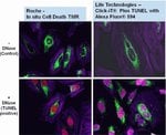
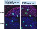
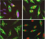
Mit dem Click-iT Plus TUNEL-Assay für den In Situ-Apoptosenachweis, der Alexa Fluor 488, 594 und 647 fluoreszierende Farbstoffoptionen bietet, können Sie mehr apoptotische Zellen in Gewebe- und Zellkulturproben nachweisen. Dieses In-situ-Apoptose-Nachweiskit ist für Gewebe- oder Zellproben optimiert und ermöglicht das Multiplexing der Farbstoffe mit anderen Farbstoffen oder Proteinen, wie GFP und RFP, und lassen sich aufgrund ihrer kleineren Größe (im Vergleich zu Antikörpern) leichter in komplexe Moleküle integrieren. Dieses TUNEL-Assay-Kit ist ebenfalls sehr flexibel und zum Testen von 1 bis 50 Proben in einem einzigen Experiment verwendbar.
Click-iT Plus TUNEL Alexa Fluor 488, 594 und 647 Assays für den In-situ-Apoptosenachweis können apoptotische Zellen in Gewebe- und Zellkulturproben nachweisen, indem sie eine kleine, hochgradig spezifische Markierung und einen hellen Fluoreszenzfarbstoff einarbeiten. Nach Einarbeitung des Markierungsanteils in DNA-Fragmente erfolgt der Nachweis über eine katalysierte „Klick“-Reaktion unter Bedingungen, die das emittierte Fluoreszenzsignal von GFP oder RFP erhalten.
Weitere Vorteile des Click-iT Plus TUNEL-Assays für den In-Situ-Apoptosenachweis:
• Optimiert für den Nachweis apoptotischer Zellen in Gewebe- oder Zellproben
• Multiplex-fähig: Optimiert für die Arbeit mit fluoreszierenden Farbstoffen oder Proteinen wie GFP und RFP
• Verbesserter TUNEL-Assay: Besserer Einbau der Markierung dank eines kleinen reaktiven Anteils
• Helle apoptotische Signale: nutzt Alexa Fluor Farbstoffe, die ein stabiles Fluoreszenzsignal ohne Photobleichung erzeugen
• Flexibilität: Assay ist für gleichzeitige Tests von 1 bis 50 Proben verwendbar
Fragmentierung der zellulären DNA ist ein Kennzeichen von Apoptose. Der TUNEL-Assay ist die am weitesten verbreitete Methode zum Nachweis fragmentierter DNA in apoptotischen Zell- oder Gewebeproben. Der TUNEL-Assay beginnt durch den Einbau modifizierter dUTP am 3'-OH Ende der fragmentierten DNA. Die dUTP-Modifikation besteht oftmals in der Kopplung eines Fluorophors. Aufgrund der Größe des Fluorophors kann das modifizierte dUTP niedrigere als erwartete Einarbeitungsraten aufweisen, was sich auf die Empfindlichkeit des TUNEL-Assays auswirken kann. Zudem weisen viele der in derzeit verfügbaren TUNEL-Assay-Kits enthaltenen Fluorophore Probleme mit Photobleichung und Fluoreszenzspektralüberlappungen auf, die beide die Empfindlichkeit und Multiplex-Möglichkeiten des Assays einschränken.
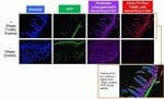
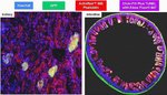
Der Click-iT Plus TUNEL-Assay soll diese Probleme umgehen. Der Assay verwendet dUTP, modifiziert mit einer Alkingruppe (eine kleine bioorthogonale funktionelle Gruppe), wodurch die Nukleotide leichter aufgenommen werden. Nach dem Einbau führt eine hochgradig spezifische „Klick“-Reaktion zwischen der Alkingruppe und einem Alexa Fluor Picolylazid-Fluoreszenzfarbstoff mit anschließendem Nachweis dieses Farbstoffs zu einem empfindlichen und spezifischen Assay für die Erkennung apoptotischer Zellen oder Gewebeproben. Aufgrund der schonenden Reaktionsbedingungen ermöglicht der Click-iT Plus TUNEL-Assay das Multiplexing mit fluoreszierenden Proteinen oder Farbstoffen.
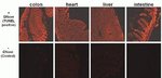
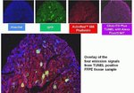
Der Click-iT Plus TUNEL-Assay wurde validiert mit mehreren verschiedenen Formalin-fixierten, Paraffin-eingebetteten Gewebetypen. In allen Fällen ist das Multiplexing mit fluoreszierenden Proteinen und Farbstoffen möglich. Auch die Aktinfärbung mit fluoreszenzmarkiertem Phalloidin ist möglich.
Der Click-iT Plus TUNEL-Assay enthält alle erforderlichen Reagenzien zum Nachweis apoptotischer Zellen in Gewebe- oder Zellproben. Die Reagenzien in diesem Kit reichen zum Testen von 50 Proben und können für die gleichzeitige Analyse von 1 bis 50 Proben verwendet werden.
Weitere Vorteile des Click-iT Plus TUNEL-Assays für den In-Situ-Apoptosenachweis:
• Optimiert für den Nachweis apoptotischer Zellen in Gewebe- oder Zellproben
• Multiplex-fähig: Optimiert für die Arbeit mit fluoreszierenden Farbstoffen oder Proteinen wie GFP und RFP
• Verbesserter TUNEL-Assay: Besserer Einbau der Markierung dank eines kleinen reaktiven Anteils
• Helle apoptotische Signale: nutzt Alexa Fluor Farbstoffe, die ein stabiles Fluoreszenzsignal ohne Photobleichung erzeugen
• Flexibilität: Assay ist für gleichzeitige Tests von 1 bis 50 Proben verwendbar
Fragmentierung der zellulären DNA ist ein Kennzeichen von Apoptose. Der TUNEL-Assay ist die am weitesten verbreitete Methode zum Nachweis fragmentierter DNA in apoptotischen Zell- oder Gewebeproben. Der TUNEL-Assay beginnt durch den Einbau modifizierter dUTP am 3'-OH Ende der fragmentierten DNA. Die dUTP-Modifikation besteht oftmals in der Kopplung eines Fluorophors. Aufgrund der Größe des Fluorophors kann das modifizierte dUTP niedrigere als erwartete Einarbeitungsraten aufweisen, was sich auf die Empfindlichkeit des TUNEL-Assays auswirken kann. Zudem weisen viele der in derzeit verfügbaren TUNEL-Assay-Kits enthaltenen Fluorophore Probleme mit Photobleichung und Fluoreszenzspektralüberlappungen auf, die beide die Empfindlichkeit und Multiplex-Möglichkeiten des Assays einschränken.
Der Click-iT Plus TUNEL-Assay soll diese Probleme umgehen. Der Assay verwendet dUTP, modifiziert mit einer Alkingruppe (eine kleine bioorthogonale funktionelle Gruppe), wodurch die Nukleotide leichter aufgenommen werden. Nach dem Einbau führt eine hochgradig spezifische „Klick“-Reaktion zwischen der Alkingruppe und einem Alexa Fluor Picolylazid-Fluoreszenzfarbstoff mit anschließendem Nachweis dieses Farbstoffs zu einem empfindlichen und spezifischen Assay für die Erkennung apoptotischer Zellen oder Gewebeproben. Aufgrund der schonenden Reaktionsbedingungen ermöglicht der Click-iT Plus TUNEL-Assay das Multiplexing mit fluoreszierenden Proteinen oder Farbstoffen.
Der Click-iT Plus TUNEL-Assay wurde validiert mit mehreren verschiedenen Formalin-fixierten, Paraffin-eingebetteten Gewebetypen. In allen Fällen ist das Multiplexing mit fluoreszierenden Proteinen und Farbstoffen möglich. Auch die Aktinfärbung mit fluoreszenzmarkiertem Phalloidin ist möglich.
Der Click-iT Plus TUNEL-Assay enthält alle erforderlichen Reagenzien zum Nachweis apoptotischer Zellen in Gewebe- oder Zellproben. Die Reagenzien in diesem Kit reichen zum Testen von 50 Proben und können für die gleichzeitige Analyse von 1 bis 50 Proben verwendet werden.
For Research Use Only. Not for use in diagnostic procedures.
Specifications
FarbeRot
BeschreibungClick-iT Plus TUNEL Assay für den In-Situ-Apoptosenachweis, Alexa Fluor™ 594 Farbstoff
Anregung/Emission590/617
Zur Verwendung mit (Geräte)Fluoreszenzmikroskop
MarkertypAlexa Fluor™ Farbstoffe
Marker oder FarbstoffAlexa Fluor™ 594
Anzahl Reaktionen50 Deckgläser
ProduktlinieClick-iT
ProdukttypTUNEL Assay
Menge1 kit
VersandbedingungTrockeneis
NachweisverfahrenFluoreszenz
FormatDeckglas
Unit SizeEach
Inhalt und Lagerung
Bei ≤ 20°C lagern und vor Licht schützen.
Häufig gestellte Fragen (FAQ)
I will be performing a cell proliferation assay using Click-iT EdU kit. At what point can I stop overnight, or do I have to perform all the steps continuously?
A control for a Click-iT EdU labeling experiment uses no EdU and the Click-iT reaction using Alexa Fluor 594 azide. The mouse heart tissue sections are showing non-specific labeling in red, seen in particular clusters of cells. They don't overlap with DAPI. What is the problem?
I need to test cells for apoptosis after they have been formaldehyde-fixed and permeabilized. What dye or conjugate do you recommend? Will Annexin V conjugates work?
Can I use Click-iT TUNEL Alexa Fluor Imaging Assays for Microscopy & HCS (Cat. No. C10246) for flow cytometry?
Can I use Click-iT Plus TUNEL Assay Kits for In Situ Apoptosis Detection (Cat. Nos. C10617, C10618, C10619) for whole mount immunofluorescence staining of zebrafish larvae?
Zitierungen und Referenzen (10)
Zitierungen und Referenzen
Abstract
CD13 deficiency leads to increased oxidative stress and larger atherosclerotic lesions.
Journal:Atherosclerosis
PubMed ID:31229835
'Atherosclerosis is an inflammatory cardiovascular disorder characterized by accumulation of lipid-loaded macrophages in the intima. Prolonged accumulation leads to apoptosis of macrophages and eventually to progression of lesion development. Prevention of macrophage accumulation within the intima has been shown to reduce lesion formation. Since CD13 mediates trafficking of macrophages to
Loss of host-derived osteopontin creates a glioblastoma-promoting microenvironment.
Journal:Neuro Oncol
PubMed ID:29016864
'Microglia and periphery-derived monocytes infiltrate human and mouse glioblastoma and their density is positively correlated with malignancy. Using microarray and RNA sequencing, we have previously shown that glioblastoma-associated microglia/monocytes (GAMs) express osteopontin/SPP1.'
Differential susceptibility of mouse strains on pancreatic injury and regeneration in cerulein-induced pancreatitis.
Journal:Int J Clin Exp Pathol
PubMed ID:31966883
'Acute pancreatitis (AP), a common disease, causes significant morbidity and mortality in clinical practice. Our objective of this study was to establish an experimental mouse AP model with cerulein treatment and to explore the susceptibility of mouse strains on the severity of pancreatic injury and the subsequent repair and regeneration.
Tanshinone IIA promotes IL2-mediated SW480 colorectal cancer cell apoptosis by triggering INF2-related mitochondrial fission and activating the Mst1-Hippo pathway.
Journal:Biomed Pharmacother
PubMed ID:30372868
IL-2-based therapy is a promising tool to treat colorectal cancer, but drug resistance always occurs in clinical practice. Mitochondrial fission is a novel target to modulate cancer development and progression. The aim of our study is to explore the effect of IL-2 combined with Tan IIA on SW480 colorectal cancer
Sirtuin-1 protects hair follicle stem cells from TNFa-mediated inflammatory stress via activating the MAPK-ERK-Mfn2 pathway.
Journal:Life Sci
PubMed ID:30292830
Stem cell transplantation is a promising tool to treat burn injuries. However, the inflammatory microenvironment in damaged skin limits the efficiency of stem cell-based therapy via poorly understood mechanisms. The aim of our study is to explore the contribution and mechanism of Sirtuin-1 (Sirt1) in TNFa-mediated inflammatory stress in hair