Search


Zitierungen und Referenzen (3)
Invitrogen™
SimplyBlue™ SafeStain
SimplyBlue SafeStain ist ein gebrauchsfertiger, schnell, empfindlicher und sicherer Coomassie G-250-Farbstoff zur Visualisierung von Proteinbanden auf Polyacrylamid-Gelen und von PVDF-Trockenmembranen.Weitere Informationen
Have Questions?
Ansicht ändern

| Katalognummer | Menge |
|---|---|
| LC6065 | 3,5 l |
| LC6060 | 1 l |
Katalognummer LC6065
Preis (EUR)
687,65
Online exclusive
732,00Ersparnis 44,35 (6%)
Each
Menge:
3,5 l
Preis (EUR)
687,65
Online exclusive
732,00Ersparnis 44,35 (6%)
Each
SimplyBlue SafeStain ist ein gebrauchsfertiger, schnell, empfindlicher und sicherer Coomassie G-250-Farbstoff zur Visualisierung von Proteinbanden auf Polyacrylamid-Gelen und von PVDF-Trockenmembranen. Dies ist vollkommen ungefährlich und erfordert keine Methanol- oder Essigsäure-Fixiermittel oder -Entfärbemittel. Es besteht kein Risiko einer Gefahrenexposition oder unangenehmer Gerüche und sorgt damit für eine gesündere Laborumgebung.
Vergleich aller Coomassie-Färbemittel ›
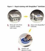
Einfaches Protokoll
Das SimplyBlue SafeStain Protokoll ist einfach durchzuführen und kann in weniger als drei Stunden abgeschlossen werden. Ein noch schnelleres Mikrowellenverfahren dauert lediglich 12 Minuten. Ein Entfärben ist nicht erforderlich, kann jedoch mit deionisiertem Wasser durchgeführt werden, um maximale Empfindlichkeit zu erreichen, insbesondere bei Downstream-Analysen wie der Massenspektrometrie oder wenn ein glasklarer Hintergrund erforderlich ist.
Vergleich aller Coomassie-Färbemittel ›
Einfaches Protokoll
Das SimplyBlue SafeStain Protokoll ist einfach durchzuführen und kann in weniger als drei Stunden abgeschlossen werden. Ein noch schnelleres Mikrowellenverfahren dauert lediglich 12 Minuten. Ein Entfärben ist nicht erforderlich, kann jedoch mit deionisiertem Wasser durchgeführt werden, um maximale Empfindlichkeit zu erreichen, insbesondere bei Downstream-Analysen wie der Massenspektrometrie oder wenn ein glasklarer Hintergrund erforderlich ist.
Nur für Forschungszwecke. Darf nicht für diagnostische Verfahren eingesetzt werden.
Specifications
DetektionsstelleIn-Gel-Detektion
NachweisverfahrenKolorimetrisch
Menge3,5 l
Haltbarkeit6 Monate
ZielmolekülProtein
FarbeBlue
Marker oder FarbstoffCoomassie
ProduktlinieSimplyBlue
ProdukttypSicheres Protein-Färbemittel
Unit SizeEach
Inhalt und Lagerung
SimplyBlue™ SafeStain wird als gebrauchsfertiges 1x-Färbereagenz geliefert. Bei Raumtemperatur lagern. Bei ordnungsgemäßer Lagerung garantiert 6 Monate lang stabil. Für dieses Produkt fallen keine Gefahrenstoff-Gebühren an.
Häufig gestellte Fragen (FAQ)
Why is Coomassie G-250 used as the tracking dye in NuPAGE LDS Sample Buffer instead of bromophenol blue?
Is the SimplyBlue SafeStain the appropriate stain for quantitation by densitometry?
The sensitivity of my SimplyBlue SafeStain seems to be decreasing over time. Why is this?
How do I destain proteins on a PVDF membrane that were stained with SimplyBlue SafeStain?
How can membranes be stained?
Zitierungen und Referenzen (3)
Zitierungen und Referenzen
Abstract
2F3 monoclonal antibody recognizes the O26 O-antigen moiety of the lipopolysaccharide of enterohemorrhagic Escherichia coli strain 4276.
Journal:Clin Diagn Lab Immunol
PubMed ID:15138178
'Enterohemorrhagic Escherichia coli (EHEC) and enteropathogenic E. coli (EPEC) organisms are groups of pathogenic strains whose infections are characterized by a typical lesion of enterocyte attachment and effacement. They are involved in enteric diseases both in humans and in animals, and EHEC strains can be responsible for hemolytic uremic syndrome
Selective fluorescent labeling of S-nitrosothiols (S-FLOS): a novel method for studying S-nitrosation.
Journal:Nitric Oxide
PubMed ID:18706513
'Protein S-nitrosation is a reversible post-translation modification critical for redox-sensitive cell signaling that is typically studied using the Biotin Switch method. This method and subsequent modifications usually require avidin binding or Western blot analysis to detect biotin labeled proteins. We describe here a modification of the Biotin Switch assay that
Vascular endothelial growth factor-C and C-C chemokine receptor 7 in tumor cell-lymphatic cross-talk promote invasive phenotype.
Journal:Cancer Res
PubMed ID:19118020
'Most carcinomas spread to distant sites through lymphatic vessels. Several preclinical and clinical studies have shown a positive correlation between the incidence of lymph node metastasis and secretion of the lymphatic growth factor vascular endothelial growth factor-C (VEGF-C) by tumor cells, suggesting tumor lymphangiogenesis as an escape mechanism. However, recent