Search


Zitierungen und Referenzen (4)
Invitrogen™
Totzell-Apoptose-Kits mit Annexin V für die Durchflusszytometrie
Beurteilen Sie die Zellviabilität mit Totzell-Apoptose-Kits mit Annexin V und konjugierten Fluoreszenzfarbstoffen wie Alexa Fluor 488, FITC, PI, PE, APC und SYTOX Green.
Have Questions?
Ansicht ändern

| Katalognummer | Menge | Marker oder Farbstoff |
|---|---|---|
| V35113 | 1 Kit | SYTOX Green, APC |
| V13241 | 50 Assays | Alexa Fluor 488, Propidiumiodid |
| V13242 | 1 Kit | FITC, Propidiumiodid |
| V35112 | 1 Kit | SYTOX Green, R-PE |
| V13245 | 250 Assays | Alexa Fluor 488, Propidiumiodid |
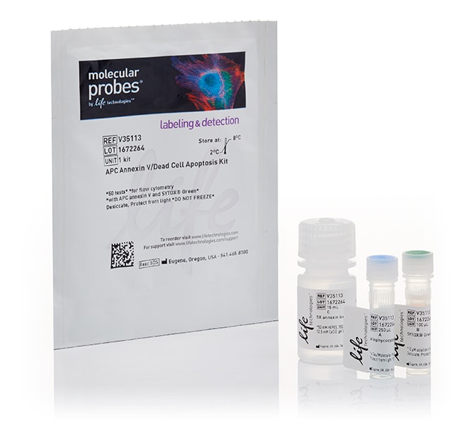
Katalognummer V35113
Preis (EUR)
806,00
1 kit
Menge:
1 Kit
Marker oder Farbstoff:
SYTOX Green, APC
Preis (EUR)
806,00
1 kit
Unterscheiden Sie ganz einfach zwischen lebenden, toten und apoptotischen Zellen während der Durchfluss-Zytometrie mit unseren Totzell-Apoptose-Kits mit Annexin V-Konjugaten, einschließlich Alexa Fluor 488, FITC, Propidium-Iodid, PE, APC und SYTOX Green. Diese Konjugate unterscheiden lebende, tote oder apoptotische Zellen durch verschiedene Färbemittel, was für die Bestätigung der Zellviabilität und Apoptose durch multiparametrische Studien unerlässlich ist.
Die Verwendung von Totzellen-Apoptose-Kits mit Annexin V für die Durchflusszytometrie bietet mehrere Vorteile bei Tests für Zellviabilität:
Hervorragende Helligkeit
Im Gegensatz zu anderen Annexin V-Kits mit niedrigeren Protein-Konzentrationen oder Reinheitsgraden ist das Alexa Fluor 488 Annexin V-Konjugat für die Durchflusszytometrie optimiert und bietet die stärkste Trennung zwischen apoptotischen und lebenden Zellen. Der Alexa Fluor 488 Farbstoff ist ein überragender, grün fluoreszierender Farbstoff mit einem Spektrum, das dem von Fluorescein (FITC) ähnelt.
Hohe Bindungseffizienz
Annexin V-Konjugate werden aus hochreinem Cys-Annexin hergestellt, was zu einer höheren Bindungseffizienz führt und so wiederum zu einer sehr genauen Charakterisierung des Apoptose-Vorgangs.
Multiparametrisch
Viele Publikationen verlangen mindestens zwei verschiedene Verfahren, um Zellen als apoptotisch zu identifizieren. Dieses Multiparameter-Kit erkennt Phosphatidylserin (PS) an der zytoplasmatischen Oberfläche der Zellmembran und die Membranintegrität mittels Propidiumiodid.
Totzell-Apoptose-Kit mit Annexin V Alexa Fluor 488 &PI
Das Totzell-Apoptose-Kit mit Annexin V Alexa Fluor 488 & Propidiumiodid (PI) (V13241, V13245) wird in der Durchfluss-Zytometrie zur Messung der frühen Apoptose verwendet, indem die Expression von Phosphatidylserin (PS) und die Membrandurchlässigkeit erkannt wird. Wenn Zellen mit Annexin V und Propidiumiodid angefärbt werden, fluoreszieren apoptotische Zellen mit PS-Expression grün (was im FITC-Kanal erkannt werden kann) und hellrot. Tote oder nekrotische Zellen fluoreszieren leuchtend rot und nicht grün, während lebende Zellen weder grün noch rot fluoreszieren.
Totzell-Apoptose-Kit mit Annexin V PE und SYTOX Green
Das Totzell-Apoptose-Kit mit Annexin V PE und SYTOX Green erkennt die PS-Externalisierung in apoptotischen Zellen während der Durchfluss-Zytometrie mittels rekombinantem Annexin V, das mit dem orangefarbenen fluoreszierenden Phycobiliprotein R-PE konjugiert ist, während abgestorbene Zellen mit dem Nukleinsäure-Farbstoff SYTOX Green nachgewiesen werden. Nach der Behandlung mit beiden Sonden fluoreszieren apoptotische Zellen orange, tote Zellen grün und lebende Zellen wenig oder gar nicht. Diese Populationen lassen sich mit einem 488 nm-Laser-Durchflusszytometer leicht in den 530/30 nm und 585/42 nm Bandpassfiltern unterscheiden.
Totzell-Apoptose-Kit mit Annexin V APC und SYTOX Green
Das Totzell-Apoptose-Kit mit Annexin V APC und SYTOX Green erkennt die PS-Externalisierung in apoptotischen Zellen während der Durchfluss-Zytometrie mittels rekombinantem Annexin V, das mit durch roten Laserstrahl angeregtes Allophycocyanin konjugiert ist, während abgestorbene Zellen mit dem Nukleinsäure-Farbstoff SYTOX Green behandelt werden. Apoptotische Zellen werden durch Annexin V-Bindung an externalisierte PS nachgewiesen. Lebende Zellen zeigen wenig oder keine Fluoreszenz, apoptotische Zellen zeigen rote Fluoreszenz und sehr wenig grüne Fluoreszenz, und späte apoptotische Zellen zeigen eine höhere Fluoreszenz von Rot und Orange. Diese Populationen lassen sich problemlos mit Hilfe eines Durchflusszytometers unterscheiden, das sowohl über eine Anregungsquelle von 488 nm als auch über eine Anregungsquelle von 633 nm verfügt (ein Argon-Ionen-Laser und ein HeNe-Laser).
Das Totzell-Apoptose-Kit mit Annexin V FITC und PI
Das Totzell-Apoptose-Kit mit Annexin V FITC und PI erkennt die PS-Externalisierung in apoptotischen Zellen mittels rekombinantem Annexin V, das mit mit grün-fluoreszierendem FITC-Färbemittel und toten Zellen mit PI konjugiert ist. Nekrotische Zellen, die sich mit PI färben, zeigen eine rote Fluoreszenz. Nach der Behandlung mit beiden Sonden fluoreszieren apoptotische Zellen grün, tote Zellen rot und grün sowie lebende Zellen wenig oder gar nicht.
For Research Use Only. Not for use in diagnostic procedures.
Specifications
BeschreibungTotzell-Apoptose-Kit mit Annexin V APC und SYTOX Green für die Durchflusszytometrie
Anregung/Emission503, 650/524, 660
Durchflusszytometer-Laserlinien633/635, 488
Zur Verwendung mit (Geräte)Fluoreszenzmikroskop, Durchflusszytometer
KitinhaltEnthält 1 Fläschchen Annexin V, APC-Konjugat (250 µl), 1 Fläschchen SYTOX Grünfärbemittel (100 µl) und 1 Flasche Annexin-Bindungspuffer (5x Lösung, 15 ml).
Marker oder FarbstoffSYTOX Green, APC
Anzahl Reaktionen50
ProdukttypApoptose-Nachweiskit
Menge1 Kit
VersandbedingungNasseis
Unit Size1 kit
Inhalt und Lagerung
Im Kühlschrank (2 bis 8 °C) aufbewahren und vor Licht schützen.
Häufig gestellte Fragen (FAQ)
How do SYTO dyes bind to DNA?
When using Dead Cell Apoptosis Kits with Annexin V for Flow Cytometry, what does a PI+ only population (hence Annexin V negative) correspond to?
I trypsinized my adherent cells and labeled with annexin V, and now my flow data is showing a high percentage of apoptotic cells even for control, untreated cells. What is the problem?
Can I detect annexin V staining in an imaging assay?
When should I stain adherent cells with annexin V for flow cytometric analysis? Before or after I trypsinize them?
Zitierungen und Referenzen (4)
Zitierungen und Referenzen
Abstract
Preclinical Evaluation of AMG 925, a FLT3/CDK4 Dual Kinase Inhibitor for Treating Acute Myeloid Leukemia.
Journal:
PubMed ID:24526162
'Acute myeloid leukemia (AML) remains a serious unmet medical need. Despite high remission rates with chemotherapy standard-of-care treatment, the disease eventually relapses in a major proportion of patients. Activating Fms-like tyrosine kinase 3 (FLT3) mutations are found in approximately 30% of patients with AML. Targeting FLT3 receptor tyrosine kinase has
Temozolomide-mediated DNA methylation in human myeloid precursor cells: differential involvement of intrinsic and extrinsic apoptotic pathways.
Journal:Clin Cancer Res
PubMed ID:23536437
'An understanding of how hematopoietic cells respond to therapy that causes myelosuppression will help develop approaches to prevent this potentially life-threatening toxicity. The goal of this study was to determine how human myeloid precursor cells respond to temozolomide (TMZ)-induced DNA damage. We developed an ex vivo primary human myeloid precursor
Peiminine serves as an adriamycin chemosensitizer in gastric cancer by modulating the EGFR/FAK pathway.
Journal:Oncol Rep
PubMed ID:29328433
Gastric cancer (GC) is one of the most common malignancies of the digestive tract. Adriamycin (ADR) has been widely utilized in various chemotherapy regimens for treating GC, yet its long-term application may increase drug resistance resulting in treatment failure. Increasing evidence shows that bioactive natural products can be used as
LncRNA HOTTIP Participates in Cisplatin Resistance of Tumor Cells by Regulating miR-137 Expression in Pancreatic Cancer.
Journal:Onco Targets Ther
PubMed ID:32280243
This study aimed to investigate the effect of HOTTIP and miR-137 on cisplatin resistance of pancreatic cancer cells, and study the mechanism of the effect of HOTTIP on the resistance to cisplatin in pancreatic cancer cells, so as to provide new targets for clinical treatment of pancreatic cancer.