Search


Citas y referencias (3)
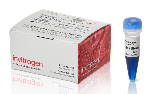
Invitrogen™
SeeBlue™ Estándar de proteínas preteñidas
El estándar preteñido SeeBlue consta de nueve polipéptidos que se resuelven en bandas nítidas y ajustadas de color azul enMás información
| Número de catálogo | Cantidad |
|---|---|
| LC5625 | 500 μl |
Número de catálogo LC5625
Precio (MXN)
-
Cantidad:
500 μl
El estándar preteñido SeeBlue consta de nueve polipéptidos que se resuelven en bandas nítidas y ajustadas de color azul en un intervalo de 4–250 kDa, lo que le permite supervisar fácilmente la electroforesis de proteínas y la eficacia de la transferencia western. El estándar de proteínas se suministra en un formato listo para su uso para la carga directa en geles; sin necesidad de calentar, reducir ni añadir un tampón de muestras antes de usarlo.
Compare y visualice todos los demás estándares y escaleras de proteínas ›
Aplicaciones
• Supervisión de la migración de proteínas durante la electroforesis en gel de poliacrilamida-SDS.
• Supervisión de la transferencia de proteínas a las membranas tras la inmunotransferencia (Western Blotting)
• Dimensionamiento de proteínas en geles de poliacrilamida-SDS e inmunotransferencia (Western blots)
Para uso exclusivo en investigación. No apto para uso en procedimientos diagnósticos.
Especificaciones
Método de detecciónColorimétrico
Compatibilidad del gelGeles Bolt™ Bis-Tris Plus, geles de tricina Novex™, geles de Tris-glicina Novex™, geles NuPAGE™ Bis-Tris
Peso molecular198, 62, 49, 38, 28, 18, 14, 6, 3 kDa
Cantidad500 μl
Listo para cargarSí
Condiciones de envíoHielo húmedo
ColorBlue
Number of Markers9
Línea de productosSeeBlue
Tipo de productoMarcadores moleculares de proteínas
Intervalo de tamañosDe 3 a 200 kDa
System TypeWestern Blotting, SDS-PAGE
Unit SizeEach
Contenido y almacenamiento
500 μL (50 aplicaciones de 10 μL cada una) suministrados en un vial de plástico. El tampón de carga consta de Tris-HCl, formamida, SDS y rojo de fenol.
Almacenar a 4 °C.
Almacenar a 4 °C.
Preguntas frecuentes
How can I obtain the date of manufacture for SeeBlue Pre-Stained Standard from the lot number?
How should the SeeBlue Pre-Stained protein standard appear after silver or Coomassie staining?
What is the expected Western transfer efficiency of the SeeBlue Prestained Standard?
Can I store the SeeBlue and SeeBlue Plus2 Pre-stained standards in the freezer to increase their shelf-life?
Why are the molecular weight values for the proteins in your prestained standards such as SeeBlue and SeeBlue Plus2 different in different gel types?
Citations & References (3)
Citations & References
Abstract
The specificity loop of T7 RNA polymerase interacts first with the promoter and then with the elongating transcript, suggesting a mechanism for promoter clearance
Journal:Proc Natl Acad Sci U S A
PubMed ID:11095736
During the early stages of transcription, T7 RNA polymerase forms an unstable initiation complex that synthesizes and releases transcripts 2-8 nt in length before disengaging from the promoter and isomerizing to a stable elongation complex. In this study, we used RNA small middle dotprotein and RNA small middle dotDNA crosslinking
Genome-scale cloning and expression of individual open reading frames using topoisomerase I-mediated ligation.
Journal:Genome Res
PubMed ID:10207160
The in vitro cloning of DNA molecules traditionally uses PCR amplification or site-specific restriction endonucleases to generate linear DNA inserts with defined termini and requires DNA ligase to covalently join those inserts to vectors with the corresponding ends. We have used the properties of Vaccinia DNA topoisomerase I to develop
beta -Glucoside Kinase (BglK) from Klebsiella pneumoniae. PURIFICATION, PROPERTIES, AND PREPARATIVE SYNTHESIS OF 6-PHOSPHO-beta -D-GLUCOSIDES.
Journal:J Biol Chem
PubMed ID:12110692
ATP-dependent beta-glucoside kinase (BglK) has been purified from cellobiose-grown cells of Klebsiella pneumoniae. In solution, the enzyme (EC ) exists as a homotetramer composed of non-covalently linked subunits of M(r) approximately 33,000. Determination of the first 28 residues from the N terminus of the protein allowed the identification and cloning