Search
Fragment Analysis Applications

Go beyond sequencing
Analysis of DNA fragments enables a variety of applications, from cell line
authentication to detection of aneuploidy. While sequencing techniques, such as next-generation sequencing (NGS) also enable these applications, researchers choose fragment analysis for features such as detection of multi-repeat regions, a faster turnaround time, and a higher sensitivity and resolution.
Fragment analysis is also more cost-effective due to its multiplexing capability; alleles for overlapping loci are distinguished by labeling locus-specific primers with different colored dyes, enabling more than 20 loci to be analyzed in a single run.

How Does Fragment Analysis Work?
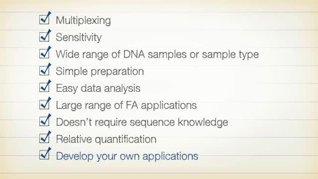
The Other Half of Your Applied Biosystems Genetic Analyzer

Brochure: DNA fragment analysis by capillary electrophoresis
Learn about our comprehensive solutions for each step of your fragment analysis workflow.
On-demand Webinar: Utility of Fragment Analysis in the Era of NGS-based Clinical Research
Speakers:
Somak Roy, MD, Cincinnati Children’s Hospital Medical Center
Steve Jackson, PhD, Thermo Fisher Scientific
")
Applications and methods
Misidentification of cell lines produces misleading results, confusion, and added costs to research. Many journals and funding agencies now require researchers to ascertain that the cell lines they use are authentic. Fragment analysis can provide a simple, inexpensive, and highly specific genetic “fingerprint” of a cell line.
Sanger sequencing is the gold standard for sequencing single genes, confirming gene variants, detecting repeat sequences, copy number variation, and single nucleotide changes. Perfect for smaller-scale projects and for confirmation of NGS results, Sanger sequencing can complement your laboratory's techniques and lead to efficient SARS-CoV-2 research.
Microsatellite markers are polymorphic DNA loci containing repeated nucleotide sequences, typically 2 to 7 nucleotides per repeat unit. The length of the repeated unit is the same for the majority of the repeats within an individual microsatellite locus, but the number of repeats for a specific locus may differ, resulting in alleles of varying length, which can be analyzed with fragment analysis by capillary electrophoresis.
The CRISPR-Cas9 system, the easiest, most precise, and most widely adopted genome editing technology, is based on the components of a simple bacterial immune system. In any genome editing experiment, the repair process is not completely efficient or accurate. Fragment analysis can be used to screen primary transformed cultures to determine editing efficiency.
Relative fluorescent quantitation or quantitative fluorescence PCR (QF-PCR) is a technique used in a variety of fragment analysis applications that requires accurate peak height or peak area comparisons across multiple samples. Research applications that utilize this technique include evaluating loss of heterozygosity (LOH), aneuploidy assays, and detecting large chromsomal deletions.
Single-nucleotide polymorphism (SNP) genotyping is a measure of genetic variation. SNP genotyping is used to research differences in genetic traits, susceptibility to disease, and response to drug therapies. A robust tool for SNP analysis is the Applied Biosystems™ SNaPshot™ Multiplex Kit. The kit allows multiplexing during single base extensions of up to 10 primer–template combinations in a single-tube, single-capillary format.
Don't see what you're looking for?
See Sanger sequencing applications ›
Relative fluorescent quantitation or quantitative fluorescence PCR (QF-PCR) is a technique used in a variety of fragment analysis applications that requires accurate peak height or peak area comparisons across multiple samples. Research applications that utilize this technique include evaluating loss of heterozygosity (LOH), aneuploidy assays, and detecting large chromsomal deletions.
Bacterial Artificial Chromosomes (BAC) have been developed to accommodate much larger pieces of DNA than plasmids can. They can be used to sequence an organism’s genome, and to model genetic diseases.
SSCP analysis detects sequence variations (single-point mutations and other small-scale changes). It is a widely used screening method that allows for the identification of different genomic variants in a large number of samples and in a broad range of organisms, from microorganisms to humans.
DNA sequence polymorphisms display different migration profiles from wild-type fragment patterns when DNA is digested with restriction fragments and separated by size using electrophoresis. RFLP analysis is useful in identifying where a specific gene for a disease lies on a chromosome and determine individuals who might be at risk for the disease or a carrier of the mutated gene.


Build your knowledge
Get bite-sized answers to your everyday Sanger sequencing and fragment analysis questions
Learn about the history of sequencing and how to pick the right platform for your research needs
Visistat reference component
For Research Use Only. Not for use in diagnostic procedures.