Outbreaks of the Zika virus disease in 2015 and 2016 sparked back-to-back global health crises, infecting millions of people, leaving thousands of children with serious birth defects, and leading to neurological problems and internal bleeding in some adults.
Zika virus belongs to the family of flaviviruses that are transmitted by mosquitos and ticks. Pregnant women can also transfer the Zika virus to their unborn babies, who often develop microcephaly (which results in abnormally small heads and sometimes smaller brains) or severe brain damage. There are currently no specific drugs to help prevent or treat Zika infections.
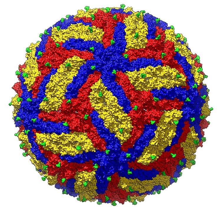
Cryo-EM structure of mature Zika virus at 3.1Å resolution. The three envelope glycoproteins are colored yellow, blue and red. (Purdue University photo/Madhumati Sevvana)
How cryo-EM is helping scientists understand the Zika virus
In 2016, scientists worldwide were searching desperately for a way to control the rapidly spreading virus. At that time, a team of researchers at Purdue University were the first to discover the structure of the Zika virus, a key step to seeing how a protein interacts with other compounds in the body.
Using cryo-EM, the team captured an image of the virus at a resolution of 3.8 Å. Two years later, the same team achieved an even higher resolution of 3.1 Å, and combined thousands of 2D images to construct a 3D model of the virus’s structure.
Purdue University researchers Michael Rossmann and Richard Kuhn have been at the forefront of discovery with the help of a technology that led to a Nobel Prize in chemistry for three scientists. Via Purdue University.
At lower resolutions, many flaviviruses appear virtually identical. The high-resolution images that scientists captured with cryo-EM enabled them to see the landscape of the Zika virus and compare its protein interactions with those of other flaviviruses. With such a detailed view of the structure, researchers were also able to identify probable drug-binding pockets, helping scientists design vaccines and anti-viral compounds that inhibit the Zika virus. Thanks, in part, to cryo-EM for solving the structure of the virus, at least two of these vaccines are now at the human test phase.
With the ability to examine structures at near atomic level, cryo-EM is helping scientists get a clearer picture of the underlying structure of the Zika virus and many other diseases. This knowledge helps researchers accelerating the development of critical vaccines that lead to the prevention of these diseases
Subscribe now to receive new Accelerating Microscopy posts straight to your inbox.
Gabriella Kiss, PhD, is a Product Marketing Manager for Single Particle Workflow at Thermo Fisher Scientific.
To learn more about cryo-EM, fill out this form to speak with an expert.
Guiding the Development of an HIV Vaccine with Structure-Based Drug Design
Novel drug development in the fight against the HIV pandemic...
Read More
Virus microscopy unravels chikungunya virus structure
Global impacts of the chikungunya virus Chikungunya is a vir...
Read More
What is cryo-EM?
Cryo-EM: cryo electron microscopy Cryo-electron microscopy (...
Read More
Glacios 2 Cryo-TEM and the Evolution of 200 kV Cryo-EM
Glacios 2 Cryo-TEM and the Evolution of 200 kV Cryo-EM Cryo-...
Read More
Leave a Reply