How dPCR Enables Cancer Research
Cell-free DNA (cfDNA)
Gaining Insight to the Genetics of Tumors
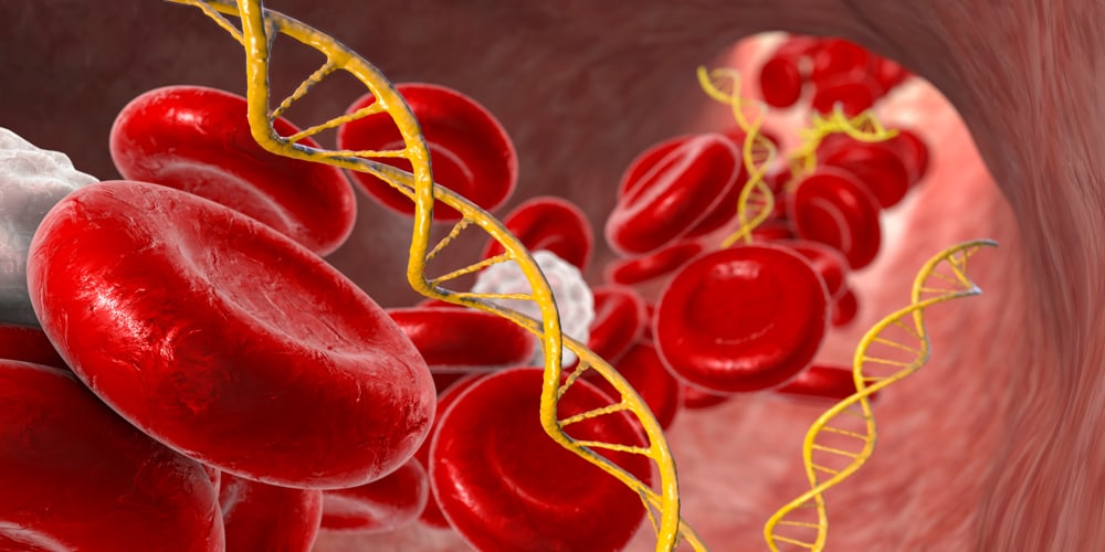
Cell-free DNA (cfDNA) is a relatively new source of information about cancer. It forms when tumor cells die or otherwise release their contents into the bloodstream or other body fluid. cfDNA is what it sounds like—DNA not extracted directly from tumor cells, but from fluids in liquid biopsy samples. cfDNA is present at very low levels compared to other circulating nucleic acids in body fluids, so it requires special methods to analyze and quantify. Although its concentration is often low, cfDNA gives special insight into the genetic composition of tumors and offers a much more accessible way to routinely monitor patients.1
Enabling Analysis With Microchambers
Digitize the Reaction, Enable Quantification
Thanks to digital PCR (dPCR), analyzing this challenging but important sample type is now more accessible than ever. dPCR splits a single bulk quantitative PCR (qPCR) reaction into hundreds or thousands of individual microchambers, such that each chamber statistically contains either one or zero copies of the target gene. This effectively digitizes the reaction and enables quantification of extremely low amounts of the target gene with higher precision and accuracy than ordinary qPCR. dPCR is emerging as an ideal technology for situations, such as cancer research, in which the need for high precision or a low abundance of the genetic target makes conventional qPCR challenging.
dPCR and Microfluidics
Unparalleled Sensitivity and Reproducibility
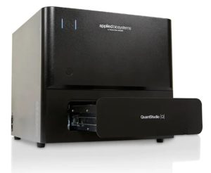
However, dPCR has limitations related to the fluidic pathways that divide a sample. Droplet-based dPCR platforms can have variable droplet sizes and other sources of variation between micro-reactions that limit the precision and reproducibility of runs for high-sensitivity assays. Advanced dPCR platforms like the Applied Biosystems™ QuantStudio™ Absolute Q™ Digital PCR System utilize microfluidics to remove these sources of variation, delivering unparalleled sensitivity and reproducibility in the dPCR space. Dueck et al. reported on these enhanced properties of microfluidic-based dPCR in a recent paper and opened a new frontier in high-precision cancer research in the process.1 cfDNA studies can be ideal cases for such technology, and Dueck et al. have put it to the test with their research on non-small cell lung carcinoma, chronic myeloid leukemia, and myelomonocytic leukemia in three distinct experiments.
Rare Mutant Quantification
EGFR T790M
Dueck et al. developed an assay to detect the T790M mutation in the EGFR gene, which is associated with resistance to tyrosine kinase inhibitors used for chemotherapy. Detecting this mutation early could help guide treatment of the associated cancers, including many non-small cell lung cancers, improving patient outcomes. The researchers carefully prepared a blend of manufactured DNA standards and background material to mimic cfDNA extracted from patients. They then used it to test whether a dPCR method optimized for a prototype microfluidics-based platform could exceed the sensitivity and accuracy of other methods for detecting this mutation. Their results showed the novel dPCR platform had the capacity to precisely quantify samples containing less than 1% of the mutant gene in a background of wild-type EGFR.
Rare Transcript Quantification
BCR-ABL1
In a similar test, Dueck et al. sought to determine how well their dPCR platform could distinguish between wild-type ABL1 and the BCR-ABL1 fusion gene that characterizes nearly 95% of chronic myeloid leukemia cases. Regularly monitoring the level of cfDNA for this gene is an important part of treating this type of cancer because it indicates when certain tyrosine kinase inhibitor therapies should be discontinued. Like other tumor genes found in cfDNA, however, it is present at extremely low levels. This is true even in severe cases, and highly sensitive methods are the only way to reliably quantify it. With clever use of multiplexing, Dueck et al. were able to distinguish between the two genotypes and quantify the fusion gene at frequencies down to 0.01% with differences between runs as low as 0.0089%. The dPCR platform thus delivered high sensitivity and reproducibility for this critical assay.
Personalized Cancer Monitoring With dPCR
Patient-specific Assay
In their final test, Dueck et al. used dPCR to investigate and monitor a patient with juvenile myelomonocytic leukemia. Previous genetic assays revealed a specific oncogenic mutation so rare that there were no commercial assays that could be used to monitor it in the patient’s cfDNA. With the use of the new and highly sensitive prototype microfluidics-based dPCR platform, Dueck et al. were able to establish a time series to monitor the patient’s cfDNA for the mutant allele and track the patient’s response to various drugs. This allowed the researchers to determine which drugs were reducing the mutant allele’s prevalence in the patient’s cfDNA.
Results like those of Dueck et al. are expected to become more common as dPCR becomes more accessible, affordable, precise, and sensitive. Platforms like the QuantStudio Absolute Q Digital PCR System are striving to bring this level of investigative power to more and more research laboratories around the world.
Learn more about the QuantStudio Absolute Q Digital PCR System
Learn More
» Mutation Detection Using Digital PCR
» Digital PCR – Powerfully simple dPCR for absolute quantification
» Applied Biosystems Solutions for Cancer Research
—
References
- Dueck ME, Lin R, Zayac A, et al. (2019) Precision cancer monitoring using a novel, fully integrated, microfluidic array partitioning digital PCR platform. Sci Rep 9(1):19606. doi:10.1038/s41598-019-55872-7.
—
For Research Use Only. Not for use in diagnostic procedures. © 2022 Thermo Fisher Scientific Inc. All Rights reserved. All trademarks are the property of Thermo Fisher Scientific and its subsidiaries unless otherwise specified.
