How does a scanning electron microscope work?
You have likely seen some of the incredible images that scanning electron microscopy (SEM) has produced of the unseen world around us. Whether it’s the geometric landscapes of crystals or the anatomy of a miniscule fruit fly, SEM has uncovered countless wonders and informed a myriad of fundamental discoveries across the life and materials sciences.

Comparison of a light microscope and a scanning electron microscope. The light microscope has been inverted for easier comparison.
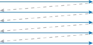
Just like our eyes can see objects due to the reflection of light off their surfaces, so too can an electron microscope capture reflected electrons to extract information from a sample surface. The smaller wavelength of electrons, however, allows electron microscopes to capture much finer details than light. The “scanning” in the name describes the fact that the relatively narrow electron beam must be moved across the sample surface to image it. This raster scan pattern is used to map the entire surface area of interest, and data is collected at frequent, discreet intervals. Think of it as systematically moving a flashlight through a dark room in order to see its contents.
Example of a zig-zag raster scan pattern.
Backscattered electron and secondary electron imaging
Scanning electron microscopes are usually designed to detect numerous signals, even beyond reflected electrons. X-rays, for instance, are also produced when the high-energy electron beam impacts the sample, and these carry additional information about the specimen. However, the two primary electron signals acquired with SEM are backscattered electrons and secondary electrons.
Backscattered electrons (BSE) are high-energy electrons from the beam that are ejected after interacting with the sample atoms. Being elastically scattered, they leave the sample with little to no energy loss. The heavier the atom (higher atomic number) the more it will backscatter electrons. BSE imaging, therefore, provides the relative composition of the sample, with brighter areas correspond to heavier atoms.

Backscattered SEM image of a powder used in additive manufacturing. The bright particles are copper (atomic number N = 29), whereas the dark grey particles are an alloy of magnesium, aluminum, and silicon (N = 12, 13, and 14 respectively).
Secondary electrons (SE), meanwhile, are the result of inelastic scattering between the electrons of the sample and the electron beam. These atomic electrons are displaced by the beam electrons and ejected from the sample. As they are relatively low energy, they can only escape the sample and reach the detector if they are ejected close to the sample surface. For this reason, secondary electron imaging is used to reveal a sample’s topography. Many electrons can escape at edges, which then appear bright, and fewer can escape at recesses, which appear darker.

Secondary electron image of carbonates, showing their morphology and topographical details.
Energy dispersive spectroscopy on the SEM
The third, and increasingly important, signal generated in an SEM is X-rays, which are produced in parallel with secondary electrons. Removal of the SE from an inner electron shell destabilizes the atom. To restore stability, an electron from a higher energy shell can release its excess energy as an X-ray photon and then fill the vacancy. The energy of this X-ray is characteristic to the specific element, and can be used to identity and quantify the elemental composition of the sample. This technique is called energy-dispersive X-ray spectroscopy (abbreviated as either EDX or EDS) and is often an integrated component of modern SEMs.
Conclusions
Scanning electron microscopy is continuously being improved, with ever higher resolution imaging becoming possible. Some high-end SEMs can even meet the resolution of transmission electron microscopes (TEMs). Additionally, many SEMs can be equipped with dedicated scanning transmission electron microscopy (STEM) detectors, which are placed below the specimen and allow for transmission electron microscopy images to be obtained without the need for a dedicated TEM.
This blog serves as a high-level introduction to scanning electron microscopes and their capabilities. If you’d like to learn more about SEM and related analytical techniques, please visit our electron microscopy webpage.
If you’d like to learn more electron microscopy basics, explore some of our other blogs in the Microscopy 101 series, including:
– Further Details on How Scanning Electron Microscopy Works
– The Anatomy of an Electron Microscope
– How Do You Make an Electron Beam? – Electron Source Fundamentals
– TEM vs SEM: What’s the Difference?

EELS vs EDS Analysis [Infographic]
Elemental analysis: EELS vs EDS Secondary signals in electro... Alex Ilitchev, PhD
Read More
How Does an Electron Microscope Work?
Since the advent of electron microscopy in the 1930s, this r... Alex Ilitchev, PhD
Read More
Optimizing the Depth of Field in an SEM
Depth of field in SEM imaging When it comes to “dep...
Read More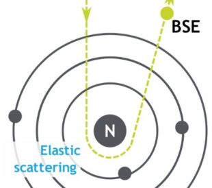
Backscattered Electrons in SEM Imaging
Unlike optical microscopes, scanning electron microsc...
Read More
Leave a Reply