Since the advent of electron microscopy in the 1930s, this revolutionary technology has progressively allowed us to capture information at micro-, nano-, and even atomic scales, surpassing the limitations of light microscopy and revealing the hidden machinery underlying all matter.
Exploring the unseen world with electron microscopes
Modern electron microscopes can clearly visualize rows and columns of discreet atoms in crystalline materials and reproduce 3D protein structures down to individual coordinated water molecules. These details have enabled countless advances in materials science, biology, and more, informing our understanding of macro-scale phenomena with molecular and atomistic insights.
With all the enlightening and often beautiful imagery produced with electron microscopy, you may wonder, how does an electron microscope work? In this blog we will explore the basic components of an electron microscope and help familiarize you with this exciting technology.

Diagram of a scanning electron microscope.
Components of an electron microscope
In general, a modern electron microscope has several key components: the electron source, electromagnetic lenses, and one or more electron detectors. The microscope begins by generating a focused beam of electrons from the source, which is then funneled by the lenses toward the sample. Once the beam electrons interact with the sample, they either reflect or pass through the sample, if it is thin enough. The detector is positioned to capture either these reflected or transmitted electrons, which carry various details about the sample due to their interaction.
Electron source
There are several types of electron sources, broadly categorized as either thermionic or field emission sources. Thermionic sources heat up a filament or crystal using an applied current until there is enough energy to emit electrons. Field emission sources apply a strong electrostatic field to a sharp metal (tungsten) tip, and the concentration of energy at the tip edge causes the ejection of electrons. Choosing a thermionic versus field emission source is largely a matter of cost and needed image quality.

Simplified illustration of electromagnetic lens structure.
Electromagnetic lenses
Similar to how glass lenses focus and direct light in an optical microscope, electromagnetic lenses control the flow of electrons through the microscope. An electromagnetic lens consists of a series of parallel electric coils that produce a magnetic field. These are connected to pole pieces, hollow metal cylinders that focus the magnetic field near the electron beam. The electrons are then pulled through the center of the pole piece by the field.
Electron detector
Detectors have evolved significantly over the history of electron microscopes. Originally, electrons were detected by having them interact directly with camera film, which responds similarly to both electrons and photons. Today, most electron microscopes use a digital camera instead, either with a scintillator that emits light or a direct electron detector. Resolution is interpreted per pixel of the detector, with some detectors reaching 4k x 4k physical pixels (or 16 million pixels total).
How electron microscope components work together
The nature of modern electron microscopes necessitates management through an external computer running control software. This ideally coordinates and monitors the source, lenses, and detectors to provide optimized imaging and data collection. Some integrated control software can even offer automation, allowing you to gather a large quantity of data in an unattended fashion.
We hope this provided a general overview of how an electron microscope works as well as its components. If you’d like to know more about electron microscopes, such as the difference between transmission and scanning electron microscopes, please check out some of our other educational blogs in the Microscopy 101 series:
– The Anatomy of an Electron Microscope
– How Do You Make an Electron Beam? – Electron Source Fundamentals
– TEM vs SEM: What’s the Difference?
For even more educational resources, visit our Electron Microscopy Learning Center >>
EELS vs EDS Analysis [Infographic]
Elemental analysis: EELS vs EDS Secondary signals in electro... Alex Ilitchev, PhD
Read More
What is Scanning Electron Microscopy?
How does a scanning electron microscope work? You have likel... Alex Ilitchev, PhD
Read More
Optimizing the Depth of Field in an SEM
Depth of field in SEM imaging When it comes to “dep...
Read More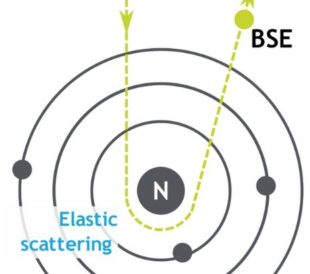
Backscattered Electrons in SEM Imaging
Unlike optical microscopes, scanning electron microsc...
Read More
This website is really good and helped me with my science project