Search


Citations & References (9)
Gibco™
Puromycin Dihydrochloride
Puromycin Dihydrochloride is an aminonucleoside antibiotic produced by Streptomyces alboniger. Puromycin works by inhibiting peptidyl transfer on both prokaryotic andRead more
Change view

| Catalog Number | Quantity |
|---|---|
| A1113803 | 10 x 1 mL |
| A1113802 | 20 mL |
Catalog number A1113803
Price (JPY)
50,400
Each
Quantity:
10 x 1 mL
Puromycin Dihydrochloride is an aminonucleoside antibiotic produced by Streptomyces alboniger. Puromycin works by inhibiting peptidyl transfer on both prokaryotic and eukaryotic ribosomes; resistance is conferred by the expression of the pac gene.
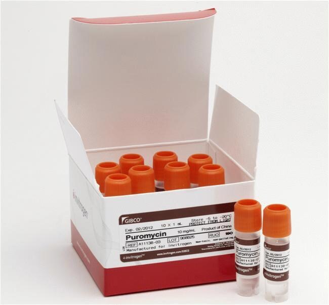
Puromycin is widely used in cell biology as a selection antibiotic agent in mammalian cell culture systems. The recommended working concentration ranges from 0.2–5.0 μg/mL, although it can be toxic to eukaryotic cells at concentrations as low as 1 μg/mL. Gibco™ Puromycin Dihydrochloride is supplied at 10 mg/mL in 20 mM HEPES buffer (pH 6.2–6.8) in 10 vials each containing 1 mL.
Other Choices and More Information
We offer a wide range of antibiotics and antimycotics in both powder and liquid formats.
See the complete list, or find products for:
• Contamination control
• Eukaryotic and bacterial selection
See recommendations for working concentrations for selection antibiotics.
Learn more about the use of antibiotics and antimycotics in cell culture, and review guidelines for decontaminating cultures.
For Research Use Only. Not for use in diagnostic procedures.
Specifications
Concentration10 mg/mL
Culture TypeMammalian Cell Culture, Insect Cell Culture
For Use With (Application)Eukaryotic Selection⁄Stable Cell Line Generation
Product LineGibco
Quantity10 x 1 mL
Shelf Life12 Months
Shipping ConditionDry Ice
FormLiquid
Product TypeAntibiotic
SterilitySterile-filtered
With AdditivesHEPES
Unit SizeEach
Contents & Storage
Storage conditions: -5 to -20°C
Shipping conditions: Frozen
Shelf life: 12 months from date of manufacture
Shipping conditions: Frozen
Shelf life: 12 months from date of manufacture
Frequently asked questions (FAQs)
Which of your antibiotics (Geneticin, Zeocin, Hygromycin B, Blasticidin, and Puromycin) can be used together for stable selection in mammalian cells?
How light-sensitive is Puromycin Dihydrochloride?
How can I decontaminate my cultures?
What antibiotics do you offer to help control or eliminate cell culture contamination?
Citations & References (9)
Citations & References
Abstract
Trop2 Promotes Multidrug Resistance by Regulating Notch1 Signaling Pathway in Gastric Cancer Cells.
Journal:Med Sci Monit
PubMed ID:31964857
'BACKGROUND Chemotherapy is widely used in gastric cancer treatment, but multidrug resistance remains a leading cause of chemotherapy failure. Trop2 is highly expressed in gastric tumor tissues and greatly influences cancer progression. However, little is known about the relationship between Trop2 and drug resistance in gastric cancer. MATERIAL AND METHODS
mTORC2 contributes to the metabolic reprogramming in EGFR tyrosine-kinase inhibitor resistant cells in non-small cell lung cancer.
Journal:Cancer Lett
PubMed ID:30036610
Non-small cell lung cancer (NSCLC) patients with activating EGFR mutations are often successfully treated with EGFR tyrosine kinase inhibitor (TKI) such as erlotinib; however, treatment resistance inevitably occurs. Given tumor metabolism of glucose and therapeutic response are intimately linked, we explored the metabolic differences between isogenic erlotinib-sensitive and -resistant NSCLC
The Plasmodium falciparum cytoplasmic translation apparatus: a promising therapeutic target not yet exploited by clinically approved anti-malarials.
Journal:Malar J
PubMed ID:30541569
The continued spectre of resistance to existing anti-malarials necessitates the pursuit of novel targets and mechanisms of action for drug development. One class of promising targets consists of the 80S ribosome and its associated components comprising the parasite translational apparatus. Development of translation-targeting therapeutics requires a greater understanding of protein
Cardiac glycosides decrease influenza virus replication by inhibiting cell protein translational machinery.
Journal:Am J Physiol Lung Cell Mol Physiol
PubMed ID:30892074
Cardiac glycosides (CGs) are used primarily for cardiac failure and have been reported to have other effects, including inhibition of viral replication. Here we set out to study mechanisms by which CGs as inhibitors of the Na-K-ATPase decrease influenza A virus (IAV) replication in the lungs. We found that CGs
Modulating eIF6 levels unveils the role of translation in ecdysone biosynthesis during Drosophila development.
Journal:Dev Biol
PubMed ID:31283922
During development, ribosome biogenesis and translation reach peak activities, due to impetuous cell proliferation. Current models predict that protein synthesis elevation is controlled by transcription factors and signalling pathways. Developmental models addressing translation factors overexpression effects are lacking. Eukaryotic Initiation Factor 6 (eIF6) is necessary for ribosome biogenesis and efficient