This blogpost serves as a high-level overview of common techniques employed in the field for cell isolation, genetic modification and approaches to cell expansion. For a more in-depth discussion, check out the accompanying chapters on cell isolation (chapter 4), engineering (chapter 5), and expansion (chapter 6) in our Cell Therapy Handbook.
Cell Isolation
At the foundation of any cell therapy development and manufacturing workflow, the quality of the starting cellular material directly impacts the final product and its efficacy of treatment. This section provides some insights into clinically relevant closed processes that can be used for the isolation of PBMCs and selection and activation of the T cell populations (CD3+).
PMBC Isolation
While traditional density gradient centrifugation successfully isolates PBMCs from red blood cells, the pellet retains contaminants such as granulocytes, monocytes, and residual red blood cells. Additionally, most density gradient centrifugation isolations are performed using an open system—which makes the procedure prone to errors, contamination, and user-to-user variability.
Another approach uses counterflow centrifugation, which separates cells based on their density as well as their size. Systems using this technology (e.g., Gibco CTS Rotea system) suspend cells in a fluidized bed by exerting a constant flow force against centrifugal forces. The suspended cells are gently concentrated without forming a densely packed pellet and can then be washed with very high recoveries. Using elutriation, dead cells can be removed to optimize viability of the population. Adjustments to centrifugal speed and flow rate allow for cells to be fractionated based on size and density, with minimal shear. Density gradient centrifugation and counterflow centrifugation produce comparable results (Figure 1).
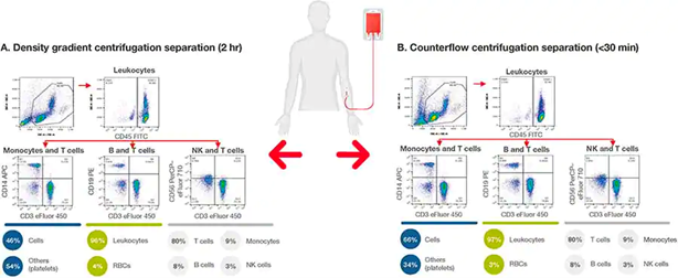
Figure 1. Closed counterflow centrifugation system versus open density gradient system for isolation of PBMCs from red blood cells. A single-donor leukopak was divided into two, and PBMCs were separated using (A) density gradient centrifugation using Ficoll polymer or (B) counterflow centrifugation using the CTS Rotea system. The CTS Rotea system can isolate PBMCs from a leukopak in less than 30 minutes, with equivalent performance to the density gradient system and the added benefit of closed processing.
T cell Isolation and Activation
Following PBMC isolation, magnetic bead isolation and activation technology is an efficient and high-fidelity approach to this step. Magnetic beads conjugated to antibodies recognize specific T cell surface markers and bind to them. When placed near a magnet, the bead-T-cell complex binds and is held while unwanted cell types can then be washed away.
Gibco CTS Dynabeads Magnetic Beads CD3/CD28 provides both the primary and co-stimulatory signals required for activation and expansion of T-cells in a single step, eliminating the need for a separate activation step. For more details on this process, see One-step isolation and activation of naive and early memory T cells with CTS Dynabeads CD3/CD28.
Genome Engineering
Gene editing technologies used to create allogeneic T cells
Numerous genome engineering approaches exist and primarily fall into three gene editing tool classifications: zinc finger nuclease (ZFN), transcription activator-like effector nuclease (TALEN), and clustered regularly interspaced short palindromic repeat (CRISPR)-associated protein 9 (CRISPR-Cas9).
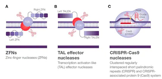
Figure 2. Gene-editing tools for allogeneic T cells.
The two genome editing technologies, ZFN and TALEN, (Figures 2A and 2B) are very specific gene-editing tools because two DNA binding proteins-linked to nucleases (left- and right-handed complexes) must be designed for each editing experiment. The genetic sequences for these tools are transferred to a cell to be expressed into functional nucleases. More recently, CRISPR-Cas9 gene-editing technology derived from Streptococcus pyogenes has become available and is vastly different from both ZFN and TALEN gene-editing technologies. The CRISPR-Cas9 system (Figure 2C) simply requires the in vitro combination of the designed gRNA and the Cas9 protein to create a single CRISPR-Cas9 ribonucleoprotein (RNP) complex that can induce the double-stranded DNA break once it is delivered into a cell.
Invitrogen TrueCut Cas9 Protein v2 is a next-generation CRISPR Cas9 protein engineered to deliver maximum editing efficiency—featuring consistently high editing efficiency in all tested cell lines including standard, immune, primary, and stem.
Delivery Systems for Gene-editing Tools
Gene-editing tools tend to be macromolecules and thus require an active delivery system to get into target cells. Currently, numerous delivery systems are available for in vivo and invitro gene editing applications. These methods are summarized in the table below.
Table 1. Delivery methods used for gene editing.
| Methods | Description | Advantages | Disadvantages | Examples |
| Lentiviral vector | DNA/RNA is packaged into the infectious viral particles and introduced into cells. | High transduction efficiency; suitable for primary immune cells; CAR T clinical usage precedent. |
Special lab environment needed; requires safety measures; labor intensive | CTS LV-MAX LentiCRISPR |
| Electroporation | Electrical pulse creates pores in the cell membrane, allowing the entry of DNA/RNA/RNP into the cell cytoplasm the nucleus. | Fast and easy; large number of cells can be transfected in minutes. | Requires special equipment; cytotoxic anions can form during the procedure, leading to cell death. | Neon Transfection System
Xenon Electroporation System |
| Nucleofection | Similar to electroporation except the tools are delivered directly into the cell nucleus using very specific conditions provided by manufacturers. | Effective for non-dividing cells; high throughput potential–multiple samples can be transfected simultaneously. | Requires special equipment; less flexible–special protocols and reagents can’t be controlled by the user. | Lonza Nucleofector System |
| Cationic lipids | Positively charged liposomes encapsulate protein/RNP and interact with negatively charged cell membrane facilitating entry into the cell cytoplasm via the endocytosis pathway. | Easy and versatile (no special equipment), not very toxic, and can be a high throughput system. | Lower transfection efficiency; not direct delivery into the nucleus; not applicable for all cell types. | Lipofectamine CRISPRMAX Cas9 Transfection Reagent |
Cell Expansion
Following activation and engineering, buffer exchange can be done using counterflow centrifuge in a closed an automated manner. During expansion, release of immunostimulatory cytokines, such as IL-2 and IFN-ɣ at desired levels can allow CD8+ cells to survive as memory T cells during expansion.
The choice of media and supplements can significantly influence the growth of the T cell population, differentiation, viability, and the CD8:CD4 ratio during expansion. It is important to select a flexible expansion medium that is compatible with other workflow processes such as T cell isolation and activation, while being amendable to various platforms ranging from static cell culture systems to larger scale dynamic bioreactors.
For more information on any of the cell therapy manufacturing techniques discussed above, check out the accompanying chapters 4,5, and 6 in our Cell Therapy Handbook.

How Process Analytical Technology Enables Real-time Quality Control in Pharmaceutical Manufacturing
Frequently Asked Questions (FAQs)... Stanislav Kasakov
Read More
Reducing Discontinuities in Pharmaceutical Scale-Up
What controlled continuity means in practice From an operati... Dirk Leister
Read More
