
This story is part of our celebration of innovation in 2025.
About 1 in 91 women will develop ovarian cancer in their lifetimes. But only around 1 in 1000 women will develop a subtype called low grade serous ovarian carcinoma (LGSOC).
The average age of LGSOC diagnosis is just 45, two decades younger than the average for the more common high-grade type. Survival outcomes skew poor and until this year, clinicians and patients have not had the benefit of drugs carrying a specific FDA approval for this disease. That’s because rare conditions like LGSOC are often underrepresented in the clinical trial landscape.
High-grade treatment options also, unfortunately, make little progress against low-grade tumors – no surprise, given that LGSOC is biologically and clinically distinct from other ovarian cancer types in ways that researchers are still uncovering.

With so few resources, how do you discover a drug needle in a haystack to give one woman in a thousand – a real-life, individual patient – hope for treatment?
In the end, much of early drug screening and discovery is just a numbers game. Improving your odds of success takes volume, speed, and scale at every level.
And few understand the oncology numbers game better than the research team led by Drs. Dane Cheasley, and Kathleen Pishas in collaboration with Professor Kaylene Simpson at the Victorian Centre for Functional Genomics (VCFG), all at the Peter MacCallum Cancer Centre in Melbourne, Australia.
If data is the haystack, Simpson and team have spent years developing an efficient process to sift and sort their way through it. Using high-content screening, automation, and advanced computational analysis, they are paving the way for a big data approach to small-scale, precision cancer medicine.
Gathering the haystack: designing the field’s largest drug screening
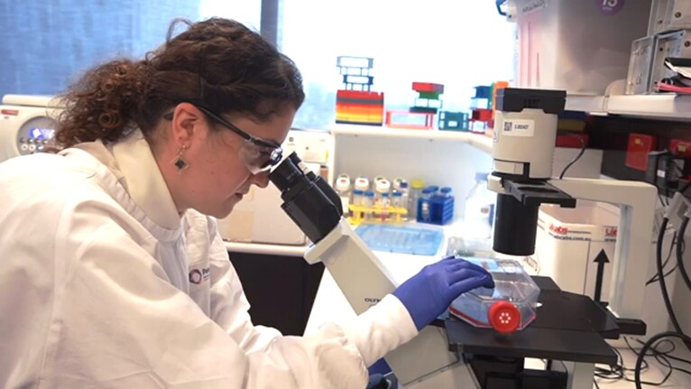
Simpson has led the VCFG since 2008, where she and her team of highly skilled experts enable colleagues like Pishas and Cheasley and others from around the world to use advanced technologies like CRISPR, RNAi and compound screening coupled with high-content imaging to answer their cancer and other disease research questions.
High-content screening (HCS), or high-content imaging, blends mainstay life science techniques like microscopy and fluorescent labeling with ultramodern innovations like high-resolution imagery, robotic sample handling and automation, and machine learning analysis.
With HCS, researchers can load 384- or 1,536-well plates with samples of treated cells that are then stained and photographed, field-by-field, well-by-well, under a laser-powered microscope. A surprising amount of the process is (literally) hands-off, as a suite of computerized machines take over the minutiae of washing, dosing, handling, and analyzing samples with incredible accuracy.
HCS is a game-changer for drug discovery because it makes short work of screening cellular responses to thousands of perturbations – simultaneously detecting and quantifying everything from changes in cell shape or protein expression to the overall health of cells and their individual parts.

This work often begins, as it did with the VCFG team, with ready-to-use chemical compound libraries of molecules curated by mode of action, regulatory approval status, and other features.
They collaborated with Compounds Australia at Griffith University who house an extensive collection of compounds for national use – and selected an expansive library of drugs in clinically advanced or approved FDA stages, complemented by smaller libraries aligned to the known genetic biology of LGSOC. This was a practical choice as much as a scientific one. Pre-approved drugs, if potent against LGSOC, could skip the time-consuming early-phase trial stage and reach patients much faster.
A second major goal of this work centered around demystifying the genetic causes of LGSOC tumors.
“About half of LGSOC tumors involve unidentified genetic drivers. The response rates to chemotherapy in those cases, especially after relapse, are abysmal and many times involve patients who are younger women,” said Cheasley. “This screen was designed to identify targeted therapy options, but also to identify new targets and possible causes of aggressive disease forms so that we can improve outcomes in the future.”
Altogether, the team performed an initial HCS screening of 6710 compounds across three patient-derived LGSOC cell lines, and one non-cancerous ovarian derived control cell line. This was subsequently narrowed down to 3,436 compounds across an additional nine patient-derived LGSOC to concentrate on drugs that were either FDA approved or clinically advanced – making it the largest quantitative screening study ever performed on LGSOC.
Patient cells tell their stories
Beyond its scale, the VCFG team’s innovative experimental approach stands out from predecessors in other ways.
The development of new treatments for LGSOC has been hindered by a longstanding lack of representative laboratory models. Ovarian cancer research has relied heavily on some of the oldest immortalized human cell lines in biology like HeLa and U2OS, limiting the clinical relevance of drug screening outcomes. Cheasley and Pishas deliberately broke away from this tradition, leveraging 12 genetically distinct, patient-derived LGSOC cell lines.
“Many screens have been done with just one or two cell lines, and those are often just the garden variety lines. They’re almost biologically meaningless for specific diseases,” said Simpson. “This whole project represents the first instance of accessing unique, patient-derived cell lines for broad drug screening to bring that big picture together.”
Another key difference in their methodology: while traditional screens focus primarily on cell viability, the VCFG team went a step further.
Using the Thermo Scientific CellInsight CX7 LZR Pro HCS Platform they captured extensive morphological data from treated cells. This morphological profiling—tracking changes in cell shape, size, and structure—provided insights into drug mechanisms and effectiveness beyond simple cell death metrics.
We kept the screening scale at a level where we can dig in beyond viability to integrate other phenotypic information and more depth,” said Simpson. “The CX7-LZR Pro platform was a game changer, so much faster than our previous CX7 generation instruments, it dramatically accelerated our research program”. “On average, I could easily process thirty 384-well plates containing LGSOC cells – that’s 11,520 wells in a single experimental run!” said Pishas.
Lastly and perhaps most unusually for this scale of screening, the team made the decision to analyze each of the 3,436 experimental compounds at three doses and to include one healthy ovarian cell line for comparison.
Taken together, these drug screen design elements have permitted the team to conduct a much more nuanced analysis than previous studies have allowed, providing data about the lowest dose necessary for therapeutic effect and ruling out drugs that would be toxic to healthy ovarian tissue.

Finding the needle: tackling big data in oncology
If you’re keeping track of the number of samples involved in this experiment, this is the point where you may have lost count.
Simpson, Cheasley, and Pishas evaluated 3,436 final drug candidates in 3 doses each across 13 cell lines, capturing cellular snapshots at a 72-hour time point. That accounts for, at minimum, more than 2.8 million images representing dozens of cell characteristics each.
While this kind of screening size is actually relatively focused in the world of HCS, it is a lot to work with. Datasets of this size can be overwhelming for even the most experienced scientists. How do you parse out the information you need, especially with so many cellular features to examine?
“What we published in Scientific Data related to cell viability based on high content imaging. But we also performed whole cell staining in our screen, like the actin and cytoskeletal staining that gave us capacity to look at cellular response to treatment,” said Simpson. “When you are working with multiple cell lines in a screen, it is actually quite difficult to come up with an analytics method that will let you compare between lines. Some may be more resistant to drug treatments than others, or some will have different genetic backgrounds.”
“The morphological features we captured through whole-cell staining and imaging gave us insight into how low-grade serous ovarian cancer cells were responding at a structural level,” said Cheasley. “It allowed us to move beyond simply measuring cell death and start interpreting how different drugs were affecting the cells, whether by disrupting the membrane, collapsing the cytoskeleton, or triggering other forms of cellular stress.”
To tackle the complex analysis, the VCFG group has leaned into the thought partnership of bioinformatics experts like their colleagues at the University of Queensland. Those researchers had previously developed a computational pipeline called UnTANGLeD (Unsupervised Trait Analysis of Networks from Gene Level Data) to link genes to more than 1,000 phenotypic traits and diseases.
Their UnTANGLeD approach had been applied to transcriptomics data, but never to image-based data outputs. After much optimizing, we have adapted UnTANGLeD to systematically interpret cellular drug response signatures in enormous datasets, sorting tested drugs into therapeutically meaningful cluster categories, across many cell lines at once. These clusters are defined by higher-level traits like DNA damage response, molecular signaling pathways, cytoskeletal changes, and more.
The clustering helps researchers quickly identify which drugs work similarly and find new uses for existing drugs. It also helps them determine next steps.
“The UnTANGLeD analysis took our primary screen to the next tier of analysis and has been really valuable. We’ve been able to hone down on the next targets that we want to look at in more detail,” said Simpson.
Though it was originally designed for transcriptomics, UnTANGLeD provided excellent results for the team’s HCS, image-based drug screening applications. As an analytic framework, it should be universal across truly all data types and screening platforms – setting the stage for use in other complex experimental questions in the future.

Translating discovery into action
In the end, the team narrowed 6710 drug compounds down to just 79 promising possibilities for LGSOC – 60 high-confidence and 19 moderate-confidence hits to pursue further in clinical study.
“The whole idea of the screening is to be ruthless in your triaging to get to a point where you’ve only got really robust targets,” said Simpson.
Some of their hits confirmed LGSOC susceptibility to drugs targeting certain cell growth signaling pathways. Others identified unknown LGSOC tumor cell vulnerabilities and novel drug class possibilities.
To bridge the gap between laboratory discoveries and clinical application, Cheasley’s team is now moving validated compounds into advanced 3D organoid models using automated Matrigel embedding protocols and sophisticated image analytics developed by Simpson’s team. These 3D organoid models better mimic the structure and environment of real human tumors.
“The hope is that by moving into 3D cultures, we can better predict clinical responses,” said Simpson. “We’re particularly keen to see how combinations with standard therapies synergize in these more realistic 3D models.”
“A major concern raised by our LGSOC cancer advocates is that, for the past 30 years, treatments have failed because they weren’t specifically designed for this rare and unique cancer type,” said Dr Kathleen Pishas. “For the first time, we have the capacity to shift the survival trajectory for these patients by validating therapies in preclinical 3D models tailored to LGSOC. Currently, there are no animal models for this disease, and most LGSOC cell lines do not grow well when transplanted into mice. This underscores the urgent need for innovative 3D culture systems to generate the foundational data needed to launch clinical trials.”
And, throwing in another highly advanced tool, Simpson’s team, together with the Molecular Genomics Core facility at Peter Mac have developed MAC-seq, a high throughput transcriptomics method that can measure transcriptional responses in 384 well plate format and integrate with concurrent phenotypic quantitation. Using this powerful technology will allow us to determine drug target specificity and cellular responses at specific time points. Largely expanding the molecular and signaling pathways of the disease response to treatment.
Shaping the future of ovarian cancer treatment
At the heart of both this screening project and the VCFG as a whole is the interesting idea that to scale down, you must first scale up.
To achieve effective therapeutics at the most personalized and precise level – drugs genetically tailored to the individual patient – researchers often must first understand how to navigate emerging technologies or wade through enormous data sets.
This integrated approach of melding advanced imaging, sophisticated computational analytics, and patient-specific strategies is already transforming drug discovery for LGSOC.
The VCFG team isn’t gatekeeping their success, either. As one of the world’s leading oncology research centers, they also aim to equip fellow researchers with the means to keep discovering.
“We have always been committed to the idea of sharing data as a resource to the broader community, and at a level where it’s fully validated,” said Simpson.
With any luck, these field-wide advances driven by ever-improving tools and methods will eventually pay off for the estimated 65,000 – 165,000 women currently living with LGSOC worldwide.
In May 2025, the first FDA-approved drug combination for LGSOC launched to market for the treatment of KRAS-mutated recurrent disease – tumor types with an already-identified genetic cause.
Simpson, Cheasley, and Pishas are still hard at work trying to expand that list.
“The ultimate goal of this project is to advance and repurpose drugs for more patients, as quickly as possible, so they can start making a difference,” said Cheasley.
Read more about the researchers and disease
- “The path from down under to global game-changer” featuring Dr. Kaylene Simpson (Thermo Fisher Scientific)
- “Peter Mac researchers seek answers for rare ovarian cancer” featuring Dr. Dane Cheasley (Peter MacCallum Cancer Centre)
- About Low-Grade Serous Ovarian Cancer (Low-Grade Serous Ovarian Cancer Initiative)
Read more about the science
- Bao, S.C., Mizikovsky, D., Pishas, K., Cowley, K.J., Zhao, Q., Marinovic, E., Carey, M., Campbell, I., Simpson, K.J., Cheasley, D. and Palpant, N.J. (2024) A robust unsupervised clustering approach for high-dimensional biological imaging data reveals shared drug-induced morphological signatures. bioRxiv. Cold Spring Harbor Laboratory. doi: 10.1101/2024.09.05.611300
- Li, X.M. et al. (2023). MAC-Seq: Coupling Low-Cost, High-Throughput RNA-Seq with Image-Based Phenotypic Screening in 2D and 3D Cell Models. In: Jenkins, B.J. (eds) Inflammation and Cancer. Methods in Molecular Biology, vol 2691. Humana, New York, NY. https://doi.org/10.1007/978-1-0716-3331-1_22
- Pishas, K.I., Cowley, K.J., Llaurado-Fernandez, M. et al. High-throughput drug screening identifies novel therapeutics for Low Grade Serous Ovarian Carcinoma. Sci Data 11, 1024 (2024). https://doi.org/10.1038/s41597-024-03869-x
- High Content Screening Overview & Sample Data (Thermo Fisher Scientific)
##
About Innovation at Thermo Fisher Scientific
In the field of life sciences, solving the world’s toughest problems is why we come to work each day. It’s what brings us together and empowers us to reach our true potential and work towards our collective goal of advancing science for the greater good.
That’s why Thermo Fisher Scientific has one of the largest R&D investments in the industry at $1 billion every year. Innovation is engraved into Thermo Fisher Scientific’s DNA, but it’s about more than developing novel technologies and products. Innovation is a mindset of iteration, evolution, and teamwork in science.
In 2025, join us in celebrating our customers – the true collaborative trailblazers who stay at the forefront of progress in a world where we need to innovate faster each day. Follow along as we explore why we are all in this journey together, at thermofisher.com/innovation.
© 2025 Thermo Fisher Scientific Inc. All rights reserved. All trademarks are the property of Thermo Fisher Scientific and its subsidiaries unless otherwise specified.
For Research Use Only. Not for use in diagnostic procedures.
References
- Gershenson, David M., Diane C. Bodurka, Karen H. Lu, Lisa C. Nathan, Ljiljana Milojevic, Kwong K. Wong, Anais Malpica, and Charlotte C. Sun. “Impact of Age and Primary Disease Site on Outcome in Women with Low-Grade Serous Carcinoma of the Ovary or Peritoneum: Results of a Large Single-Institution Registry of a Rare Tumor.” Journal of Clinical Oncology 33, no. 24 (August 20, 2015): 2675–82. https://doi.org/10.1200/jco.2015.61.0873.
- “Gynecologic & Ovarian Cancer Recurrence.” Ovarian Cancer Research Alliance (OCRA). Accessed June 25, 2025. https://ocrahope.org/for-patients/recurrence/.
- “Low-Grade Serous Ovarian Cancer Guide.” Low-Grade Serous Ovarian Cancer Initiative, April 15, 2025. https://lgsoc.org/lgsoc-guide/.
- Manning‐Geist, Beryl L., Ryan M. Kahn, David Nemirovsky, Jeffrey Girshman, Anya Laibangyang, Sushmita Gordhandas, Alexia Iasonos, et al. “Chemotherapy Response in Low‐grade Serous Ovarian Carcinoma at a Comprehensive Cancer Center: Readdressing the Roles of Platinum and Cytotoxic Therapies.” Cancer 129, no. 13 (March 23, 2023): 2004–12. https://doi.org/10.1002/cncr.34753.
- “Ovarian Cancer Statistics: How Common Is Ovarian Cancer.” Ovarian Cancer Statistics | How Common is Ovarian Cancer | American Cancer Society. Accessed June 25, 2025. https://www.cancer.org/cancer/types/ovarian-cancer/about/key-statistics.html.

Thermo Fisher Scientific Partners with AIM Biotech on Development of Microphysiological Systems
The adoption of New Approach Methodologies (NAMs) in drug di...
Read More
5 Solutions for Overcoming Research Challenges in Vaccine Development
What are the main challenges in vaccine development? The mai...
Read More
Deoxyribonuclease I in Advanced Molecular Biology Applications
Deoxyribonuclease I (DNase I) is a vital enzyme that acts as...
Read More
Spatial Omics Meets Neuroscience: Dual ISH-IHC Brain Mapping Case Study
Spatial omics biology is reshaping how researchers study com... Dana D'Amico
Read More
Leave a Reply