
Have you ever wondered how microscopes were created? They have come a long way since the first version was introduced centuries ago, as have the crucial discoveries scientists are now able to uncover. In particular, cryo-electron microscopes (cryo-EMs) are now to scientists what scalpels are to surgeons: tools of action, not just observation. As Nobel Laureate Dr. Alan Finkel said, “without microscopy, there is no modern science.”

Cryo-EM image depicting the interaction between a brain cell (blue) and a synthetic material (green), mimicking the extracellular environment.
Humans are inherently curious, and our fascination with the unknown drives us to dig into some of life’s most difficult challenges. Today’s advanced microscopes help scientists see the structure of viruses and proteins in 3D. Scientists now use these advanced microscopes and new technologies to study neurodegenerative diseases, such as Alzheimer’s, Parkinson’s, and Huntington’s, as well as cancers, HIV, malaria, the Zika virus, and many other illnesses. A better understanding of how proteins and viruses function can help researchers speed the path to more effective treatments and therapies.
The beginning of microscopy
The evolution of the microscope started almost as soon as the instrument was developed in the 1500s. As scientists in the 16th century began making strides in biology and chemistry, they knew they would need to see more than the naked eye would allow. Their solution was to use glass to bend light, thus creating the magnifying glass. But what would happen if they put multiple lenses together? In the late 1500s, two Dutch spectacle makers—Zacharias Janssen and his father, Hans—took it upon themselves to find out. They stacked several lenses in a tube and realized that, together, they made items appear much larger than under a single magnifying glass. Because the maximum magnification was only 9x, they were more of a novelty than a scientific instrument.

Sketch of early microscopical nature discovery by Antonie Van Leeuwenhoek in 1825.
Antony Van Leeuwenhoek, a Dutch scientist and one of the pioneers of microscopy, became the first to create and use a microscope for scientific purposes. His handheld, single-lens microscopes were made by grinding and then polishing glass balls into curved lenses, finding that the curvature allowed him to see objects up to 270x larger than with the naked eye. By comparison, microscopes using flat lenses could only see up to 50x magnification. He used his new microscope to analyze insects and eventually discover bacteria, earning him the title “father of the microscope.”
To build on the power of a single-lens microscope, scientists had to find a way to reduce the focal length of the microscope while maintaining the lens diameter. If the lens was reduced too much, it would be difficult to see through and the images could become blurry. The solution: compound microscopes.
A compound microscope uses two or more lenses to enlarge an image to a higher magnification. Its basic structure consists of two parts:
- The objective, which is the lens closest to the object being viewed
- The eyepiece, which is the lens closest to the eye
Combined, these new compound microscopes allowed researchers to see single-cell organisms, yeast, and other building blocks of life in unprecedented detail, despite the images being slightly distorted because of the low-quality glass and the imperfectly shaped lenses. At their core, even today’s massive microscopes are considered compound microscopes.
How the transmission electron microscope (TEM) was invented
In 1900, visible light microscopes reached their theoretic limit of resolution. Physics dictates that light microscopes are limited to magnification of 500x to 1,000x and a resolution of 0.2 micrometers (2,000 angstroms). Four years later, Carl Zeiss overcame this limitation and introduced the first commercial UV microscope, which had a resolution twice as powerful as a visible light microscope. Yet, it wasn’t until nearly 30 years later that researchers found a way to blast through these limitations altogether.
In 1931, two German scientists, Ernst Ruska and Max Knoll, found a way to achieve a resolution greater than that of light. They realized that they could transmit electrons through a specimen to form an image. This discovery led to the first transmission electron microscope (TEM), in which electrons are shot directly at the sample and pass through it to create the image.

Transmission electron micrograph images of Palaemonetes pugio (grass shrimp) embryos showing the development of an embryonic coat, from 1933.
Rise of the scanning electron microscope (SEM) and digital imaging
Ten years later, Ruska created a similar yet different approach using a focused beam of electrons to scan a sample’s surface in a rectangular pattern to deliver information about its topography and composition. Unlike TEM, the image from this new scanning electron microscope (SEM) was created after the microscope collected and counted the scattered electrons.
In 1986, scientists in Japan introduced the digital microscope, which many claim revolutionized microscopy. They created a way to transfer the image from under the microscope to a computer for instant analysis. Today, microscopes have built-in, high-definition monitors, eliminating the need for an external computer to view images.
Breakthroughs in cryo-EM and 3D structure analysis
The major challenge with microscopy, even up to recent years, was recreating the fuzzy 2D images as sharp 3D structures. Over the course of nearly two decades, three researchers—Jacques Dubochet, Joachim Franck, and Richard Henderson—created a technique for generating a 3D structure of a protein at an atomic level using an electron microscope. Their technique used vitrification to cool a sample to cryogenic temperatures, thereby allowing the biomolecules to retain their shape in a vacuum. This approach, called cryo-electron microscopy (cryo-EM), was awarded the Nobel Prize in Chemistry in 2017.

Image of the birth of a carbon nanotube from a cobalt ferrite nanoparticle obtained using a Thermo Scientific™ Krios™ 3Gi Cryo-TEM.
Looking back, the microscope has come a long way. From single-lens magnifying glasses to today’s massive electron microscopes, this technology is changing science. Thanks to the advancement of microscopes, we’re able to see samples at the atomic level and up to 10,000,000x more than the human eye can see.
To learn more about microscopy, contact an expert.

Optimizing the Depth of Field in an SEM
Depth of field in SEM imaging When it comes to “dep...
Read More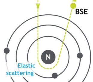
Backscattered Electrons in SEM Imaging
Unlike optical microscopes, scanning electron microsc...
Read More
EDS Analysis with SEM: How Does it Work?
From searching for food contaminants to identifying machine ...
Read More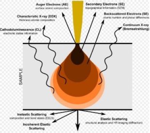
SEM: Types of Electrons and the Information They Provide
Electron microscopes are versatile instruments that can prov...
Read More
Dear sirs.
Your explanation is simple, clear and in depth.
Thanks too much.