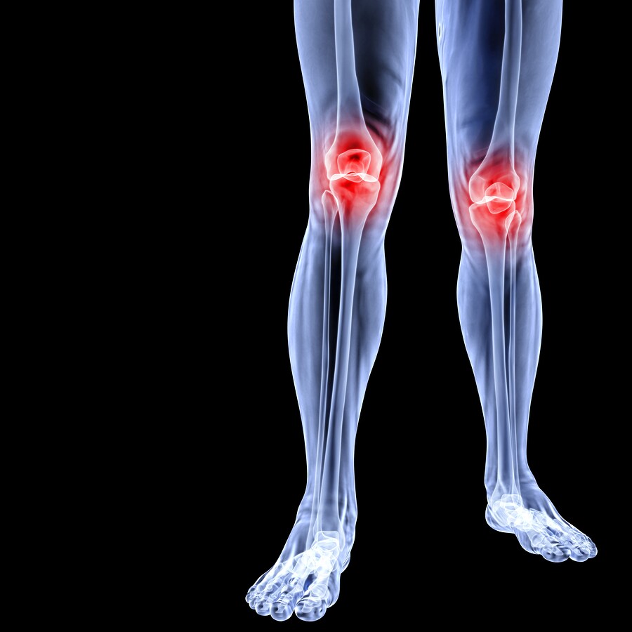
 Recently, researchers evaluated the proteome-level factors influencing the development of osteoarthritis (OA). Ritter et al. (2013) conducted a previous study utilizing liquid chromatography–tandem mass spectrometry (LC-MS/MS) to reveal 18 differentially expressed proteins in OA-affected synovial fluids (SF). Now, these researchers have combined gel electrophoresis and MS to probe more deeply into the OA SF proteome. The team compared their results with mRNA-level profiling to confirm differential expression and to articulate the particular pathways and tissue types that likely contribute to the protein-level changes accompanying OA.
For the electrophoresis-based study, the researchers labeled SF samples from OA patients and healthy controls and subjected them to two-dimensional difference-in-gel electrophoresis (DIGE) followed by MS (Thermo Scientific). After filtering the data, the research team identified 43 proteins from the first gel and 42 proteins from the second gel that demonstrated differential expression. Nineteen of the identified proteins were present in both gels. When the researchers combined the data for both experiments, 66 differentially expressed proteins emerged as clinically significant between the healthy controls and advanced OA samples. The research team further compared the expression of these proteins between samples from early and advanced OA patients and found 100% overlap, indicating the early onset of identifiable proteome-level changes in OA SF.
Ritter et al. then utilized bioinformatics to determine which pathways were affected by proteome-level dysregulation in OA SF. They found significant changes in the cellular and molecular functions associated with antigen presentation, cell-to-cell signaling and interaction, lipid metabolism, molecular transport, and small molecule biochemistry. The affected pathways were acute-phase response signaling, the complement system, the coagulation system, and the extrinsic and intrinsic prothrombin activation pathways.
The research team developed quantitative selective reaction monitoring (SRM) assays to validate their findings. They evaluated SF for 10 specific peptides associated with five of the differentially expressed proteins identified using 2-D DIGE, including afamin, clusterin, insulin-like growth factor binding protein complex acid-labile subunit (IGFALS), lumican, and pigment epithelium-derived factor. The team confirmed significant upregulation for all 10 peptides in both early and advanced OA samples.
Finally, the researchers evaluated both synovial tissue and cartilage for mRNA-level confirmation. They used expression data sets from OA-affected synovial tissue biopsy samples to probe for mRNA corresponding with 64 of the 66 differentially expressed proteins that had been identified using DIGE. The research team found that 11 of the 14 complement pathway proteins were concordantly expressed in both synovial tissue and SF. Fewer than one-third of the coagulation and acute-phase SF proteins demonstrated concordance with synovial tissue. Several other proteins, including lumican, plasma protease C1 inhibitor, and apolipoprotein A-1 were discordant and thus unlikely to be synovial derivatives.
Ritter et al. compared the protein expression from the SF findings to existing cartilage microarray data. They found concordance in the upregulation of matrix proteins cartilage acidic protein 1 and lumican, and concordant downregulation of transaldolase and glutathione peroxidase 3 in both OA cartilage and SF. Five of the 14 complement pathway proteins concordantly expressed in the cartilage tissue samples, while two of the nine coagulation system factors were concordant. When comparing the microarray transcripts and the 66 differentially expressed proteins identified with DIGE, the cartilage samples evidenced differential expression in 17 of the 66 transcripts, 10 of which were concordant. The synovial tissue samples indicated differential expression for 15 of the 66 transcripts, with 11 transcripts demonstrating concordance. The research team asserts that several of those transcripts belong to the complement and acute-phase response pathways. These findings indicate that both the synovium and cartilage may play a role in the regulation of differentially expressed proteins in OA SF.
Overall, Ritter et al. used DIGE and MS to identify a comprehensive list of differentially expressed proteins for OA SF and healthy controls, with detection of 66 proteins of clinical significance between the two groups. They also identified the three dominant pathways impacted: the acute-phase response signaling pathway, the complement pathway and the coagulation pathway. This information offers insight into the role the proteome plays in the development of OA and may prove beneficial in the search for biomarkers and therapeutic targets for the early identification, monitoring and treatment of OA.
References
Ritter, S., et al. (2013) “Proteomic Analysis of Synovial Fluid From the Osteoarthritic Knee: Comparison With Transcriptome Analyses of Joint Tissues,” Arthritis & Rheumatism, 65(4) (pp. 981–92).

Novel Autoantibody Targets in Systemic Sclerosis
Systemic sclerosis (SSc), also known as scleroderma, is an a...
Read More
Is Juvenile Idiopathic Arthritis an Autoimmune Disease?
Juvenile idiopathic arthritis (JIA) is a common rheumatologi...
Read More
Proteomics Investigates Monocyte Differentiation
Monocytes are a type of white blood cell and a vital pa...
Read More
TAILing the Terminome: Underlying Mechanisms Regulate Immune Response
A previous blog post offered a complete workflow for identif...
Read More
Leave a Reply