Search

Thermo Scientific™
PageRuler™ ungefärbte Proteinleiter, Broad Range
Die Thermo Scientific PageRuler ungefärbte Breitspektrum-Proteinleiter ist eine Mischung aus 11 Proteinen (5 bis 250 kDa) zur Verwendung als GrößenstandardsWeitere Informationen
Have Questions?
Ansicht ändern

| Katalognummer | Menge |
|---|---|
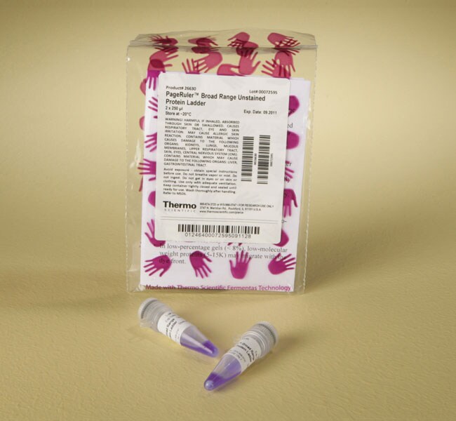
| 26630X4 | 8 x 250 μL |
| 26630 | 2 x 250 μl |
Katalognummer 26630X4
Preis (EUR)
275,65
온라인 행사
332,00Ersparnis 56,35 (17%)
Each
Menge:
8 x 250 μL
Preis (EUR)
275,65
온라인 행사
332,00Ersparnis 56,35 (17%)
Each
Die Thermo Scientific PageRuler ungefärbte Breitspektrum-Proteinleiter ist eine Mischung aus 11 Proteinen (5 bis 250 kDa) zur Verwendung als Größenstandards in der Proteinelektrophorese (SDS-PAGE) und beim Western Blotting. Die Proteinleiter wird in einem gebrauchsfertigen Format geliefert für die direkte Auftragung auf Gelen. Vor der Verwendung muss kein Probenpuffer erwärmt, reduziert oder hinzugefügt werden.
Alle anderen Proteinstandards und -leitern anzeigen und vergleichen ›
Produktmerkmale
• Referenzbanden – die 100, 50 und 20 kDa-Banden sind intensiver und erleichtern die Orientierung
• Getaggt – jedes Protein enthält eine integrierte Strep-tag™ II-Sequenz und kann mit Strep-Tactin™ Konjugaten oder einem Antikörper gegen die Strep-tag™ II-Sequenz auf Western Blots nachgewiesen werden
Anwendungen
• Genaue Größenbestimmung von Proteinen auf SDS-Polyacrylamid-Gelen und Western Blots
For Research Use Only. Not for use in diagnostic procedures.
Specifications
Molekulargewicht250, 150, 100, 70, 50, 40, 30, 20, 15, 10, 5 kDa
Menge8 x 250 μL
Sofort einsatzbereitJa
Number of Markers11
ProduktliniePageRuler
ProdukttypProteinleiter
Größenbereich5 bis 250 kDa
Stain TypeUngefärbt
System TypeSDS-PAGE
Unit SizeEach
Inhalt und Lagerung
Inhalt: acht 250 μL-Fläschchen
Lagerpuffer: 62,5 mmol Tris-H3PO4 (pH 7,5 bei 25 °C), 1 mmol EDTA, 2 % SDS, 10 mmol DTT, 1 mmol NaN3, 0,01 % Bromophenolblau und 33 % Glycerin
Lagerung: Nach Erhalt bei -20 °C lagern
Lagerpuffer: 62,5 mmol Tris-H3PO4 (pH 7,5 bei 25 °C), 1 mmol EDTA, 2 % SDS, 10 mmol DTT, 1 mmol NaN3, 0,01 % Bromophenolblau und 33 % Glycerin
Lagerung: Nach Erhalt bei -20 °C lagern