Search


Citations et références (10)
Invitrogen™
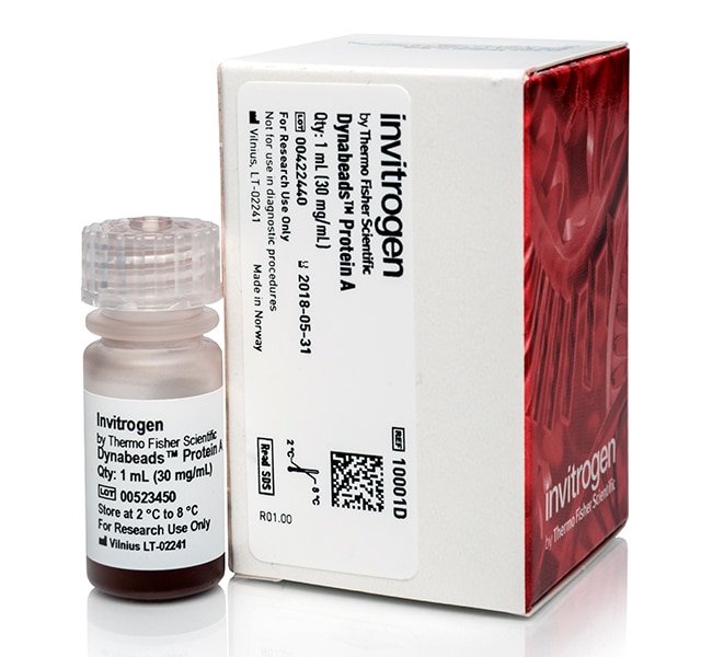
Protéine A pour immunoprécipitation Dynabeads™
Les protéines A Dynabeads sont des microbilles uniformes et superparamagnétiques de 2,8 μm avec des protéines A recombinantes (∼ 45Afficher plus
Have Questions?
Modifier l'affichage

| Référence | Quantité |
|---|---|
| 10001D | 1 ml |
| 10002D | 5 ml |
| 10008D | 50 ml |
Référence 10001D
Prix (EUR)
193,65
Offre exceptionnelle en ligne
245,00Économisez 51,35 (21%)
Each
Quantité:
1 ml
Prix (EUR)
193,65
Offre exceptionnelle en ligne
245,00Économisez 51,35 (21%)
Each
Les protéines A Dynabeads sont des microbilles uniformes et superparamagnétiques de 2,8 μm avec des protéines A recombinantes (∼ 45 kDa) couplées par covalence à la surface. Les protéines A Dynabeads offrent une meilleure alternative à la résine de Sepharose ou à la résine d’agarose pour l’immunoprécipitation (IP), et des protocoles manuels et automatisés sont disponibles.
• IP en moins de 40 minutes
• Haut rendement de protéines cibles avec faible consommation d’anticorps
• Liaison non spécifique très faible avec un rapport signal-bruit élevé
• Aucune colonne, opérations de centrifugation ni aucun pré-nettoyage chronophage nécessaire
• Grande reproductibilité et haut débit compatibles avec les instruments KingFisher
La séparation manuelle de Dynabeads est rapide et facile à exécuter
Le protocole manuel est simple et peut être exécuté en moins de 40 minutes. Tout d’abord, l’anticorps de la protéine cible est incubé avec les protéines A Dynabeads dans un tube pendant 10 minutes. L’excès d’anticorps est lavé en plaçant le tube dans un aimant DynaMag et en enlevant le surnageant. Les microbilles liées aux anticorps peuvent ensuite être utilisées pour diverses applications en aval, notamment l’IP, le Co-IP, la chromatine IP (ChIP), l’ARN IP (RIP), la purification à petite échelle de l’IgG et la purification des protéines. Les matériaux liés sont facilement rassemblés à l’aide d’un aimant DynaMag, en utilisant les propriétés magnétiques uniques des Dynabeads. La protéine A recombinante sur les billes ne contient aucun site de liaison d’albumine, l’albumine n’est donc pas co-purifiée pendant la procédure. L’IP est rapide et offre un rendement élevé, une reproductibilité élevée et très peu de liaison non spécifique, un pré-nettoyage n’est donc pas nécessaire.
La séparation Dynabeads automatisée permet d’augmenter le débit et de réduire le temps de manipulation
Si vous travaillez avec plusieurs échantillons en parallèle, le nombre d’étapes de nettoyage et le temps de manipulation augmente proportionnellement avec le nombre d’échantillons. Le pipetage et les autres manipulations manuelles ont tendance à être moins cohérents que l’automatisation lorsqu’ils travaillent avec de nombreux échantillons à la fois. Pour mieux gérer un nombre d’échantillons moyen à haut débit, réduire le temps de manipulation et garantir une reproductibilité élevée, nous avons développé des protocoles IP pour les instruments KingFisher Flex et KingFisher Duo Prime. Les protocoles automatisés répliquent les protocoles manuels, obtenant un rendement protéique cible aussi élevé, la même liaison non spécifique faible et la même reproductibilité élevée. Que vous travailliez avec 10 ou 96 échantillons, la durée du protocole IP est inférieure à 40 minutes, quel que soit le cas. Il vous suffit de charger les réactifs sur les plaques, d’appuyer sur le bouton “Start” (Démarrage) et avant la fin de votre préparation de l’analyse en aval, l’IP est terminée. Une certaine optimisation (par ex., les temps d’incubation) peut être nécessaire en fonction de votre anticorps et de l’abondance et/ou de la spécificité de votre protéine cible.
• Utilisez l’instrument KingFisher Duo pour un débit faible à moyen (1 à 12 échantillons/cycle)
• Utilisez l’instrument KingFisher Flex pour un débit élevé (12 à 96 échantillons/cycle)
Voir les protocoles automatisés
Regarder une vidéo sur l’instrument KingFisher Flex
La séparation non agressive provoque un stress physique minimal des protéines
La technologie de séparation magnétique utilisée par les protéines A Dynabeads est rapide et non agressive, ce qui provoque un stress physique minimal pour vos protéines cibles. Cela permet l’isolement et la concentration des composites labiles qui peuvent autrement se dissocier ou être endommagés par les protéases pendant les longues durées d’incubation. La conformation des protéines natives et les complexes protéiques de grande taille sont préservés.
Force de liaison et capacité
Les protéines A Dynabeads permettent d’isoler la plupart des immunoglobulines (Ig) de mammifères. La quantité d’lg capturées dépend de la concentration d’Ig dans l’échantillon de départ et du type et de la source d’Ig. 100 μl de protéine A Dynabeads– peuvent isoler approximativement 25 à 30 μg d’lgG humaine à partir d’un échantillon contenant 20 à 200 μg d’IgG/ml. La liaison prédominante à Fc permet une orientation optimale de l’lg. Les anticorps se lient à la surface externe lisse des microbilles, et ne sont donc pas piégés dans de grands pores comme les microbilles à base de Sepharose/agarose. Tous les anticorps sont disponibles pour la liaison protéique, afin de n’avoir besoin que de faibles quantités d’anticorps, tout en obtenant toujours le même rendement élevé de protéines cibles. La surface lisse des microbilles est également la raison qui explique le manque de liaison non spécifique des protéines Dynabeads.
Pour en savoir plus sur les protéines Dynabeads
• La protéine A Dynabeads est également disponible sous la forme de tampons “prêts à utiliser” inclus
• Voir les guides, les données et les références de sélection d’immunoprécipitation
• Voir les aimants de séparation des Dynabeads
• Voir les autres produits Dynabeads utiles à d’autres applications
Achat par les OEM
Pour acheter les protéines A et G Dynabeads sur la base d’un OEM, contactez notre département des sous-licences à des ventes aux OEM.
*Sepharose est une marque commerciale de GE Healthcare Bio-Sciences AB.
• IP en moins de 40 minutes
• Haut rendement de protéines cibles avec faible consommation d’anticorps
• Liaison non spécifique très faible avec un rapport signal-bruit élevé
• Aucune colonne, opérations de centrifugation ni aucun pré-nettoyage chronophage nécessaire
• Grande reproductibilité et haut débit compatibles avec les instruments KingFisher
La séparation manuelle de Dynabeads est rapide et facile à exécuter
Le protocole manuel est simple et peut être exécuté en moins de 40 minutes. Tout d’abord, l’anticorps de la protéine cible est incubé avec les protéines A Dynabeads dans un tube pendant 10 minutes. L’excès d’anticorps est lavé en plaçant le tube dans un aimant DynaMag et en enlevant le surnageant. Les microbilles liées aux anticorps peuvent ensuite être utilisées pour diverses applications en aval, notamment l’IP, le Co-IP, la chromatine IP (ChIP), l’ARN IP (RIP), la purification à petite échelle de l’IgG et la purification des protéines. Les matériaux liés sont facilement rassemblés à l’aide d’un aimant DynaMag, en utilisant les propriétés magnétiques uniques des Dynabeads. La protéine A recombinante sur les billes ne contient aucun site de liaison d’albumine, l’albumine n’est donc pas co-purifiée pendant la procédure. L’IP est rapide et offre un rendement élevé, une reproductibilité élevée et très peu de liaison non spécifique, un pré-nettoyage n’est donc pas nécessaire.
La séparation Dynabeads automatisée permet d’augmenter le débit et de réduire le temps de manipulation
Si vous travaillez avec plusieurs échantillons en parallèle, le nombre d’étapes de nettoyage et le temps de manipulation augmente proportionnellement avec le nombre d’échantillons. Le pipetage et les autres manipulations manuelles ont tendance à être moins cohérents que l’automatisation lorsqu’ils travaillent avec de nombreux échantillons à la fois. Pour mieux gérer un nombre d’échantillons moyen à haut débit, réduire le temps de manipulation et garantir une reproductibilité élevée, nous avons développé des protocoles IP pour les instruments KingFisher Flex et KingFisher Duo Prime. Les protocoles automatisés répliquent les protocoles manuels, obtenant un rendement protéique cible aussi élevé, la même liaison non spécifique faible et la même reproductibilité élevée. Que vous travailliez avec 10 ou 96 échantillons, la durée du protocole IP est inférieure à 40 minutes, quel que soit le cas. Il vous suffit de charger les réactifs sur les plaques, d’appuyer sur le bouton “Start” (Démarrage) et avant la fin de votre préparation de l’analyse en aval, l’IP est terminée. Une certaine optimisation (par ex., les temps d’incubation) peut être nécessaire en fonction de votre anticorps et de l’abondance et/ou de la spécificité de votre protéine cible.
• Utilisez l’instrument KingFisher Duo pour un débit faible à moyen (1 à 12 échantillons/cycle)
• Utilisez l’instrument KingFisher Flex pour un débit élevé (12 à 96 échantillons/cycle)
Voir les protocoles automatisés
Regarder une vidéo sur l’instrument KingFisher Flex
La séparation non agressive provoque un stress physique minimal des protéines
La technologie de séparation magnétique utilisée par les protéines A Dynabeads est rapide et non agressive, ce qui provoque un stress physique minimal pour vos protéines cibles. Cela permet l’isolement et la concentration des composites labiles qui peuvent autrement se dissocier ou être endommagés par les protéases pendant les longues durées d’incubation. La conformation des protéines natives et les complexes protéiques de grande taille sont préservés.
Force de liaison et capacité
Les protéines A Dynabeads permettent d’isoler la plupart des immunoglobulines (Ig) de mammifères. La quantité d’lg capturées dépend de la concentration d’Ig dans l’échantillon de départ et du type et de la source d’Ig. 100 μl de protéine A Dynabeads– peuvent isoler approximativement 25 à 30 μg d’lgG humaine à partir d’un échantillon contenant 20 à 200 μg d’IgG/ml. La liaison prédominante à Fc permet une orientation optimale de l’lg. Les anticorps se lient à la surface externe lisse des microbilles, et ne sont donc pas piégés dans de grands pores comme les microbilles à base de Sepharose/agarose. Tous les anticorps sont disponibles pour la liaison protéique, afin de n’avoir besoin que de faibles quantités d’anticorps, tout en obtenant toujours le même rendement élevé de protéines cibles. La surface lisse des microbilles est également la raison qui explique le manque de liaison non spécifique des protéines Dynabeads.
Pour en savoir plus sur les protéines Dynabeads
• La protéine A Dynabeads est également disponible sous la forme de tampons “prêts à utiliser” inclus
• Voir les guides, les données et les références de sélection d’immunoprécipitation
• Voir les aimants de séparation des Dynabeads
• Voir les autres produits Dynabeads utiles à d’autres applications
Achat par les OEM
Pour acheter les protéines A et G Dynabeads sur la base d’un OEM, contactez notre département des sous-licences à des ventes aux OEM.
*Sepharose est une marque commerciale de GE Healthcare Bio-Sciences AB.
Usage exclusivement réservé à la recherche. Ne pas utiliser pour des procédures de diagnostic.
Spécifications
Certifications/ConformitéISO9001 and ISO13485
Concentration30 mg/mL
DescriptionRecombinant Protein A covalently bound to Dynabeads
Diamètre (métrique)2,8 μm
À utiliser avec (application)Immunoprécipitation
À utiliser avec (équipement)KingFisher™ Duo Prime, KingFisher™ Flex
FormatMicrobilles en suspension
Compatibilité à haut débitCompatible avec le haut débit
Type de ligandProtéine A
MatériauPolystyrène
Pureté ou qualitéFor Research Use Only
Quantité1 ml
Durée de conservation24 mois à compter de la date de fabrication
Conditions d’expéditionTempérature ambiante
Suffisant pour20 Tests
Fonctionnalité de surfaceProtein A
CibleAntibodies
UniformitéMonosized 2.8 mm (CV <5%)
Gamme de produitsDynabeads
TypeMicrobilles superparamagnétiques de protéine A
Unit SizeEach
Contenu et stockage
Contenu : 1 ml de protéine A Dynabeads™
Conservation : 2°C à 8°C.
Conservation : 2°C à 8°C.
Foire aux questions (FAQ)
My Dynabeads magnetic beads are not pelleting well with the magnet. Do you have any suggestions for me?
I have a long double-stranded DNA fragment I would like to isolate. What product do you recommend?
Can I use Dynabeads magnetic beads to isolate single-stranded DNA templates?
What is the magnetic susceptibility for Dynabeads magnetic beads?
How can I determine coupling efficiency of Dynabeads magnetic beads?
Citations et références (10)
Citations et références
Abstract
Nuclear tumor necrosis factor receptor-associated factor 6 in lymphoid cells negatively regulates c-Myb-mediated transactivation through small ubiquitin-related modifier-1 modification.
Journal:J Biol Chem
PubMed ID:18093978
'Tumor necrosis factor receptor-associated factor 6 (TRAF6) is an adaptor/scaffold protein that mediates several important signaling pathways, including the tumor necrosis factor-R:NF-kappaB pathway, involved in immune surveillance, inflammation, etc. Because most studies of TRAF6 function have focused primarily on its role as an adaptor molecule in signaling pathways in the
The barrier-to-autointegration factor is a component of functional human immunodeficiency virus type 1 preintegration complexes.
Journal:J Virol
PubMed ID:12663813
'Retroviral integration in vivo is mediated by preintegration complexes (PICs) derived from infectious virions. In addition to the integrase enzyme and cDNA substrate, PICs contain a variety of viral and host cell proteins. Whereas two different cell proteins, high-mobility group protein A1 (HMGA1) and the barrier-to-autointegration factor (BAF), were identified
Constitutive expression of functionally active cyclin-dependent kinases and their binding partners suggests noncanonical functions of cell cycle regulators in differentiated neurons.
Journal:Cereb Cortex
PubMed ID:17050646
'Neurodegeneration in Alzheimer''s disease and various experimental lesion paradigms are associated with an unscheduled upregulation of cell cycle-related proteins, indicating a link between cell cycle reactivation and neuronal death. Recent evidence, however, suggests that at least some of the canonical cell cycle regulators are constitutively expressed in differentiated neurons of
A DOUBLETIME kinase binding domain on the Drosophila PERIOD protein is essential for its hyperphosphorylation, transcriptional repression, and circadian clock function.
Journal:Mol Cell Biol
PubMed ID:17452449
'A common feature of animal circadian clocks is the progressive phosphorylation of PERIOD (PER) proteins from hypo- to hyperphosphorylated species, events that are highly dependent on casein kinase 1 epsilon (termed DOUBLETIME [DBT] in Drosophila melanogaster) and necessary for normal clock progression. Drosophila PER (dPER) functions in the negative limb
Parallel activation of phosphatidylinositol 4-kinase and phospholipase C by the extracellular calcium-sensing receptor.
Journal:J Biol Chem
PubMed ID:11907035
'The calcium-sensing receptor (CaR) is a G protein-coupled receptor that regulates physiological processes including Ca(2+) metabolism, Na(+), Cl(-), K(+), and H(2)0 balance, and the growth of some epithelial cells through diverse signaling pathways. Although many effects of CaR are mediated by the heterotrimeric G proteins Galpha(q) and Galpha(i), not all