Search Thermo Fisher Scientific
Far–Western Blot Analysis
Far–western blot analysis is an alternative method to analyze protein–protein interactions that entails probing proteins that have been separated by gel electrophoresis with tagged bait proteins, and then detecting the interacting proteins (prey) through multiple methods. In recent years, far–western blot analysis has been used to determine receptor–ligand interactions and screen libraries for interacting proteins. With this method of analysis, it is possible to study the effect of post-translational modifications on protein–protein interactions, examine interaction sequences using synthetic peptides as probes, and identify protein–protein interactions without using antigen-specific antibodies.
View and select products
Studying protein–protein interactions by far-western blotting
Far–western blot vs. western blot
The far–western blot technique is similar to western blotting; in a western blot, an antibody is used to detect the corresponding antigen on a membrane, while in a classical far-western analysis, a labeled or antibody-detectable "bait" protein is used to probe and detect a target "prey" protein on the membrane. The sample (usually a lysate) containing the unknown prey protein is separated by sodium dodecyl sulfate–polyacrylamide gel electrophoresis (SDS-PAGE) or native PAGE and then transferred to a membrane. After transfer, the membrane is blocked and then probed with a known bait protein, which usually is applied in pure form; alternatively, this reaction can be performed in-gel, as described later in this review. Following the reaction of the bait with the prey protein, a detection system, dependent upon the bait protein used, identifies the band that corresponds to the prey protein.
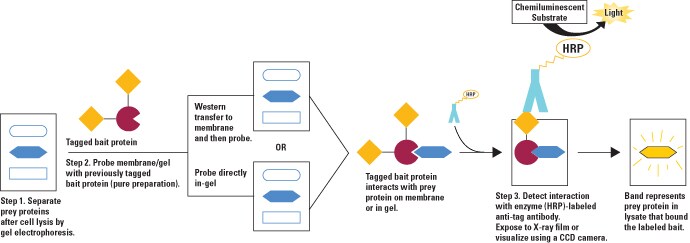
Diagram of far–western blot to analyze protein–protein interactions. In this example, a tagged bait protein is used to probe either the transfer membrane or a gel for the prey protein. Once bound, enzyme (horseradish peroxidase; HRP)-conjugated antibody that targets the bait tag is used to label the interaction, which is then detected by enzymatic chemiluminescence. This general approach can be adjusted, as shown in the table below, by using untagged bait protein that is detected by antibody, biotinylated bait protein that is detected by enzyme-conjugated streptavidin, or radiolabeled bait protein that is detected by exposure to film.
Comparison of western blot and far–western blot methods
| Step | Western blotting | Far-western blotting |
| Gel electrophoresis | Native or denaturing (usually) | Native (usually) or denaturing |
| Transfer system | Optimal membrane and transfer system determined empirically | Optimal membrane and transfer system determine empirically |
| Blocking buffer | Optimal blocking system determined empirically | Optimal blocking system determined empirically |
| Detection strategies † (Arrows (→) denote the sequence of steps of the detection strategy) | Unlabeled primary antibody→ Enzyme-labeled secondary antibody→ Substrate reagent | Unlabeled bait protein→ Enzyme-labeled bait-specific antibody→ Substrate reagent |
| Enzyme-labeled secondary antibody→ Substrate reagent | Radiolabeled bait protein→ Exposure to film | |
| Biotinylated antibody→ Enzyme-labeled streptavidin→ Substrate reagent | Biotinylated bait protein→ Enzyme-labeled streptavidin→ Substrate reagent | |
| Fusion-tagged bait protein→ Tag-specific antibody→ Enzyme-labeled secondary antibody→ Substrate reagent | ||
| †Labeled antibodies are generally labeled with an enzyme (either horseradish peroxidase or alkaline phosphatase). By contrast, bait proteins generally are not enzyme labeled because a large enzyme label is likely to sterically hinder unknown binding sites between bait and prey proteins. Other labeling and detection schemes are possible. | ||
Protein Transfer Technical Handbook
This 23-page handbook provides an in-depth description of protein transfer, which is a vital step in western blotting. The transfer of proteins involves separation in a gel by electrophoresis to a solid support matrix so that specific proteins can be identified using immunodetection techniques. We offer a complete array of products to support rapid and efficient protein transfer for western blotting. Our portfolio of high quality protein transfer products unites membranes, buffers, stains, and molecular weight markers alongside a comprehensive choice of transfer devices designed to suit your needs and enable better quality western blot results.

Critical factors of far–western blot analysis
Importance of native prey protein structure in far-western analysis
Far-western blotting procedures must be performed with care and attention to preserving the native conformation and interaction conditions for the proteins under study. Denatured proteins may not interact with their respective binding partner(s), resulting in a failure to identify an interaction. Alternatively, proteins presented in non-native conformations may interact in novel, artificial ways, resulting in false positive interactions. The prey protein in particular is subjected to preparative processing steps for far–western blotting that can have significant effects on the detection of protein–protein interactions. This is not to imply that the identification of valid interactions is not possible, but only to stress the importance of appropriate validation and the use of controls in all far-western analyses.
Gel electrophoresis
Protein separation by SDS-PAGE (i.e., denaturing conditions with or without a reducing agent) offers more information about the molecular weight, the presence of disulfide bonds and the subunit composition of a prey protein, but may also render the protein unrecognizable by the bait protein. In these cases, electrophoresis under native conditions (i.e., nondenaturing and without reducing agent) should be used.


Precast vs. handcast protein gels for SDS-PAGE. Polyacrylamide gels can be purchased precast and ready to use (left) or prepared from reagents in the lab using a gel-casting system (right). Pictured here are the Novex WedgeWell 4-20% Tris-Glycine Mini Gels (left) and the Invitrogen SureCast Gel Handcast System. Although no universal standard exists among manufacturers, the term “mini gel” typically refers to gels that are 1 mm thick and approximately 8 x 8 cm. Cassette dimensions and compatibility between tanks varies. Most Invitrogen mini gel cassettes, including those shown here, are compatible with the Invitrogen Mini Gel Tank.
Protein transfer
After gel separation, proteins are electrophoretically transferred from the gel to a membrane. The type of membrane (e.g., nitrocellulose or PVDF) used for the protein transfer is critical, as some proteins bind selectively or preferably to a particular membrane. The protein transfer rate is inversely proportional to the molecular weight of the protein. In some cases, transfer conditions alter the conformation of the protein and destroy or sterically hinder the interaction site on the protein.

Polyacrylamide gel electrophoresis in progress. Prepared gel cassettes are added to a gel tank, in this case the Invitrogen Mini Gel Tank, which holds two mini gels at a time. After wells are loaded with protein samples, the gels submerged in a conducting running buffer, and electrical current is applied, typically for 20 to 40 minutes. Run times vary according to the size and percentage of the gel and particular gel chemistry.
For far-western analysis, it is essential that at least the interaction domain of the prey protein is not disrupted by the transfer or is able to re-fold on the membrane to form a three-dimensional structure comprising an intact interaction site. Generally, when SDS is eliminated during the transfer process, proteins generally renature with greater efficiency and are, therefore, more easily detected by far-western blotting.
In the event that the protein is unable to re-fold to create an intact binding site, guanidinium hydrochloride may be added to denature the membrane-bound proteins, and after the reagent is removed the proteins are provided another change to fold properly. An alternative to this approach, if renaturation does not occur, is to perform the far-western analysis in-gel, without the need to transfer proteins to a membrane (see ‘In-gel far-western detection’ below).
Blocking buffer
After transferring proteins to the membrane, western blotting procedures require that unreacted binding sites on the membrane be blocked with a non-relevant protein solution. By blocking all remaining binding sites on the membrane, a blocking buffer reduces nonspecific binding and aids in protein renaturation. A variety of different protein blockers may be used, and no one blocking protein solution will work for all blotting experiments. Any given protein blocker may crossreact or otherwise disrupt the target protein–protein interaction.
The determination of an effective blocking buffer must be made empirically. Bovine serum albumin (BSA) is often used as a starting point for many membrane-probing reactions. Insufficient blocking may result in high background, whereas prolonged blocking could result in a weak or masked signal. Renaturation of the protein also occurs during the blocking step, so it is important to optimize the blocking conditions to obtain the best signal-to-noise ratio for each application. A variety of blocking buffers ranging from milk or normal serum to highly purified proteins have been used to block free sites on a membrane. The blocking buffer should improve the sensitivity of the assay by reducing background interference and improving the signal-to-noise ratio. The proper choice of blocker for a given blot depends on the antigen itself and on the type of detection label used. For example, in applications where alkaline phosphatase conjugates are used, a blocking buffer in TBS should be selected because PBS interferes with alkaline phosphatase.

Comparison of SuperBlock Blocking Buffer and milk. Two-fold serial dilutions of HeLa cell lysate (20, 10, 5, 2.5, 1.25, 0.625, and 0.3125 µg) were separated by SDS-PAGE and transferred to nitrocellulose (panels A–C) or PVDF (panels D–E) membranes. Membranes were blocked for 1 hour with 5% nonfat milk in Tris-buffered saline and 0.05% Thermo Scientific Tween 20 detergent, or Thermo Scientific SuperBlock Blocking Buffer in phosphate-buffered saline with 0.05% Tween 20 detergent. Blots were processed for 5 minutes using Thermo Scientific SuperSignal West Pico Chemiluminescent Substrate (Cat. No. 34080) and exposed to film. The results show that SuperBlock Blocking Buffer is superior to milk for detection of target proteins.
Binding and wash conditions
Protein–protein interactions vary depending on the nature of the interacting proteins. The strength of the interactions may depend on the pH, salt concentration and the presence of certain co-factors during incubation with the bait protein. Whatever the necessary conditions, they must be maintained throughout the procedure to preserve the interaction until it can be detected. This may influence the formulation of wash buffers used between probing steps.
Detection methods
As indicated in the above table, multiple approaches can be used to detect far–western blot protein–protein interactions depending on the presence of a label or tag on the bait protein, and include:
- Direct detection of prey protein with a radioactive bait protein
- Indirect detection with an antibody specific to the bait protein
- Indirect detection with an antibody specific to the tag of a fusion-tagged bait protein
- Indirect detection with a biotinylated bait protein and enzyme-conjugated avidin or streptavidin
Each method has its own advantages and disadvantages, as described below.
There are several methods to generate radioactive isotope labels on bait proteins. The isotope P-32 is commonly used to label fusion-tagged protein probes at phosphorylation sites on the tag. This method of phosphorylation has little effect on the protein–protein interaction, because the phosphorylation site is located in the fusion tag portion of the protein. Another radioactive method is to directly label bait protein at endogenous phosphorylation sites. However, this technique can only be used if P-32 labeling does not interfere with the interaction of the bait and prey proteins. Radioactive detection has also been used with probes made by the incorporation of S-35-methionine during in vitro translation. One disadvantage of this method is that it can only be used for protein probes that have multiple methionine or cysteine residues.
Although radioactive isotopes generally do not interfere with interactions, isotopic detection methods have several disadvantages, including health hazards and disposal issues. GST- or histidine-tagged recombinant bait proteins are often detected with antibodies that are specific to the tag; antibodies to both of these popular fusion tags are commercially available. When recombinant techniques cannot be used to create fusion-tagged bait proteins and bait-specific antibodies are not available, bait proteins can be biotinylated and detected with labeled avidin or streptavidin.
Whatever non-isotopic labeling used, the last probing step usually involves an antibody or streptavidin probe that is conjugated to an enzyme to generate a chemiluminescent response that is localized to the protein interaction on the membrane or in the gel. Horseradish peroxidase (HRP) and alkaline phosphatase (AP) are the most popular enzyme labels used for this purpose, with HRP being the most versatile. As with traditional western blotting, sensitivity in far-western blotting depends largely on the enzyme–substrate system used for detection.
Although lysate containing the bait protein can be used for probing membranes, this can result in high background; therefore, it is preferable to purify the bait protein before probing. The example below compares the use of two reagents used for chromogenic western blot detection.
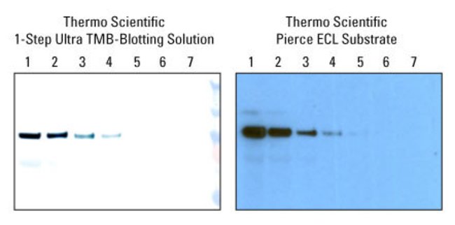
Chromogenic western blot detection. Thermo Scientific 1-Step Ultra TMB-Blocking Solution provides sensitivity similar to Thermo Scientific Pierce ECL Western Blotting Substrate. These reagents detect horseradish peroxidase (HRP) enzyme activity.
Controls
When identifying protein–protein interactions by far–western blot analysis, it is important to include appropriate controls to distinguish true protein–protein interaction bands from nonspecific artifacts. For example, experiments involving detection with recombinant GST fusion proteins should be replicated with GST alone. A bait protein with a mutation in the predicted interaction domain can be processed as a control to determine the specificity of the protein–protein interaction. A non-relevant protein can also be processed alongside the prey protein sample as a negative control; ideally, this protein would be similar in size and charge to the protein under investigation and would not interact with the bait protein.
In approaches that use a secondary system to detect the prey protein, such as enzyme-labeled streptavidin with a biotinylated bait protein, it is important to include a duplicate control membrane that is probed only with the labeled streptavidin to discover any bands resulting from endogenous biotin in the sample or nonspecific binding of the labeled streptavidin. When a fusion tag is used with a corresponding antibody, it is critical to probe one of the control membranes with the labeled antibody alone. This control helps to confirm that the relevant band is not the result of nonspecific binding of the labeled secondary antibody. To obtain meaningful results, appropriate test and control experiments should be subjected to gel electrophoresis, transfer and probing in parallel with the test sample.
In-gel far-western detection
Because of restrictions associated with the transfer process, blocking and the possibility of nonspecific binding of bait proteins to unrelated bands on the membrane, it is sometimes advantageous to perform far-western detection within the gel. In this procedure, prey protein samples are separated in either native or denaturing gels. Following electrophoresis, the gels are pre-treated with 50% isopropyl alcohol and water to remove SDS from the gel and allow the prey protein to renature. The gel is then incubated with the bait protein, and the target protein–protein interaction is detected by one of the methods described above.
The same controls and experimental conditions necessary for optimization of membrane-based far–western blots apply to in-gel detection. The blocking step can be eliminated with in-gel detection, but the bait protein and the labeled detection protein must be diluted in blocking buffer to reduce nonspecific binding. Also, higher amounts of prey and bait proteins are often required for in-gel detection compared to membrane detection using equivalent chemiluminescent substrate.
Importance of native prey protein structure in far-western analysis
Far-western blotting procedures must be performed with care and attention to preserving the native conformation and interaction conditions for the proteins under study. Denatured proteins may not interact with their respective binding partner(s), resulting in a failure to identify an interaction. Alternatively, proteins presented in non-native conformations may interact in novel, artificial ways, resulting in false positive interactions. The prey protein in particular is subjected to preparative processing steps for far-western blotting that can have significant effects on the detection of protein–protein interactions. This is not to imply that the identification of valid interactions is not possible, but only to stress the importance of appropriate validation and the use of controls in all far-western analyses.
Recommended reading
- Burgress RR et al. (2000) Mapping protein-protein interaction domains using ordered fragment ladder far-western analysis of hexahistidine-tagged fusion proteins. Methods Enzymol 328:141–157.
- Edmondson DG, Dent SYR (2001) Identification of protein interactions by far western analysis. Current Protocols in Protein Science 19.7.1–19.7.10.
- Golemis E (2002) Protein-protein interactions: A molecular cloning manual. Cold Spring Harbor (NY): Cold Spring Harbor Laboratory Press. p ix, 682.
- Reddy VM, Kumar B (2000) Interaction of Mycobacterium avium complex with human respiratory epithelial cells. J Infect Dis 181:1189–1193.
For Research Use Only. Not for use in diagnostic procedures.