 The Africanized honeybee (AHB) originated with a cross-breeding experiment between Apis mellifera scutellata and European drones over half a century ago. It has since been found in parts of South, Central and North America. Recently, Resende et al. used a label-free quantification approach to compare the proteome and phosphoproteome of AHB venom with that of two European subspecies, namely Apis mellifera ligustica and A. m. carnica.1
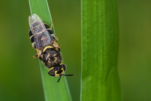
Africanized honeybees are notoriously aggressive and referred to as “killer bees” for very good reasons. The venom packed inside the AHB’s stinger is loaded with histamine, dopamine, serotonin, melittin and phospholipases that can result in adverse health effects, including potentially fatal toxic and allergic reactions. Another critical feature of AHB venom is that its exact molecular composition may vary throughout the year.2 The strong immunological, physiological and neurological responses triggered in response to the toxins found in AHB venom have long spurred interest in researchers, especially regarding the potential for developing novel pharmaceuticals, including successful antivenin treatments.
The research team began by capturing and immobilizing AHBs via quick freezing at −20°C. They dissected the sting apparatus and removed the venom reservoirs. They also lyophilized and stored the venom sacs at −20°C. They manually extracted venom samples by squeezing the venom reservoirs against the vials and solubilizing their content in water. The team purchased A. m. ligustica venom from a private supplier, while an apiary donated the samples of A. m. carnica venom.
Using a BCA assay, the researchers quantified the protein amounts in AHB venom. After alkylating and digesting the venom samples with trypsin, they injected the samples into a Dionex UltiMate 3000 nano-HPLC system (Thermo Scientific). The team obtained mass spectrometry (MS) and collision induced dissociation data using an LTQ Orbitrap Velos hybrid ion trap-Orbitrap mass spectrometer (Thermo Scientific).
The team generated peak lists of MS2 spectra .raw files using Trans-Proteomic Pipeline (version 4.3.1 [34]) and Mascot (version 2.2.2, Matrix Science) software. They compared the MS data alongside database entries for A. mellifera extracted from the NCBI database. Out of 51 proteins identified, the researchers found that 42 were common to all three subspecies. A gene ontology analysis of these proteins revealed unknown functions for 43% of them.
The researchers identified many novel proteins with likely roles in the envenoming process and/or venom self-protection. Most notably, the researchers identified the phosphorylated forms of the toxins melittin and icarapin, the latter of which is also an allergen. They determined icarapin was phosphorylated at the 205Ser residue, which is located in close proximity to its known antigenic site. Melittin was phosphorylated in all venoms at the 10Thr and 18Ser residues.
The researchers posited that the phosphorylated melittin and icarapin might also exert an allergenic response that differed from that of the native peptide; they found that phosphorylation reduced melittin activity as compared to the native peptide. With further testing, the team compared the toxicities of the phosphorylated/unphosphorylated forms. 18Ser phosphorylated melittin, the major of its two phosphorylated forms, was less toxic compared to the native peptide. Resende et al. found that melittin possesses the ability to lyse erythrocyte membranes. It also can participate in cell signaling on the membrane surface of other cells and stimulate inflammatory cells. Taken together, this work emphasizes the importance of characterizing post-translational modifications in the study of venoms.
References
1. Resende, V.M., et al. (2013, September) “Proteome and phosphoproteome of Africanized and European honeybee venoms,” Proteomics, 13 (pp. 2638–48), doi: 10.1002/pmic.201300038.
2. Schumacher, M.J., et al. (1992) “Biochemical variability of venoms from individual European and Africanized honeybees (Apis mellifera),” Journal of Allergy and Clinical Immunology, 90 (pp. 59–65).
Post Author: Emily Humphreys. Emily has previous research experience in eye development, infectious diseases, and aging. While she enjoyed the thrill of research, She has since traded bench work for science journalism. Emily has been a regular contributor to Accelerating Science since 2012.

Assessing Ion Interference Using a Triple Knockout Standard Diagnostic Tool
Researchers use isobaric tandem mass tag (TMT) reagents as a...
Read More
Identifying Metal Species with Electrospray Ionization and Tandem Mass Spectrometry
Elemental transition metals iron (Fe), zinc (Zn), nickel (Ni...
Read More
Setting BPA Free: Bisphenol Fragmentation Pathways
Bisphenols are common additives in everyday products like pl...
Read More
Creepy Science: Black Widow Silk
Spiders possess a singular ability to strike irrational fear...
Read More
Leave a Reply