By Parveen Dawood and Sarvani Emani
Learn more about the TNFR pathway and CRISPR-validated* Invitrogen antibodies.
What is the significance of the TNFR pathway and what are the key proteins?
The TNFR pathway is one of the most studied oncogenic and apoptotic pathways. Tumor necrosis factor-alpha (TNF-alpha) is a proinflammatory cytokine which plays a key role in carcinogenesis, cancer progression, metastasis and immunity. Signaling mediated by TNF through its receptors TNFR1 and TNFR2, is essential for cell proliferation, differentiation, apoptosis, modulation of immune responses and induction of inflammation.
Binding of the trimeric TNF-alpha to the TNFR1 results in trimerization of the receptor and recruitment of TNFR1-associated death domain protein (TRADD) and receptor-interacting protein (RIP). TRADD further recruits another adaptor protein, TNF-receptor-associated factor 2 (TRAF2). This TNF-alpha receptor complex activates transforming growth factor-β-activated kinase 1 (TAK1) in a RIP dependent manner. TAK1 leads to the aggregation of a downstream kinase complex – IKK complex. This complex is composed of the catalytic components- IKKα and IKKβ – and a regulatory protein, NF-κB essential modifier (NEMO). Inhibitory κB (IκB) tightly regulates nuclear factor-kappaB transcription factor (NF-κBp60 and NF-κBp50). Phosphorylation of IκB by the IKK complex results in the release of the NF-κB transcription factor, followed by ubiquitin-dependent degradation of IκB by the 26S proteosome complex. NF-κB translocates to the nucleus and activates genes involved in cell proliferation and anti-apoptosis. In addition to NF-κB activation, RIP has been suggested as a component of the p38 pathway. TAK1 is known to activate MKK3 which then phosphorylates p38. Phospho-p38 is then imported into the nucleus where it phosphorylates specific transcription factors that activate several stress and growth related genes (Figure 1).
Figure 1 – TNFR pathway showing downstream effectors

What methods are used to address antibody specificity?
Testing antibody performance against genetically modified samples is one way to verify that an antibody recognizes a specific target. Measuring the relevant signal in control cells versus cells in which the target gene has been knocked out using CRISPR-Cas9 technology is used to assess specificity of the antibody. Elimination of target protein expression prevents antibody binding and demonstrates that the antibody is specific. Example of this strategy are shown in Figure 2,3 and 4.
Figure 2
Western blot analysis of TRADD (Fig. 1a) was performed using whole cell extracts of ME-180 CAS9 Control (lane 1), ME-180 CAS9 Scrambled (lane 2), ME-180 TNFR1 KO (lane 3) and ME-180 TRADD KO (lane 4). TRADD was detected at ~ 32 kDa using Anti- TRADD Rabbit Polyclonal Antibody (Product # PA5-17465, 1:1000 dilution). Loss of signal in CRISPR mediated knockout (KO) confirms antibody specificity.
Western blot analysis of IKKα (Fig. 1b) was performed using whole cell extracts of ME-180 CAS9 Control (lane 1), ME-180 CAS9 Scrambled (lane 2), ME-180 TNFR1 KO (lane 3), ME-180 TRADD KO (lane 4) and ME-180 IKKα KO (lane 5). IKKα was detected at ~ 80 kDa using Anti-IKK alpha Rabbit Polyclonal Antibody (Product # PA5-17803, 1:1000 dilution). Loss of signal in CRISPR mediated knockout (KO) confirms antibody specificity.

CRISPR mediated knock out of NF-κBp65 and NF-κBp50 were also performed to address specificity of antibodies against respective unmodified proteins (Fig. 3 and Fig. 4).
Figure 3
Western blot analysis of NFkB p65 was performed using whole cell extracts of HeLa control (lane 1), HeLa NFkB p65 knockout (lane 2). NFkB p65 was detected at 65 kDa using NFkB p65 mouse monoclonal antibody (Product # 33-9900, 1 µg/mL). Loss of signal in CRISPR mediated knockout (KO) confirms that antibody is specific to NFkB p65.

Figure 4
Western blot analysis of NFkB p50 was performed using whole cell extracts of HeLa control (lane 1), HeLa NFkB p50 knockout (lane 2). NFkB p50 was detected at 105/50 kDa using Anti-NFkB p50 Monoclonal Antibody (Product # MA5-15860, 1:1000 dilution). Loss of signal in CRISPR mediated knockout (KO) confirms that antibody is specific to NFkB p50.

How is CRISPR-CAS9 used to assess the specificity of antibodies against downstream effectors in the pathway?
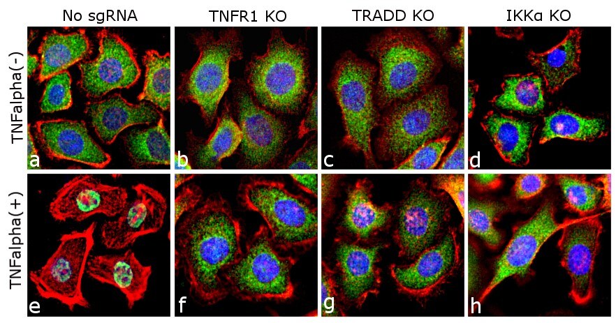
CRISPR-Cas9 is a powerful tool to study pathway dynamics and signaling cascades. Specificity of antibodies against activated downstream targets can be determined by knocking out key upstream proteins in a pathway. Treatment of ME-180 cells with TNFα results in recruitment of TRADD, which further leads to the activation of all downstream targets. NF-κB is released and translocates to the nucleus and p38 is phosphorylated. In contrast, when upstream mediators like TNFR1, TRADD or IKKα are knocked out, the activation cascade is blocked, even when treated with TNF-alpha. Figure 5 highlights the effect of knocking out TNFR1, TRADD or IKK-alpha on the nuclear translocation of NF-κB. Downstream events like phosphorylation is arrested due to knock out of the upstream protein, like TRADD. This is shown in figure 6 where there is a loss of phosphorylation of p38 in TRADD knock-out ME-180 cells when compared to control cells upon induction with TNF-alpha.
Figure 5
Specificity of anti-NF-κB p65 (Product # 710048) was demonstrated using CRISPR-Cas9 based TNFR1, TRADD and IKK alpha knockout (KO) cell lines. TNFalpha induced nuclear translocation of NF-κB, a downstream target in the TNFR1, TRADD and IKK alpha was observed in control cell line (panels a,e) and not in the TNFR1, TRADD and IKK alpha knockout cell lines (panels b-d; f-h). Immunofluorescence analysis of NFkB p65 was done on 70% confluent log phase ME-180 Cas9 cells. The cells were either mock treated or treated with TNF-alpha (50 ng/mL for 20 min). The cells were subsequently labeled with NFkB p65 (Green) Rabbit Oligoclonal Antibody (710048) at 1µg/mL in 0.1% BSA. Nuclei (Blue) were stained with SlowFade® Gold Antifade Mountant DAPI (S36938). F-actin (Red) was stained with Rhodamine Phalloidin (Product # R415, 1:300). TNF alpha induced nuclear translocation of NFkB, a downstream target in the TNFR1, TRADD and IKK alpha was observed in control cell line (panels a,e) and not in the TNFR1, TRADD and IKK alpha knockout (KO) cell lines (panels b-d; f-h). The images were captured at 40X magnification.

Figure 6
Western blot analysis of phosphorylation of p38 was performed with whole cell extracts of ME-180 CAS9 Control (lane1), ME-180 CAS9 Control treated with TNFα (50ng/mL for 20mins) (Lane 2), ME-180 TRADD KO (Lane 3) and ME-180 TRADD KO treated with TNFα (50ng/mL for 20mins) (Lane 4). Phospho p38 was detected at ~ 38 kDa using Phospho-p38 MAPK alpha (Thr180, Tyr182) Monoclonal Antibody (F.52.8) (MA5-15177). Upon induction with TNFα, phosphorylation of the downstream targets of TRADD (p38) was observed in control cell line lanes (1,2) and not in the TRADD knockout cell lines lanes (3,4).

CRISPR-validated Invitrogen antibodies recommended for the TNFR pathway
*The use or any variation of the word “validation” refers only to research use antibodies that were subject to functional testing to confirm that the antibody can be used with the research techniques indicated. It does not ensure that the product(s) was validated for clinical or diagnostic uses.
For Research Use Only. Not for Use in Diagnostic Procedures.


Leave a Reply