Search


Citations & References (5)
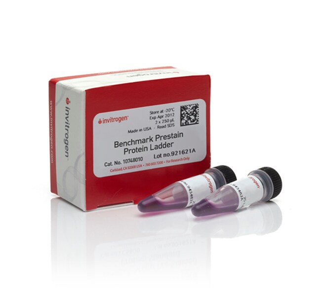
Invitrogen™
BenchMark™ Pre-stained Protein Ladder
Benchmark 染色済みタンパク質ラダーは、電気泳動のモニタリングや、ウェスタンブロッティングでのゲルから膜への転写に使用する、見かけ分子量 6~180 kDa 10種類のタンパク質を含みます。タンパク質標準物質はすぐに使用できるフォーマットで提供され、ゲルに直接ロードできます詳細を見る
| 製品番号(カタログ番号) | 数量 |
|---|---|
| 10748010 | 2x250 μL |
製品番号(カタログ番号) 10748010
価格(JPY)
31,000
Each
数量:
2x250 μL
Benchmark 染色済みタンパク質ラダーは、電気泳動のモニタリングや、ウェスタンブロッティングでのゲルから膜への転写に使用する、見かけ分子量 6~180 kDa 10種類のタンパク質を含みます。タンパク質標準物質はすぐに使用できるフォーマットで提供され、ゲルに直接ロードできます。使用前に加熱、還元、またはサンプルバッファーを追加する必要はありません。
その他のあらゆるタンパク質標準物質およびラダーの比較と閲覧›
アプリケーション
•SDSポリアクリルアミドゲル電気泳動中のタンパク質の移動のモニタリング
• ウェスタンブロッティング後のタンパク質の膜転写のモニタリング
• SDS-PAGEゲルおよびウェスタンブロット上のタンパク質のサイジング
その他のあらゆるタンパク質標準物質およびラダーの比較と閲覧›
アプリケーション
•SDSポリアクリルアミドゲル電気泳動中のタンパク質の移動のモニタリング
• ウェスタンブロッティング後のタンパク質の膜転写のモニタリング
• SDS-PAGEゲルおよびウェスタンブロット上のタンパク質のサイジング
研究用にのみ使用できます。診断用には使用いただけません。
仕様
検出法比色法
ゲル適合性Novex™トリス-グリシンゲル
分子量180、115、82、64、49、37、26、19、15、6 kDa
数量2x250 μL
ロード可能状態可
出荷条件氷水またはドライアイスでの出荷が承認されています
Number of Markers10
製品ラインBenchMark
製品タイプタンパク質ラダー
サイズ範囲6~180 kDa
Stain Type2色:ブルー、ピンク
System Typeウェスタンブロッティング、SDS-PAGE
Unit SizeEach
組成および保存条件
それぞれ 250 μLの2本のバイアルは、50 mMトリス-HCl(pH 6.8)、5 mM EDTA、10 mM DTT、1%(w/v)SDS、10%(w/v)グリセロールで構成されるローディングバッファーに含まれています。-20°Cで保存凍結や解凍を繰り返し行わないでください。
よくあるご質問(FAQ)
What is the amount of protein in BenchMark Pre-Stained Ladders?
I used one of your pre-stained protein standards for a western transfer and I noticed that the intensity of the band faded from the membrane during the transfer process. Why is this?
I used one of your protein standards for a western transfer and noticed that some of the lower-molecular weight protein bands passed through the membrane. How can I resolve this issue?
I used one of your protein standards for a western transfer and noticed that some of the higher-molecular weight bands transferred very poorly to the membrane. Can you offer some tips?
I used one of your pre-stained standards on a Tris-Glycine gel and noticed that the molecular weights of the proteins were different than on a NuPAGE Bis-Tris gel. What is the reason for this?
引用および参考文献 (5)
引用および参考文献
Abstract
2F3 monoclonal antibody recognizes the O26 O-antigen moiety of the lipopolysaccharide of enterohemorrhagic Escherichia coli strain 4276.
Journal:Clin Diagn Lab Immunol
PubMed ID:15138178
'Enterohemorrhagic Escherichia coli (EHEC) and enteropathogenic E. coli (EPEC) organisms are groups of pathogenic strains whose infections are characterized by a typical lesion of enterocyte attachment and effacement. They are involved in enteric diseases both in humans and in animals, and EHEC strains can be responsible for hemolytic uremic syndrome
In the Absence of the First Membrane-spanning Segment of Subunit 4(b), the Yeast ATP Synthase Is Functional but Does Not Dimerize or Oligomerize.
Journal:J Biol Chem
PubMed ID:11799128
'The N-terminal portion of the mitochondrial b-subunit is anchored in the inner mitochondrial membrane by two hydrophobic segments. We investigated the role of the first membrane-spanning segment, which is absent in prokaryotic and chloroplastic enzymes. In the absence of the first membrane-spanning segment of the yeast subunit (subunit 4), a
The formation of highly soluble oligomers of alpha-synuclein is regulated by fatty acids and enhanced in Parkinson's disease.
Journal:Neuron
PubMed ID:12597857
Accumulation of misfolded proteins as insoluble aggregates occurs in several neurodegenerative diseases. In Parkinson's disease (PD) and dementia with Lewy bodies (DLB), alpha-synuclein (alpha S) accumulates in insoluble inclusions. To identify soluble alpha S oligomers that precede insoluble aggregates, we probed the cytosols of mesencephalic neuronal (MES) cells, normal and
The typically mitochondrial DNA-encoded ATP6 subunit of the F1F0-ATPase is encoded by a nuclear gene in Chlamydomonas reinhardtii.
Journal:J Biol Chem
PubMed ID:11744727
The atp6 gene, encoding the ATP6 subunit of F(1)F(0)-ATP synthase, has thus far been found only as an mtDNA-encoded gene. However, atp6 is absent from mtDNAs of some species, including that of Chlamydomonas reinhardtii. Analysis of C. reinhardtii expressed sequence tags revealed three overlapping sequences that encoded a protein with
Bee Venom Phospholipase Inhibits Malaria Parasite Development in Transgenic Mosquitoes.
Journal:J Biol Chem
PubMed ID:12167627
Malaria kills millions of people every year, and new control measures are urgently needed. The recent demonstration that (effector) genes can be introduced into the mosquito germ line to diminish their ability to transmit the malaria parasite offers new hope toward the fight of the disease (Ito, J., Ghosh, A.,