Search


Citations & References (6)
Invitrogen™
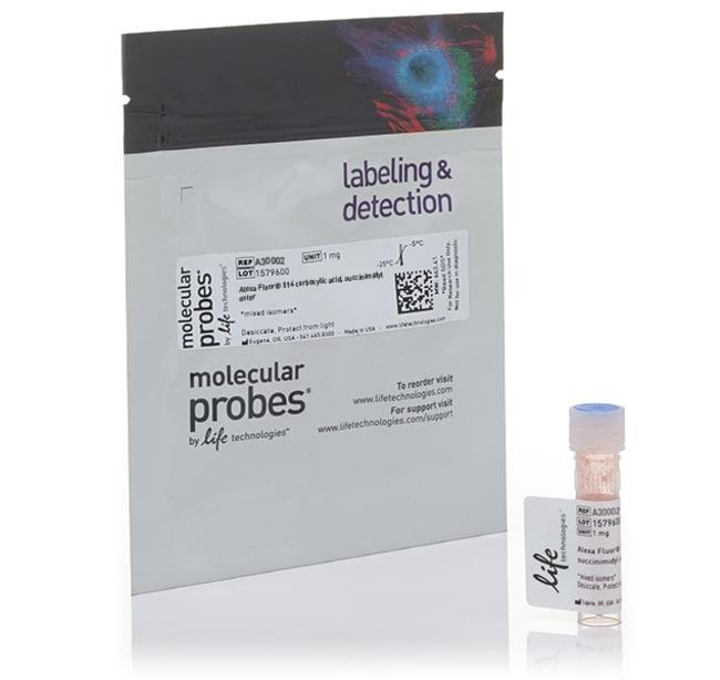
Alexa Fluor™ 514 NHS Ester (Succinimidyl Ester)
Alexa Fluor™ 514は、明るい緑色蛍光色素です。Alexa Fluor™ 514色素は、イメージングやフローサイトメトリーで安定したシグナルを生成するために使用され、水溶性でpH 4~10の影響を受けません詳細を見る
| 製品番号(カタログ番号) | 数量 |
|---|---|
| A30002 または、製品番号A-30002 | 1 mg |
製品番号(カタログ番号) A30002
または、製品番号A-30002
価格(JPY)
88,000
Each
数量:
1 mg
Alexa Fluor™ 514は、明るい緑色蛍光色素です。Alexa Fluor™ 514色素は、イメージングやフローサイトメトリーで安定したシグナルを生成するために使用され、水溶性でpH 4~10の影響を受けません。反応性色素の組成に加えて、細胞の標識および検出用に最適化されたさまざまな抗体、ペプチド、タンパク質、トレーサー、および増幅基質に結合したAlexa Fluor™ 514色素を提供しています。
Alexa Fluor™ 514のNHSエステル(またはスクシンイミジルエステル)は、タンパク質または抗体にこの色素を結合させるための最も一般的なツールです。NHSエステルは、タンパク質の一級アミン(R-NH2)、アミン修飾オリゴヌクレオチド、およびその他のアミン含有分子を標識するのに使用できます。結果として生じるAlexa Fluor™コンジュゲートコンジュゲートは、スペクトル的に類似した他のフルオロフォアコンジュゲートよりも明るい蛍光と、より高い光安定性を発揮します。
このAlexaFluor™ NHSエステルの詳細情報:
フルオロフォア標識:Alexa Fluor™ 514色素
反応基:NHSエステル
反応性:タンパク質およびリガンドの一級アミン、アミン修飾オリゴヌクレオチド
コンジュゲートの励起/発光:517/542 nm
減衰係数:80,000 cm-1M-2
標準的なコンジュゲーション反応
アミン反応性試薬は、事実上すべてのタンパク質またはペプチドとコンジュゲートすることができます(提供されているプロトコルはIgG抗体用に最適化されています)。任意のタンパク質量に合わせて反応規模を調整できますが、最適な結果を得るにはタンパク質の濃度を2 mg/mL以上にする必要があります。反応試薬とタンパク質の3つのモル比を使用して、3つの異なる標識度を試すことが推奨されます。
Alexa Fluor™ NHS Esterは通常、高品質の無水ジメチルホルムアミド(DMF)またはジメチルスルホキシド(DMSO)(D12345)に溶解し、0.1~0.2 Mの重炭酸ナトリウムバッファー(pH 8.3)中で室温で1時間反応させます。末端アミンのpKaはリジンのエプシロンアミノ基のpKaよりも低いため、中性pHに近いバッファーを使用するとアミン末端をより選択的に標識できます。
コンジュゲート精製
標識抗体は通常、Sephadex™ G-25、BioGel™ P-30、または同等のゲルろ過カラムを使用して、遊離Alexa Fluor™色素から分離できます。タンパク質のサイズが大きい場合や小さい場合は、適切な分子量分画のゲルろ過培地を選択するか、または透析により精製してください。当社は、さまざまな量の抗体コンジュゲート用に最適化された精製キットを複数提供しています:
0.5~1 mg用Antibody Conjugate Purification Kit(A33086)
20~50 µg用Antibody Conjugate Purification Kit(A33087)
50~100 µg用Antibody Conjugate Purification Kit(A33088)
タンパク質および抗体の標識に関する詳細
当社は、 お客様の出発物質および実験のセットアップに適したMolecular Probes™抗体およびタンパク質標識キットを幅広く提供しています。その他の選択肢は、当社の抗体標識キットをご覧になるか、当社の標識化学選択ツールをご利用ください。標識キットの詳細については、Molecular Probes™ハンドブックのタンパク質および核酸の標識キット—セクション 1.2をご覧ください。
当社では、お客様に合わせてカスタムコンジュゲートを実施
当社のオンラインカタログで目的の製品が見つからない場合は、カスタム抗体またはタンパク質コンジュゲートをご用意ください。当社のカスタム結合サービスは効率的かつ機密性が確保され、品質が保証されています。当社はISO 9001:2000認証を取得しています。
研究用にのみ使用できます。診断用には使用いただけません。
仕様
化学反応性アミン
発光542 nm
励起517 nm
標識または色素Alexa Fluor™ 514
製品タイプ色素
数量1 mg
反応性部分活性エステル、スクシンイミジルエステル
出荷条件室温
標識タイプAlexa Fluor色素
製品ラインAlexa Fluor
Unit SizeEach
組成および保存条件
フリーザー(-5∼-30度)に保存し、遮光してください。
よくあるご質問(FAQ)
I am labeling a protein with Alexa Fluor 488 SDP ester. The manual recommends using a sodium bicarbonate buffer at pH 8.3. Can I use a different buffer instead?
I am not going to use all of my Alexa Fluor succinimidyl ester reactive dye. Can I just make it up in DMSO and store aliquots at -20 degrees C?
引用および参考文献 (6)
引用および参考文献
Abstract
Toxicity of organic fluorophores used in molecular imaging: literature review.
Journal:Mol Imaging
PubMed ID:20003892
'Fluorophores are potentially useful for in vivo cancer diagnosis. Using relatively inexpensive and portable equipment, optical imaging with fluorophores permits real-time detection of cancer. However, fluorophores can be toxic and must be investigated before they can be administered safely to patients. A review of published literature on the toxicity of
Robust approaches to quantitative ratiometric FRET imaging of CFP/YFP fluorophores under confocal microscopy.
Journal:J Microsc
PubMed ID:19196425
'Ratiometric quantification of CFP/YFP FRET enables live-cell time-series detection of molecular interactions, without the need for acceptor photobleaching or specialized equipment for determining fluorescence lifetime. Although popular in widefield applications, its implementation on a confocal microscope, which would enable sub-cellular resolution, has met with limited success. Here, we characterize sources
Glioblastoma cellular architectures are predicted through the characterization of two-cell interactions.
Journal:
PubMed ID:24733941
To understand how pairwise cellular interactions influence cellular architectures, we measured the levels of functional proteins associated with EGF receptor (EGFR) signaling in pairs of U87EGFR variant III oncogene receptor cells (U87EGFRvIII) at varying cell separations. Using a thermodynamics-derived approach we analyzed the cell-separation dependence of the signaling stability, and
O-glycosylation as a novel control mechanism of peptidoglycan hydrolase activity.
Journal:
PubMed ID:23760506
Acm2, the major autolysin of Lactobacillus plantarum, is a tripartite protein. Its catalytic domain is surrounded by an O-glycosylated N-terminal region rich in Ala, Ser, and Thr (AST domain), which is of low complexity and unknown function, and a C-terminal region composed of five SH3b peptidoglycan (PG) binding domains. Here,
Programmable in situ amplification for multiplexed imaging of mRNA expression.
Journal:Nat Biotechnol
PubMed ID:21037591
In situ hybridization methods enable the mapping of mRNA expression within intact biological samples. With current approaches, it is challenging to simultaneously map multiple target mRNAs within whole-mount vertebrate embryos, representing a significant limitation in attempting to study interacting regulatory elements in systems most relevant to human development and disease.