Search


Citations & References (12)
Invitrogen™
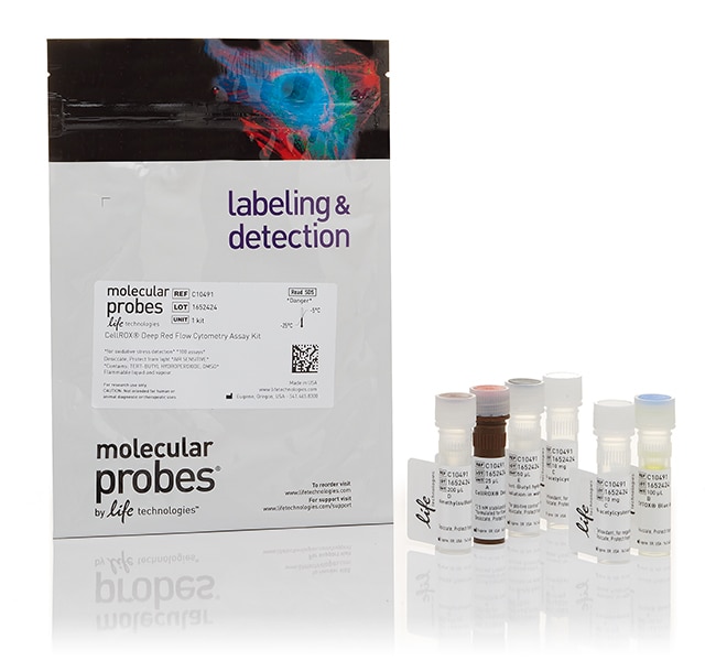
CellROX™ Deep Red Flow Cytometry Assay Kit
CellROX™ディープレッドフローサイトメトリーアッセイキットは、生細胞内の活性酸素種(ROS)のフローサイトメトリー検出を可能にします。このキットには、新しい蛍光性CellROX™CellROXディープレッド試薬のほか、SYTOX™死細胞染色剤、N-アセチルシステイン(陰性対照用の抗酸化剤)とtert-ブチルヒドロペルオキシド溶液詳細を見る
| 製品番号(カタログ番号) | 数量 |
|---|---|
| C10491 | 100 Assays |
製品番号(カタログ番号) C10491
価格(JPY)
67,700
Each
数量:
100 Assays
CellROX™ディープレッドフローサイトメトリーアッセイキットは、生細胞内の活性酸素種(ROS)のフローサイトメトリー検出を可能にします。このキットには、新しい蛍光性CellROX™CellROXディープレッド試薬のほか、SYTOX™死細胞染色剤、N-アセチルシステイン(陰性対照用の抗酸化剤)とtert-ブチルヒドロペルオキシド溶液(ROSの誘導因子であるTBHP)が含まれます。
すべてのCellROX™試薬およびキットの選択ガイドをご覧ください。
CellROX™ディープレッドフローサイトメトリーアッセイキットの特長:
•ROSの存在下で酸化されるフローサイトメトリー用に処方された蛍光性プローブ
•他のレーザー線で励起された蛍光体との重なりが少ない多色相溶性で、—他の試薬との多重化が容易
•単純なプロトコル—細胞は、完全培地や他の適切なバッファーで細胞を染色することができ、血清を含まない培地は必要ありません
CellROX™ディープレッド検出試薬は細胞透過性があり、還元状態では非蛍光または非常に弱い蛍光を発します。酸化すると、試薬は強い蛍光シグナルを示し、その吸収/発光極大値は644/665 nmで、細胞質に局在したままになります。付属のSYTOX™ブルーディープ細胞染色剤と併用すると、酸化ストレスを受けた細胞と受けていない細胞をフローサイトメトリーで確実に区別することができます。
研究用にのみ使用できます。診断用には使用いただけません。
仕様
細胞タイプ哺乳類細胞、真核細胞
検出法蛍光
染色剤タイプCellROX™ディープレッド試薬
フォーマットチューブ
数量100 Assays
出荷条件湿氷
溶解性DMSO(ジメチルスルホキシド)
EmissionCellROX™ディープレッド:644⁄665、SYTOX™ブルー:444⁄480
使用対象(アプリケーション)フローサイトメトリー
使用対象 (装置)フローサイトメーター
製品ラインCellROX
製品タイプ試薬
Unit SizeEach
組成および保存条件
1バイアルのCellROX™ディープレッド試薬(25 µL)、1バイアルのSYTOX™ブルーデッド細胞染色剤(100 µL)、2バイアルのN-アセチルシステイン(バイアルあたり10 mg)、1バイアルのtert-ブチルヒドロペルオキシド(50 µL、70%水溶液)、および1バイアルのDMSO(200 µL)を含みます。
キットは光から保護し、-5~-30℃で保存してください。CellROX™試薬は、空気に敏感です。
キットは光から保護し、-5~-30℃で保存してください。CellROX™試薬は、空気に敏感です。
よくあるご質問(FAQ)
I want to assay cells for reactive oxygen species using carboxy-H2DCFDA, but I want to do so with a plate reader instead of microscope. Will it work?
I have GFP-transfected cells and need to label for reactive oxygen species. Can I use H2DCFDA?
I need a formaldehyde-fixable reactive oxygen species detection assay. Is H2 DCFDA fixable?
What dyes can I use to detect reactive oxygen species (ROS) in my bacteria?
What cellular processes can be analyzed with a flow cytometer?
引用および参考文献 (12)
引用および参考文献
Abstract
Critical Role for the NLRP3 Inflammasome during Acute Lung Injury.
Journal:
PubMed ID:24795455
'The inflammasome is a key factor in innate immunity and senses soluble pathogen and danger-associated molecular patterns as well as biological crystals (urate, cholesterol, etc.), resulting in expression of IL-1ß and IL-18. Using a standard model of acute lung injury (ALI) in mice featuring airway instillation of LPS, ALI was
A role for apoptosis-inducing factor in T cell development.
Journal:J Exp Med
PubMed ID:22869892
Apoptosis-inducing factor (Aif) is a mitochondrial flavoprotein that regulates cell metabolism and survival in many tissues. We report that aif-hypomorphic harlequin (Hq) mice show thymic hypocellularity and a cell-autonomous thymocyte developmental block associated with apoptosis at the ß-selection stage, independent of T cell receptor ß recombination. No abnormalities are observed
Piperlongumine treatment inactivates peroxiredoxin 4, exacerbates endoplasmic reticulum stress, and preferentially kills high-grade glioma cells.
Journal:
PubMed ID:24879047
Piperlongumine, a natural plant product, kills multiple cancer types with little effect on normal cells. Piperlongumine raises intracellular levels of reactive oxygen species (ROS), a phenomenon that may underlie the cancer-cell killing. Although these findings suggest that piperlongumine could be useful for treating cancers, the mechanism by which the drug
Dual roles for splice variants of the glucuronidation pathway as regulators of cellular metabolism.
Journal:
PubMed ID:24141015
Transcripts of the UGT1A gene, encoding half of human UDP-glucuronosyltransferase (UGT) enzymes, undergo alternative splicing, resulting in active enzymes named isoforms 1 (i1s) and novel truncated isoforms 2 (i2s). Here, we investigated the effects of depleting endogenous i2 on drug response and attempted to unveil any additional biologic role(s) for
Skeletal muscle stem cells adopt a dormant cell state post mortem and retain regenerative capacity.
Journal:Nat Commun
PubMed ID:22692546
The accessibility to stem cells from healthy or diseased individuals, and the maintenance of their potency are challenging issues for stem cell biology. Here we report the isolation of viable and functional skeletal myogenic cells from humans up to 17 days, and mice up to 14 days post mortem, much