Search


Citations & References (10)
Invitrogen™
Click-iT™ Plus TUNEL Assay Kits for In Situ Apoptosis Detection
Click-iT Plus TUNELアッセイキットは、細胞および組織サンプル中のアポトーシスを検出します。これにより、容易な色素結合を実現し、GFPおよびRFPによるマルチプレックス化を可能にします。


製品番号(カタログ番号) C10618
価格(JPY)
140,900
Each
色:
Red
標識または色素:
Alexa Fluor™ 594
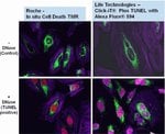
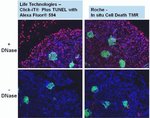
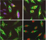
In Situアポトーシス検出用Click-iT Plus TUNELアッセイではAlexa Fluor 488、594、および647蛍光色素オプションが利用できるため、組織および培養細胞サンプル中のアポトーシス細胞をより多く検出できます。このin situアポトーシス検出キットは組織または細胞サンプル用に最適化されており、これらの色素はGFPやRFPなど他の色素やタンパク質とのマルチプレックス化が可能であり、より小さなサイズ(抗体と比較した場合)のため複雑な分子に容易に組み込むことができます。このTUNELアッセイキットは、非常に柔軟性が高く、1回の実験で1∼50サンプルを検査する場合に使用できます。
in situアポトーシス検出用のClick-iT Plus TUNEL Alexa Fluor 488、594、および647アッセイは、小、高特異性標識の部分と明るい蛍光色素を組み合わせることで、組織および培養細胞サンプル中のアポトーシス細胞の検出を可能にします。標識の部分をDNA断片に組み込んだ後、GFPまたはRFPから放出される蛍光シグナルを保存するために十分な穏やかな条件を用いて、触媒的な「クリック」反応により検出を行います。
Click-iT Plus TUNEL Assay for In Situ Apoptosis Detectionでは、さらに以下の利点が得られます:
• 組織サンプルまたは細胞サンプル中のアポトーシス細胞の検出に最適化
• マルチプレックスにより、GFPやRFPなどの蛍光色素や蛍光タンパク質との併用に最適化
• 改良されたTUNEL Assay—反応部位が小さいため、ラベルの取り込みが容易
•明るいアポトーシス性シグナル—Alexa Fluor色素により、反応性の強い明るい光標識が小さく、安定した非光退色蛍光シグナル
• 柔軟性—一度に1~50サンプルを検査するようアッセイを構成
細胞DNAの断片化がアポトーシスの特徴。TUNELアッセイは、アポトーシス細胞中または組織サンプルの断片化DNAを検出するためにもっとも広く使用されている方法です。TUNELアッセイは、断片化されたDNAの3’-OH末端に修飾dUTPを組み込むことから始まります。dUTP修飾は多くの場合、蛍光色素分子の付加であることが多いです。蛍光色素分子サイズにより、修飾dUTPは予想される取り込み速度よりも低く表示されることがあり、これがTUNELアッセイ感度に影響を与える可能性があります。さらに、現在市販されているTUNELアッセイキット向け蛍光色素の多くには、光退色や蛍光スペクトルのオーバーラップの問題があり、いずれもアッセイの感度と多重化能力を低下させます。
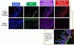
Click-iT Plus TUNELアッセイは、こうした問題を解消するために開発されました。このアッセイでは、ヌクレオチドがより容易に取り込まれるようにするアルキン基(小さな生体直交性官能基)で修飾されたdUTPを使用します。取り込み後、アルキン基とAlexa Fluorピコリルアジド蛍光色素の間で高度に特異的なクリック反応が起こり、その後にその色素が検出されます。これにより、アポトーシス細胞を検出するための高感度かつ特異的なアッセイが可能となります。Click-iT Plus TUNELアッセイは反応条件が穏やかなため、蛍光タンパク質または色素によるマルチプレックスを可能にします。
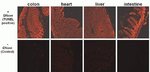
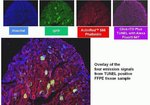
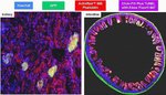
Click-iT Plus TUNELアッセイは、さまざまなホルマリン固定パラフィン包埋組織タイプでバリデーション済みです。いずれの場合も、蛍光タンパク質および蛍光色素でのマルチプレックス能力が保持されていました。さらに、蛍光標識ファロイジンを使用してアクチンを染色する能力も保持されました。
Click-iT Plus TUNELアッセイには、組織サンプルまたは細胞サンプルからアポトーシス細胞を検出するために必要なすべての試薬が含まれています。このキットに含まれる試薬は、50サンプルの検査に使用でき、一度に1~50サンプルの検査を行うように構成できます。
Click-iT Plus TUNEL Assay for In Situ Apoptosis Detectionでは、さらに以下の利点が得られます:
• 組織サンプルまたは細胞サンプル中のアポトーシス細胞の検出に最適化
• マルチプレックスにより、GFPやRFPなどの蛍光色素や蛍光タンパク質との併用に最適化
• 改良されたTUNEL Assay—反応部位が小さいため、ラベルの取り込みが容易
•明るいアポトーシス性シグナル—Alexa Fluor色素により、反応性の強い明るい光標識が小さく、安定した非光退色蛍光シグナル
• 柔軟性—一度に1~50サンプルを検査するようアッセイを構成
細胞DNAの断片化がアポトーシスの特徴。TUNELアッセイは、アポトーシス細胞中または組織サンプルの断片化DNAを検出するためにもっとも広く使用されている方法です。TUNELアッセイは、断片化されたDNAの3’-OH末端に修飾dUTPを組み込むことから始まります。dUTP修飾は多くの場合、蛍光色素分子の付加であることが多いです。蛍光色素分子サイズにより、修飾dUTPは予想される取り込み速度よりも低く表示されることがあり、これがTUNELアッセイ感度に影響を与える可能性があります。さらに、現在市販されているTUNELアッセイキット向け蛍光色素の多くには、光退色や蛍光スペクトルのオーバーラップの問題があり、いずれもアッセイの感度と多重化能力を低下させます。
Click-iT Plus TUNELアッセイは、こうした問題を解消するために開発されました。このアッセイでは、ヌクレオチドがより容易に取り込まれるようにするアルキン基(小さな生体直交性官能基)で修飾されたdUTPを使用します。取り込み後、アルキン基とAlexa Fluorピコリルアジド蛍光色素の間で高度に特異的なクリック反応が起こり、その後にその色素が検出されます。これにより、アポトーシス細胞を検出するための高感度かつ特異的なアッセイが可能となります。Click-iT Plus TUNELアッセイは反応条件が穏やかなため、蛍光タンパク質または色素によるマルチプレックスを可能にします。
Click-iT Plus TUNELアッセイは、さまざまなホルマリン固定パラフィン包埋組織タイプでバリデーション済みです。いずれの場合も、蛍光タンパク質および蛍光色素でのマルチプレックス能力が保持されていました。さらに、蛍光標識ファロイジンを使用してアクチンを染色する能力も保持されました。
Click-iT Plus TUNELアッセイには、組織サンプルまたは細胞サンプルからアポトーシス細胞を検出するために必要なすべての試薬が含まれています。このキットに含まれる試薬は、50サンプルの検査に使用でき、一度に1~50サンプルの検査を行うように構成できます。
For Research Use Only. Not for use in diagnostic procedures.
仕様
色Red
概要Click-iT Plus TUNEL Assay for In Situ Apoptosis Detection、Alexa Fluor™ 594色素
励起/発光590/617
使用対象 (装置)蛍光顕微鏡
標識タイプAlexa Fluor™色素
標識または色素Alexa Fluor™ 594
反応数50個のカバースリップ
製品ラインClick-iT
製品タイプTUNELアッセイ
数量1 kit
出荷条件ドライアイス
検出法蛍光
フォーマットカバースリップ
Unit SizeEach
組成および保存条件
≤20℃で保存し、遮光してください。
よくあるご質問(FAQ)
Click-iT® EdU のコントロールとして、EdUを投与しなかったサンプルをAlexa Fluor 594 azide で染色した。 サンプルはマウスの心臓組織切片だが、ある特定の細胞集団で非特異的な赤色の染色が見られる。 それらはDAPIの染色と重ならない。何が起こっているか?
固定・透過処理したサンプルでアポトーシスの確認をしたい。 どの試薬がおすすめか? 蛍光標識 Annexin V は使用できるか?
I will be performing a cell proliferation assay using Click-iT EdU kit. At what point can I stop overnight, or do I have to perform all the steps continuously?
A control for a Click-iT EdU labeling experiment uses no EdU and the Click-iT reaction using Alexa Fluor 594 azide. The mouse heart tissue sections are showing non-specific labeling in red, seen in particular clusters of cells. They don't overlap with DAPI. What is the problem?
I need to test cells for apoptosis after they have been formaldehyde-fixed and permeabilized. What dye or conjugate do you recommend? Will Annexin V conjugates work?
引用および参考文献 (10)
引用および参考文献
Abstract
CD13 deficiency leads to increased oxidative stress and larger atherosclerotic lesions.
Journal:Atherosclerosis
PubMed ID:31229835
'Atherosclerosis is an inflammatory cardiovascular disorder characterized by accumulation of lipid-loaded macrophages in the intima. Prolonged accumulation leads to apoptosis of macrophages and eventually to progression of lesion development. Prevention of macrophage accumulation within the intima has been shown to reduce lesion formation. Since CD13 mediates trafficking of macrophages to
Loss of host-derived osteopontin creates a glioblastoma-promoting microenvironment.
Journal:Neuro Oncol
PubMed ID:29016864
'Microglia and periphery-derived monocytes infiltrate human and mouse glioblastoma and their density is positively correlated with malignancy. Using microarray and RNA sequencing, we have previously shown that glioblastoma-associated microglia/monocytes (GAMs) express osteopontin/SPP1.'
Differential susceptibility of mouse strains on pancreatic injury and regeneration in cerulein-induced pancreatitis.
Journal:Int J Clin Exp Pathol
PubMed ID:31966883
'Acute pancreatitis (AP), a common disease, causes significant morbidity and mortality in clinical practice. Our objective of this study was to establish an experimental mouse AP model with cerulein treatment and to explore the susceptibility of mouse strains on the severity of pancreatic injury and the subsequent repair and regeneration.
Tanshinone IIA promotes IL2-mediated SW480 colorectal cancer cell apoptosis by triggering INF2-related mitochondrial fission and activating the Mst1-Hippo pathway.
Journal:Biomed Pharmacother
PubMed ID:30372868
IL-2-based therapy is a promising tool to treat colorectal cancer, but drug resistance always occurs in clinical practice. Mitochondrial fission is a novel target to modulate cancer development and progression. The aim of our study is to explore the effect of IL-2 combined with Tan IIA on SW480 colorectal cancer
Sirtuin-1 protects hair follicle stem cells from TNFa-mediated inflammatory stress via activating the MAPK-ERK-Mfn2 pathway.
Journal:Life Sci
PubMed ID:30292830
Stem cell transplantation is a promising tool to treat burn injuries. However, the inflammatory microenvironment in damaged skin limits the efficiency of stem cell-based therapy via poorly understood mechanisms. The aim of our study is to explore the contribution and mechanism of Sirtuin-1 (Sirt1) in TNFa-mediated inflammatory stress in hair