Search
Gránulos de referencia de tamaño y calibración del citómetro de flujo

- Confianza —Ayuda a garantizar un rendimiento óptimo diario del instrumento
- Fiabilidad —Permite una variación mínima para una adquisición uniforme de los datos
- Compatibilidad —gama completa de herramientas para cualquier instrumento
La calibración del instrumento es fundamental para recopilar y analizar datos experimentales precisos. os gránulos de alineación del citómetro de flujo y los gránulos de configuración de clasificación celular están diseñadas para ayudar a garantizar que su citómetro de flujo tenga un rendimiento óptimo, su diseño experimental sea sólido y los datos que recopile y analice sean precisos. Además, nuestros kits de calibración de tamaño y referencia de tamaño sirven como referencias de tamaño confiables para los usuarios de citómetros de flujo.
En esta página:
- Calibración del instrumento/de instrumentos
- Calibración ATC
- Referencia de tamaño de partículas submicrométrico
- Calibración de clasificador de células
Guía de selección de gránulos de calibración de citómetro de flujo
| Partículas de calibración de varios colores | Gránulos de alineación del citómetro de flujo Alignflow | ||||||
|---|---|---|---|---|---|---|---|
| Uso | Calibración de rutina de citómetros de flujo. | ||||||
| Contenido | Mezcla única de partículas con 8 intensidades fluorescentes diferentes. | Fluorescencia única, partículas específicas para láser disponibles en tres versiones (por excitación por láser). | |||||
| Tipo de laser | UV a láser rojo | Láser UV | láser DPSS | (láser rojo) | |||
| Excitación | 365-650 nm | 350-370 nm | 488 nm | 633 nm | |||
| Emisión | 400-680 nm | 400-470 nm | 515-660 nm | 645-680 nm | |||
| ID de gránulo | 3.0–3.4 micras | 2,5 micras | 6,0 micras | 2,5 micras | 6,0 micras | 2,5 micras | 6,0 micras |
| N.º de cat. | A34305 | A16502 | A16505 | A16500 | A16503 | A16501 | A16504 |
| Kit de calibración de tamaño para citometría de flujo | |
|---|---|
| Uso | Estime el tamaño de las células en una muestra experimental comparando las señales FSC con las de las microesferas de referencia. |
| Contenido | Contiene seis suspensiones de microesferas de poliestireno sin teñir (1,0 micras a 15 micras de diámetro). |
| USP Tipo | Cualquier antígeno de |
| N.º de cat. | F13838 |
| Kit de referencia de tamaño de partículas submicrométrico para citometría de flujo | |
|---|---|
| Uso | Seleccione cualquiera de las siguientes opciones:
|
| Contenido | Kit de 6 suspenciones de microesferas fluorescentes verdes (0,02 micrones a 2,0 micrones de diámetro). |
| USP Tipo | láser DPSS |
| Láser | 488 nm |
| Emisión | 515 nm |
| N.º de cat. | F13839 |
| Gránulos de clasificación de células | ||||
|---|---|---|---|---|
| Uso | Calibración de rutina de los clasificadores de citometría de flujo. Los gránulos se pueden usar para verificar la configuración del clasificador de células, como el retraso y la eficiencia (pérdida de células durante la clasificación). | |||
| Contenido | Microesferas de 6 micrones (+/– 10%) infundidas con colorante fluorescente que se han optimizado para su uso con láseres UV, azul, verde / amarillo y rojo. | |||
| USP Tipo | UV | láser DPSS | Láser verde / amarillo | (láser rojo) |
| Láser | Rojo (405 nm) | 488 nm | 532/561 nm | 633 nm |
| Emisión | 460 nm | 515 nm | 575 nm | 680 nm |
| Coincidencias de fluoróforos | DAPI, Hoechst dyes | FITC, Alexa Fluor 488 | R-PE, tetramethylrhodamine | Cy 5 |
| N.º de cat. | C16505 | C16508 | C16509 | C16507 |
Partículas de calibración de varios colores
Partículas de calibración de varios colores Las partículas de calibración se pueden usar para la calibración de rutina de los citómetros de flujo utilizando longitudes de onda que varían de 365 a 650 nm.

Figure 1. Histogram analysis of particle fluorescence at 488 nm excitation and 530/30 nm emission.

Figure 2. Particle fluorescence plotted at 488 nm excitation and 530/30 nm emission versus 633 nm excitation and 670/14 nm emission.
Gránulos de alineación del citómetro de flujo Alignflow
Gránulos de alineación del citómetro de flujo Alignflow de Invitrogen son referencias confiables para alinear, enfocar y calibrar citómetros de flujo. Estas microesferas de poliestireno teñidas con fluorescencia son altamente uniformes con respecto al tamaño y la intensidad de fluorescencia (Figura 3) , y están diseñadas para replicar aproximadamente el tamaño, la longitud de onda de emisión y la intensidad de las muestras biológicas. Debido a que los tintes están contenidos dentro de la matriz de la microesfera, en lugar de sobre la superficie de los gránulos , los gránulos AlignFlow tienen una excelente estabilidad fotoquímica y física, proporcionando señales de referencia confiables para la configuración del instrumento. Los tintes fluorescentes han sido cuidadosamente seleccionados para una excitación óptima por fuentes de láser comúnmente utilizadas en citometría de flujo.
Los gránulos AlignFlow están disponibles en tres versiones: para excitación de 350–370 nm con láseres UV, para excitación de 488 nm con láseres azules y para excitación de 633 nm con láseres rojos. Ambos están disponibles en dos tamaños 2.5 μm de diámetro y 6.0 μm de diámetro. Consulte la tabla de abajo para la selección de productos.

Figura 3. Gránulos de alineación del citómetro de flujo Alignflow La emisión de fluorescencia amplia se detecta en los tres canales. Note la variación excepcionalmente pequeña de la intensidad de fluorescencia de los gránulos. Contribuido por Carleton Stewart, Roswell Park Cancer Institute.
Calibración ATC
El kit de calibración de tamaño para citometría de flujo Invitrogen proporciona un conjunto de suspensiones de microesferas no fluorescentes que sirven como referencias de tamaño confiables para los usuarios de citometría. El kit contiene seis suspensiones de microesferas de poliestireno sin teñir, cada una con un diámetro conocido, determinada por microscopía electrónica de transmisión. El tamaño de las células en una muestra experimental se puede estimar comparando las señales de dispersión directa (FSC) con las de las microesferas de referencia. Las microesferas funcionan como marcadores de tamaño reproducibles (Figura 4) y se pueden mezclar con la muestra experimental o usarse en corridas paralelas.

Figura 4. Kit de calibración del tamaño de citometría para flujo de Invitrogen. Se muestra el análisis de histograma de los valores del canal de registro de intensidad de dispersión hacia adelante (FSC) de las seis muestras de microesferas de poliestireno suministradas en el Kit de calibración de tamaño para citometría de flujo . Las mediciones de FSC se realizaron en un citómetro de flujo Becton Dickinson FACScan ™ utilizando e xitaciónc a 488 nm.
Referencia de tamaño de partículas submicrométrico
El kit de referencia de tamaño de partículas submicrométrico de citometría de flujo Invitrogen proporciona un conjunto de suspensiones de microesferas de fluorescencia verde que sirven como referencias de tamaño confiable para usuarios de citometría de flujo. El kit contiene seis suspensiones de microesferas de poliestireno, cada una con un diámetro conocido determinado por microscopía electrónica de transmisión. El perfil de excitación y emisión de todas los gránulos es similar al colorante Invitrogen Alexa Fluor 488 o las células teñidas con FITC (la excitación y los máximos de emisión son 505 nm y 515 nm, respectivamente).
El tamaño (o rango de tamaño) de las biopartículas en una muestra experimental puede estimarse comparando su FSC con las de las microesferas de referencia (Figura 5) . Las microesferas de cada componente funcionan como marcadores de tamaño reproducibles y se pueden usar individualmente (un tamaño), premezcladas (dos a seis tamaños), entremezcladas con la muestra experimental o en series paralelas. Este kit se puede usar para verificar el rendimiento del instrumento y establecer parámetros que sean adecuados para analizar partículas de submicrón. Por ejemplo, el kit se puede utilizar para comprobar:
- Límite de resolución y rango dinámico de medición de tamaño de partícula
- Sensibilidad de los tubos fotomultiplicadores de dispersión lateral y frontal.
- Nivel de ruido del valor inicial del instrumento
- Alineación y estabilidad ópticas y por láser
- stabilidad del sistema de fluídica

Figura 5. Kit de referencia de tamaño de partículas submicrométrico para citometría de flujo Señales de (el kit tiene seis tamaños de partícula; solo se usaron cinco aquí) se adquirieron cinco partículas de tamaño diferente del Referencia de tamaño de partículas submicrométrico utilizando excitación de 488 nm y 530/30 nm Filtro de emisión de paso de banda (BP) en el citómetro de enfoque acústico Attune. Los diámetros de las cinco microesferas fluorescentes verdes diferentes se identifican en una gráfica de partículas fluorescentes versus dispersión lateral.
Gránulos de clasificación de células
Los gránulos de clasificación de células Invitrogen son estándares confiables para la configuración y calibración de instrumentos de clasificación de citometría de flujo. Los gránulos tienen un diámetro de 6 μm (± 10%) y, por lo tanto, aproximan el tamaño, la longitud de onda de emisión y la intensidad de muchas muestras biológicas. En consecuencia, los gránulos se pueden usar para verificar los ajustes del clasificador de células, como el retraso de la caída y la eficiencia (pérdida de células durante la clasificación). Los gránulos también se pueden usar para calibrar la fuente láser, la óptica y el flujo de un citómetro de flujo sin desperdiciar material experimental valioso y sensible.

Resources
Fluorophore and reagent selection guide for flow cytometry
Download Flow Cytometry Protocols Handbook
Spectral Flow Cytometry Fundamentals
Invitrogen eBioscience Resources—Selection guides, Best Protocols, product performance and more.
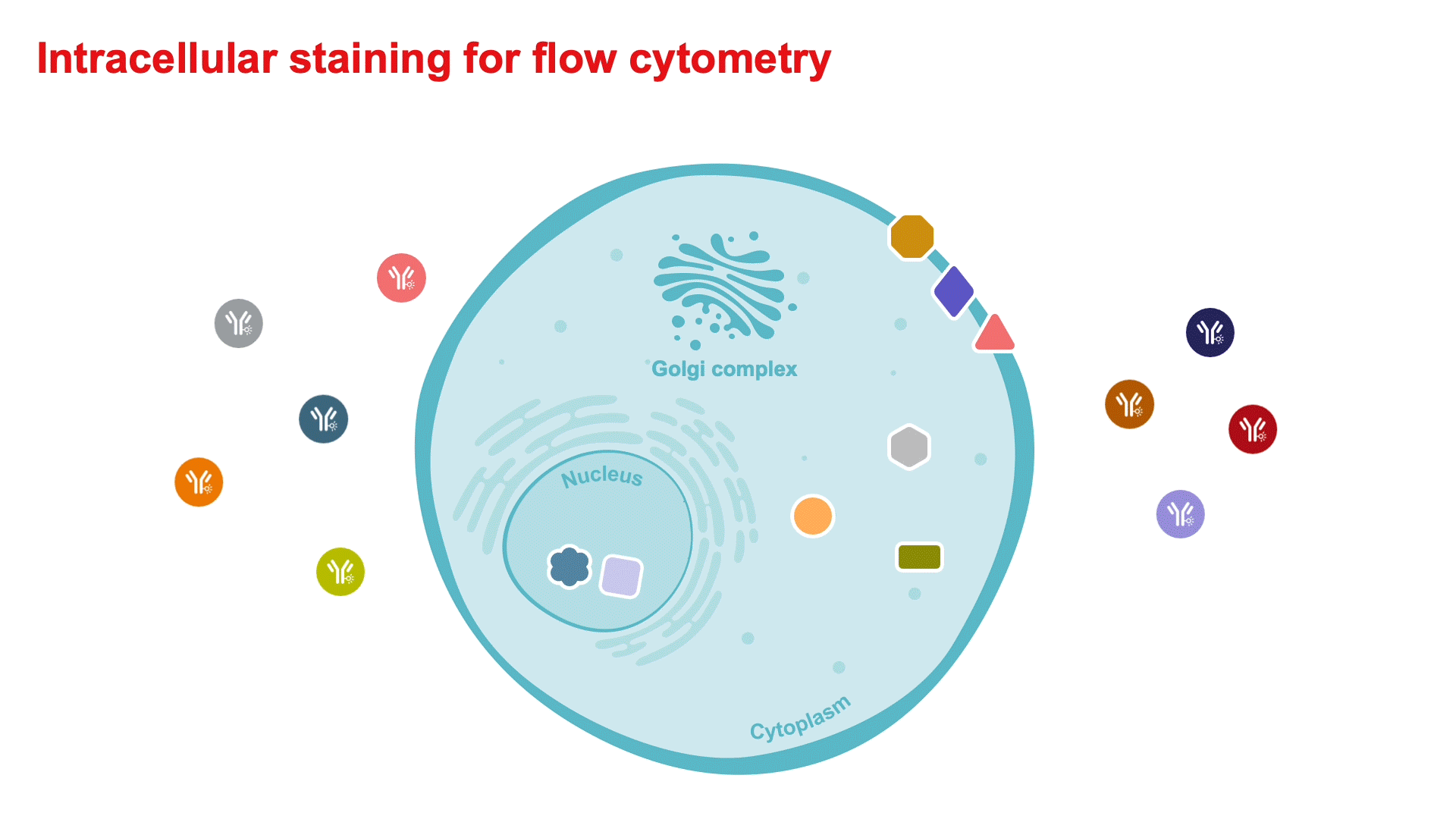
Intracellular Staining for Flow Cytometry How-To Video—for detecting cytokines and intranuclear markers.
Flow Cytometry Learning Center—Access flow cytometry educational resources for better experiment planning and execution.
Flow Cytometry Panel Builder—Design your flow cytometry panel with this online tool for a simplified, customizable experience to fit your needs.
5 Steps Resources
Support
Flow Cytometry Support Center—Find technical support recommendations for your flow cytometry workflows, including tips for experimental setup and in-depth troubleshooting help.
Flow Cytometry Panel Design Support—Work with one of our technical sales specialists to discuss your experimental needs and guide you through the process.