Search


Citations & References (1)
Thermo Scientific™
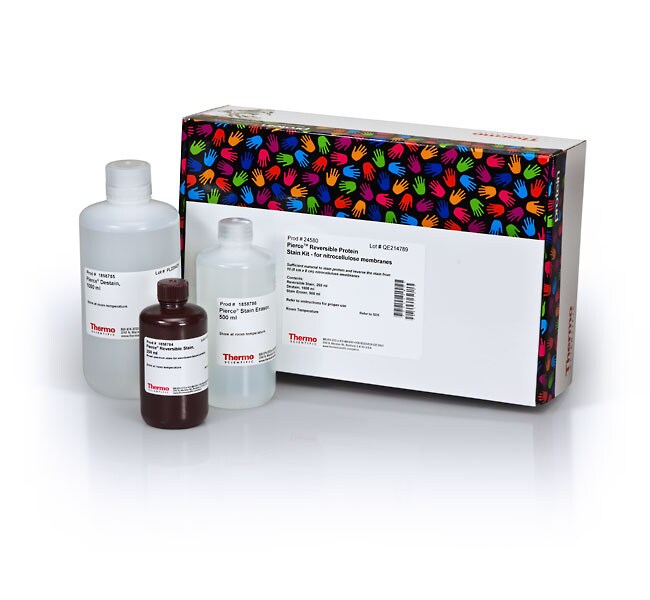
Pierce™ Reversible Protein Stain Kit for Nitrocellulose Membranes
ニトロセルロース膜用Pierceリバーシブルタンパク質染色剤キットは、ポリアクリルアミドゲルから転写した後のニトロセルロース膜上でのタンパク質検出のための、ポンソーS染色剤に代わる迅速かつ高感度な代替品です。特長:•高感度—高親和性で全タンパク質染色、検出下限はバンドあたり25~50 ng• 特異的—タンパク質のみを検出し、他の電気泳動成分やサンプル成分と結合したり、相互作用したりしません。•詳細を見る
| 製品番号(カタログ番号) | 数量 |
|---|---|
| 24580 | 1.5 L |
製品番号(カタログ番号) 24580
価格(JPY)
41,700
Each
数量:
1.5 L
ニトロセルロース膜用Pierceリバーシブルタンパク質染色剤キットは、ポリアクリルアミドゲルから転写した後のニトロセルロース膜上でのタンパク質検出のための、ポンソーS染色剤に代わる迅速かつ高感度な代替品です。
特長:
•高感度—高親和性で全タンパク質染色、検出下限はバンドあたり25~50 ng
• 特異的—タンパク質のみを検出し、他の電気泳動成分やサンプル成分と結合したり、相互作用したりしません。
• プロトコル—5分以内に染色され、15分以内に完全に消えて、反転染色されます。
• 安定性—コンポーネントは室温で安定しており、冷蔵庫のスペースを節約し、平衡化のステップが不要です。
すべてのメンブレン用染色剤を比較›
このメンブレン染色剤キットは、ニトロセルロース膜上のタンパク質を、非破壊、可逆、高信頼性、高感度のメソッドで染色および検出することができます。この方法による検出下限は、1バンドあたり25~50 ngです(従来のポンソーS染色の5倍以上の感度)。この染色液は、膜表面およびタンパク質転写試薬との非特異的な相互作用を最小限に抑えます。染色プロトコルは、シンプルで迅速で、色あせないターコイズブルーのバンドが得られ、将来の参照のために簡単に撮影することができます。15分以内に簡単にリバース染色できます。染色剤はタンパク質を変化させず、完全に除去されるため、その後のウエスタンブロット検出は影響を受けません。また、染色剤は、膜から切除および溶出したタンパク質のN末端配列解析にも対応しています。
研究用途にのみご使用ください。診断目的には使用できません。
仕様
概要Pierceニトロセルロース膜用可逆性タンパク質染色剤キット
検出位置インブロット検出
検出法比色法
数量1.5 L
標的分子タンパク質
標識または色素独自のミックス
製品ラインPierce
製品タイプリバーシブルタンパク質染色キット
Unit SizeEach
組成および保存条件
対応範囲:ミニゲル(SDS-PAGE)から取り出した10枚のニトロセルロースメンブレン(8 cm x 8 cm)
• リバーシブル染色剤、250 mL
• 脱染剤、2 x 500 mL
• 染色イレーサー、250 mL
室温で保管してください
• リバーシブル染色剤、250 mL
• 脱染剤、2 x 500 mL
• 染色イレーサー、250 mL
室温で保管してください
よくあるご質問(FAQ)
How can I increase the signal intensity when I am using SuperSignal West Femto Maximum Sensitivity Substrate?
How can I increase the signal intensity when I am using SuperSignal West Dura Extended Duration Substrate?
引用および参考文献 (1)
引用および参考文献
Abstract
A high-affinity reversible protein stain for Western blots.
Journal:Anal Biochem
PubMed ID:15158487
'We describe a reversible staining technique, using MemCode, a reversible protein stain by which proteins can be visualized on nitrocellulose and polyvinylidine fluoride (PVDF) membranes without being permanently fixed to the membrane itself. This allows subsequent immunoblot analysis of the proteins to be performed. The procedure is applicable only to