Search


Citations & References (8)
Invitrogen™
Click-iT™ Plus TUNEL Assay Kits for In Situ Apoptosis Detection
Click-iT Plus TUNELアッセイキットは、細胞および組織サンプル中のアポトーシスを検出します。これにより、容易な色素結合を実現し、GFPおよびRFPによるマルチプレックス化を可能にします。


製品番号(カタログ番号) C10619
価格(JPY)
140,900
Each
色:
遠赤色
標識または色素:
Alexa Fluor™ 647
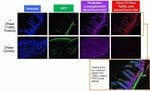
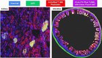
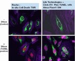
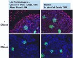
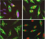
In Situアポトーシス検出用Click-iT Plus TUNELアッセイではAlexa Fluor 488、594、および647蛍光色素オプションが利用できるため、組織および培養細胞サンプル中のアポトーシス細胞をより多く検出できます。このin situアポトーシス検出キットは組織または細胞サンプル用に最適化されており、これらの色素はGFPやRFPなど他の色素やタンパク質とのマルチプレックス化が可能であり、より小さなサイズ(抗体と比較した場合)のため複雑な分子に容易に組み込むことができます。このTUNELアッセイキットは、非常に柔軟性が高く、1回の実験で1∼50サンプルを検査する場合に使用できます。
in situアポトーシス検出用のClick-iT Plus TUNEL Alexa Fluor 488、594、および647アッセイは、小、高特異性標識の部分と明るい蛍光色素を組み合わせることで、組織および培養細胞サンプル中のアポトーシス細胞の検出を可能にします。標識の部分をDNA断片に組み込んだ後、GFPまたはRFPから放出される蛍光シグナルを保存するために十分な穏やかな条件を用いて、触媒的な「クリック」反応により検出を行います。
Click-iT Plus TUNEL Assay for In Situ Apoptosis Detectionでは、さらに以下の利点が得られます:
• 組織サンプルまたは細胞サンプル中のアポトーシス細胞の検出に最適化
• マルチプレックスにより、GFPやRFPなどの蛍光色素や蛍光タンパク質との併用に最適化
• 改良されたTUNEL Assay—反応部位が小さいため、ラベルの取り込みが容易
•明るいアポトーシス性シグナル—Alexa Fluor色素により、反応性の強い明るい光標識が小さく、安定した非光退色蛍光シグナル
• 柔軟性—一度に1~50サンプルを検査するようアッセイを構成
細胞DNAの断片化がアポトーシスの特徴。TUNELアッセイは、アポトーシス細胞中または組織サンプルの断片化DNAを検出するためにもっとも広く使用されている方法です。TUNELアッセイは、断片化されたDNAの3’-OH末端に修飾dUTPを組み込むことから始まります。dUTP修飾は多くの場合、蛍光色素分子の付加であることが多いです。蛍光色素分子サイズにより、修飾dUTPは予想される取り込み速度よりも低く表示されることがあり、これがTUNELアッセイ感度に影響を与える可能性があります。さらに、現在市販されているTUNELアッセイキット向け蛍光色素の多くには、光退色や蛍光スペクトルのオーバーラップの問題があり、いずれもアッセイの感度と多重化能力を低下させます。
Click-iT Plus TUNELアッセイは、こうした問題を解消するために開発されました。このアッセイでは、ヌクレオチドがより容易に取り込まれるようにするアルキン基(小さな生体直交性官能基)で修飾されたdUTPを使用します。取り込み後、アルキン基とAlexa Fluorピコリルアジド蛍光色素の間で高度に特異的なクリック反応が起こり、その後にその色素が検出されます。これにより、アポトーシス細胞を検出するための高感度かつ特異的なアッセイが可能となります。Click-iT Plus TUNELアッセイは反応条件が穏やかなため、蛍光タンパク質または色素によるマルチプレックスを可能にします。
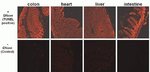
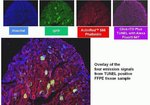
Click-iT Plus TUNELアッセイは、さまざまなホルマリン固定パラフィン包埋組織タイプでバリデーション済みです。いずれの場合も、蛍光タンパク質および蛍光色素でのマルチプレックス能力が保持されていました。さらに、蛍光標識ファロイジンを使用してアクチンを染色する能力も保持されました。
Click-iT Plus TUNELアッセイには、組織サンプルまたは細胞サンプルからアポトーシス細胞を検出するために必要なすべての試薬が含まれています。このキットに含まれる試薬は、50サンプルの検査に使用でき、一度に1~50サンプルの検査を行うように構成できます。
Click-iT Plus TUNEL Assay for In Situ Apoptosis Detectionでは、さらに以下の利点が得られます:
• 組織サンプルまたは細胞サンプル中のアポトーシス細胞の検出に最適化
• マルチプレックスにより、GFPやRFPなどの蛍光色素や蛍光タンパク質との併用に最適化
• 改良されたTUNEL Assay—反応部位が小さいため、ラベルの取り込みが容易
•明るいアポトーシス性シグナル—Alexa Fluor色素により、反応性の強い明るい光標識が小さく、安定した非光退色蛍光シグナル
• 柔軟性—一度に1~50サンプルを検査するようアッセイを構成
細胞DNAの断片化がアポトーシスの特徴。TUNELアッセイは、アポトーシス細胞中または組織サンプルの断片化DNAを検出するためにもっとも広く使用されている方法です。TUNELアッセイは、断片化されたDNAの3’-OH末端に修飾dUTPを組み込むことから始まります。dUTP修飾は多くの場合、蛍光色素分子の付加であることが多いです。蛍光色素分子サイズにより、修飾dUTPは予想される取り込み速度よりも低く表示されることがあり、これがTUNELアッセイ感度に影響を与える可能性があります。さらに、現在市販されているTUNELアッセイキット向け蛍光色素の多くには、光退色や蛍光スペクトルのオーバーラップの問題があり、いずれもアッセイの感度と多重化能力を低下させます。
Click-iT Plus TUNELアッセイは、こうした問題を解消するために開発されました。このアッセイでは、ヌクレオチドがより容易に取り込まれるようにするアルキン基(小さな生体直交性官能基)で修飾されたdUTPを使用します。取り込み後、アルキン基とAlexa Fluorピコリルアジド蛍光色素の間で高度に特異的なクリック反応が起こり、その後にその色素が検出されます。これにより、アポトーシス細胞を検出するための高感度かつ特異的なアッセイが可能となります。Click-iT Plus TUNELアッセイは反応条件が穏やかなため、蛍光タンパク質または色素によるマルチプレックスを可能にします。
Click-iT Plus TUNELアッセイは、さまざまなホルマリン固定パラフィン包埋組織タイプでバリデーション済みです。いずれの場合も、蛍光タンパク質および蛍光色素でのマルチプレックス能力が保持されていました。さらに、蛍光標識ファロイジンを使用してアクチンを染色する能力も保持されました。
Click-iT Plus TUNELアッセイには、組織サンプルまたは細胞サンプルからアポトーシス細胞を検出するために必要なすべての試薬が含まれています。このキットに含まれる試薬は、50サンプルの検査に使用でき、一度に1~50サンプルの検査を行うように構成できます。
For Research Use Only. Not for use in diagnostic procedures.
仕様
色遠赤色
概要Click-iT Plus TUNEL Assay for In Situ Apoptosis Detection、Alexa Fluor™ 647色素
励起/発光650/665
使用対象 (装置)蛍光顕微鏡
標識タイプAlexa Fluor™色素
標識または色素Alexa Fluor™ 647
反応数50個のカバースリップ
製品ラインClick-iT
製品タイプTUNELアッセイ
数量1 kit
出荷条件ドライアイス
検出法蛍光
フォーマットカバースリップ
Unit SizeEach
組成および保存条件
≤20℃で保存し、遮光してください。
よくあるご質問(FAQ)
固定・透過処理したサンプルでアポトーシスの確認をしたい。 どの試薬がおすすめか? 蛍光標識 Annexin V は使用できるか?
What is the fluorescence excitation and emission maxima of Alexa Fluor 647 dye?
I will be performing a cell proliferation assay using Click-iT EdU kit. At what point can I stop overnight, or do I have to perform all the steps continuously?
I need to test cells for apoptosis after they have been formaldehyde-fixed and permeabilized. What dye or conjugate do you recommend? Will Annexin V conjugates work?
Can I use Click-iT Plus TUNEL Assay Kits for In Situ Apoptosis Detection (Cat. Nos. C10617, C10618, C10619) for whole mount immunofluorescence staining of zebrafish larvae?
引用および参考文献 (8)
引用および参考文献
Abstract
Spontaneous calcium waves in the developing enteric nervous system.
Journal:Dev Biol
PubMed ID:28528728
The enteric nervous system (ENS) is an extensive network of neurons in the gut wall that arises from neural crest-derived cells. Like other populations of neural crest cells, it is known that enteric neural crest-derived cells (ENCCs) influence the behaviour of each other and therefore must communicate. However, little is
Arundic Acid Prevents Developmental Upregulation of S100B Expression and Inhibits Enteric Glial Development.
Journal:Front Cell Neurosci
PubMed ID:28280459
S100B is expressed in various types of glial cells and is involved in regulating many aspects of their function. However, little is known about its role during nervous system development. In this study, we investigated the effect of inhibiting the onset of S100B synthesis in the development of the enteric
The impact of detergents on the tissue decellularization process: A ToF-SIMS study.
Journal:Acta Biomater
PubMed ID:27993639
Biologic scaffolds are derived from mammalian tissues, which must be decellularized to remove cellular antigens that would otherwise incite an adverse immune response. Although widely used clinically, the optimum balance between cell removal and the disruption of matrix architecture and surface ligand landscape remains a considerable challenge. Here we describe
Angiopoietin-1 deficiency increases renal capillary rarefaction and tubulointerstitial fibrosis in mice.
Journal:PLoS One
PubMed ID:29293543
Presence of tubulointerstitial fibrosis is predictive of progressive decline in kidney function, independent of its underlying cause. Injury to the renal microvasculature is a major factor in the progression of fibrosis and identification of factors that regulate endothelium in fibrosis is desirable as they might be candidate targets for treatment
Dual roles of hydrogen peroxide in promoting zebrafish renal repair and regeneration.
Journal:Biochem Biophys Res Commun
PubMed ID:31248596
Acute renal injury (AKI) is a serious disorder of renal failure or renal damage that occurs within hours or days. At present, there is no approved pharmaceutical treatment for AKI. Zebrafish is an excellent model for studying the repair of AKI because of its remarkable ability to repair kidney injury.