Search


Citations & References (13)
Invitrogen™
pHrodo™ Red and Green AM Intracellular pH Indicator Dyes
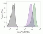
Quantify intracellular pH with pHrodo Red and Green AM intracellular pH indicator dyes, which permeate cell membranes and fluoresce in acidic cytosolic environments. Both the pHrodo Red and Green AM indicator dyes are compatible with HCS, fluorescence microplate, flow cytometry, and HTS platforms.


製品番号(カタログ番号) P35372
価格(JPY)
74,400
Each
色:
オレンジ
Measure and quantify intracellular pH with pHrodo Red and Green AM indicator dyes, which are fluorogenic probes that traverse the cell membrane and remain within the intracellular space upon cleavage by nonspecific esterases. pHrodo Red and Green AM indicator dyes become increasingly fluorescent as the pH drops and can be used to quantify cytosolic pH in the range of 4–9. These indicator dyes can also be used in multiplex experiments within HCS, flow cytometry, microplate fluorometry, and HCS applications.
pHrodo Red (Cat. No. P35372) and Green (Cat. No. P35373) AM intracellular pH indicators are fluorogenic probes used to measure intracellular pH in live cells. They are weakly fluorescent at neutral pH but become increasingly fluorescent as the pH decreases. These reagents can be used to quantify cellular cytosolic pH in the range of 4–9 with a pKa of ∼6.5 and with excitation/emission spectra of 560/585 nm (pHrodo Red) or 509/533 nm (pHrodo Green).
Both pHrodo Red and pHrodo Green pH indicator dyes are photostable and appropriate for use in multiplexing experiments. pHrodo Red easily multiplexes with a wide variety of blue, green, and far red dyes and reporters such as GFP, Fuo-4, calcein, NucBlue, CellEvent Caspase 3/7 green, Mitosox Green, and Mitotracker Deep Red, among many others. pHrodo Green easily multiplexes with a wide variety of blue, red, and far red dyes reporters such as Mitosox Red, CellEvent Caspase 3/7 Red, NucBlue, RFPs, and Mitotracker Deep Red, among many others.
The pHrodo Red and Green dyes have been modified with AM ester groups, resulting in an uncharged molecule that can permeate cell membranes. Once inside the cell, the lipophilic blocking groups of the dyes are cleaved by nonspecific esterases, resulting in compounds that are retained within the intracellular space. The fluorescence intensity of the probe then becomes an indicator of intracellular pH. Subsequent use of the Intracellular pH Calibration Buffer Kit (Cat. No. P35379) allows this intracellular pH to be quantified.
pHrodo Red and Green AM intracellular pH indicator dyes are compatible with various platforms, such as traditional fluorescence microscopy, high content screening (HCS), flow cytometry, and microplate-based fluorimetry or high throughput screening (HTS). These reagents are also compatible with widefield or confocal fluorescence microscopy, flow cytometry, fluorescence plate readers, and high content instruments.
For Research Use Only. Not for human or animal therapeutic or diagnostic use.
仕様
概要pHrodo™ Red AM Intracellular pH Indicator
検出法蛍光
染色剤タイプ蛍光色素ベース
形状液体
数量50 μL
溶解性DMSO(ジメチルスルホキシド)
細胞内局在細胞質
色オレンジ
Emission可視
使用対象(アプリケーション)Cell Analysis
使用対象 (装置)蛍光顕微鏡, フローサイトメーター, フルオロメータ, ハイコンテント装置
製品ラインpHrodo
製品タイプpHインジケータ
Unit SizeEach
組成および保存条件
2℃~8℃で保存し、乾燥させ、遮光してください。
引用および参考文献 (13)
引用および参考文献
Abstract
HIV-1 and morphine regulation of autophagy in microglia: Limited interactions in the context of HIV-1 infection and opioid abuse.
Journal:
PubMed ID:25355898
'Microglia are the predominant resident central nervous system (CNS) cell type productively infected by human immunodeficiency virus (HIV) type-1, and play a key role in the progression of HIV-associated dementia (HAD). Moreover, neural dysfunction and progression to HAD are accelerated in opiate drug abusers. In the present study, we examined
Neuroprotective effects of mGluR II and III activators against staurosporine- and doxorubicin-induced cellular injury in SH-SY5Y cells: New evidence for a mechanism involving inhibition of AIF translocation.
Journal:
PubMed ID:25661514
There are several experimental data sets demonstrating the neuroprotective effects of activation of group II and III metabotropic glutamate receptors (mGluR II/III), however, their effect on neuronal apoptotic processes has yet to be fully recognized. Thus, the comparison of the neuroprotective potency of the mGluR II agonist LY354740, mGluR III
Macrophage Metabolism of Apoptotic Cell-Derived Arginine Promotes Continual Efferocytosis and Resolution of Injury.
Journal:Cell Metab
PubMed ID:32004476
Histone chaperone FACT complex coordinates with HIF to mediate an expeditious transcription program to adapt to poorly oxygenated cancers.
Journal:Cell Rep
PubMed ID:35108543
Single cell RNA-sequencing identifies a metabolic aspect of apoptosis in Rbf mutant.
Journal:Nat Commun
PubMed ID:30479347