Product References
Poly-L-ornithine blocks the inhibitory effects of fibronectin on oligodendrocyte differentiation and promotes myelin repair.
Neural regeneration research
Xiong YJ,Soomro SH,Huang ZH,Yu PP,Ping J,Fu H
Published figure using Ki-67 monoclonal antibody (Product # 14-5698-82) in Immunocytochemistry
Sat Apr 01 00:00:00 EDT 2023
Mitochondrial pyruvate metabolism regulates the activation of quiescent adult neural stem cells.
Science advances
Petrelli F,Scandella V,Montessuit S,Zamboni N,Martinou JC,Knobloch M
14-5698-82 was used in Immunohistochemistry to highlight the importance of metabolism for NSPC regulation and identify an important pathway through which mitochondrial pyruvate import controls NSPC quiescence and activation.
Wed Mar 01 00:00:00 EST 2023
Lrig1-expressing epidermal progenitors require SCD1 to maintain the dermal papilla niche.
Scientific reports
Lim SBH,Wei S,Tan AH,van Steensel MAM,Lim X
14-5698-82 was used in Immunohistochemistry to suggest that this takes place via autocrine Wnt signalling and paracrine Hedgehog signalling.
Fri Mar 10 00:00:00 EST 2023
Voluntary running exercise modifies astrocytic population and features in the peri-infarct cortex.
IBRO neuroscience reports
Yamaguchi N,Sawano T,Nakatani J,Nakano-Doi A,Nakagomi T,Matsuyama T,Tanaka H
14-5698-82 was used in Immunohistochemistry to suggest that exercise modifies the composition of astrocytic population and their phenotype.
Thu Jun 01 00:00:00 EDT 2023
CD133/Prom1 marks proximal mouse oviduct epithelial progenitors and adult epithelial cells with a low generative capacity.
Biology open
Ford MJ,Harwalkar K,Kazemdarvish H,Yamanaka N,Yamanaka Y
14-5698-82 was used in Immunohistochemistry-immunofluorescence to investigate the role of CD133/Prom1 expression in the oviduct (fallopian tube) epithelium and its association with stem/progenitor cells.
Fri Sep 15 00:00:00 EDT 2023
EZH2 controls epicardial cell migration during heart development.
Life science alliance
Jiang H,Bai L,Song S,Yin Q,Shi A,Zhou B,Lian H,Chen H,Xu CR,Wang Y,Nie Y,Hu S
14-5698-82 was used in Immunohistochemistry to provide new insight into the function of EZH2 in cell migration and epicardial development.
Thu Jun 01 00:00:00 EDT 2023
Oncogenic BRAFV600E induces microglial proliferation through extracellular signal-regulated kinase and neuronal death through c-Jun N-terminal kinase.
Neural regeneration research
Ye Q,Srivastava P,Al-Kuwari N,Chen X
14-5698-82 was used in Immunocytochemistry-immunofluorescence to identify distinct consequences mediated by distinct downstream effectors in dividing glial cells and in neurons following the same BRAF mutational activation and a causal link between BRAF-activated microglia and neuronal cell death that does not require physical proximity.
Sat Jul 01 00:00:00 EDT 2023
Transformation of primary murine peritoneal mast cells by constitutive KIT activation is accompanied by loss of Cdkn2a/Arf expression.
Frontiers in immunology
Capellmann S,Sonntag R,Schüler H,Meurer SK,Gan L,Kauffmann M,Horn K,Königs-Werner H,Weiskirchen R,Liedtke C,Huber M
Published figure using Ki-67 monoclonal antibody (Product # 14-5698-82) in Flow Cytometry
Wed Apr 19 00:00:00 EDT 2023
BALB.NCT-Cpox is a unique mouse model of hereditary coproporphyria.
Molecular genetics and metabolism reports
Kang X,Shimada S,Miyahara H,Higuchi K,Mori M
14-5698-82 was used in Immunohistochemistry to indicate that BALB.NCT-Cpox nct mice serve as the suitable animal model to help gain insight into the pathogenesis and therapy of HCP.
Thu Jun 01 00:00:00 EDT 2023
Increased serum extracellular vesicle miR-144-3p and miR-486a-3p in a mouse model of adipose tissue regeneration promote hepatocyte proliferation by targeting Txnip.
PloS one
Niitsu Y,Komiya C,Takeuchi A,Hara K,Horino M,Aoki J,Okazaki R,Murakami M,Tsujimoto K,Ikeda K,Yamada T
14-5698-82 was used in Immunocytochemistry and Immunohistochemistry to investigate alterations in serum EV-miRNAs in iFIRKO mice.
Mon May 08 00:00:00 EDT 2023
Organoid models of fibrolamellar carcinoma mutations reveal hepatocyte transdifferentiation through cooperative BAP1 and PRKAR2A loss.
Nature communications
Rüland L,Andreatta F,Massalini S,Chuva de Sousa Lopes S,Clevers H,Hendriks D,Artegiani B
14-5698-82 was used in Immunohistochemistry-immunofluorescence to demonstrate and characterise a CRISPR-engineered human hepatocyte organoids, which recreates different FLC backgrounds, including the predominant genetic alteration, the DNAJB1-PRKACA fusion, as well as a recently reported background of FLC-like tumors, encompassing inactivating mutations of BAP1 and PRKAR2A.
Wed May 03 00:00:00 EDT 2023
Serotonin Transporter Activity in Mouse Oocytes Is a Positive Indicator of Follicular Growth and Oocyte Maturity.
International journal of molecular sciences
Alyoshina NM,Tkachenko MD,Nikishina YO,Nikishin DA
14-5698-82 was used in Immunohistochemistry to indicate that the mechanism of specific membrane transport of serotonin normally ensures the accumulation of serotonin in maturing oocytes, and can be considered as a promising positive marker of their mature status.
Sat Jul 08 00:00:00 EDT 2023
Engineered human hepatocyte organoids enable CRISPR-based target discovery and drug screening for steatosis.
Nature biotechnology
Hendriks D,Brouwers JF,Hamer K,Geurts MH,Luciana L,Massalini S,López-Iglesias C,Peters PJ,Rodríguez-Colman MJ,Chuva de Sousa Lopes S,Artegiani B,Clevers H
14-5698-82 was used in Immunohistochemistry to present FatTracer, a CRISPR screening platform to identify steatosis modulators and putative targets using APOB-/- and MTTP-/- organoids.
Wed Nov 01 00:00:00 EDT 2023
Phenotypic and transcriptional characterization of oligodendrocyte precursor cells in a 3D culture.
Biomaterials science
Nakano S,Uyeda A,Matsunaga YT,Muramatsu R
14-5698-82 was used in Immunocytochemistry to identify the effect of culture dimension as well as the complexity of the scaffold on OPC responses at the cellular and molecular levels.
Tue Apr 11 00:00:00 EDT 2023
Activation of Vitamin D/VDR Signaling Reverses Gemcitabine Resistance of Pancreatic Cancer Cells Through Inhibition of MUC1 Expression.
Digestive diseases and sciences
Wei D,Wang L,Liu Y,Hafley MA,Tan L,Lorenzi PL,Yang P,Zuo X,Bresalier RS
14-5698-82 was used in Immunohistochemistry to demonstrate a previously unidentified vitamin D/VDR-MUC1 signaling axis involved in the regulation of gemcitabine resistance in pancreatic ductal adenocarcinoma (PDA) and suggests that combinational therapies that include targeted activation of vitamin D/VDR signaling may improve the outcomes of patients with PDA.
Sat Jul 01 00:00:00 EDT 2023
Dynamic states of cervical epithelia during pregnancy and epithelial barrier disruption.
iScience
Cooley A,Madhukaran S,Stroebele E,Colon Caraballo M,Wang L,Akgul Y,Hon GC,Mahendroo M
14-5698-82 was used in Immunohistochemistry-immunofluorescence to find heterogeneous subpopulations of cervical epithelia displaying spatial and temporal specificity to maintain dynamic states of homeostasis in pregnancy and labour.
Fri Feb 17 00:00:00 EST 2023
The importance of nuclear RAGE-Mcm2 axis in diabetes or cancer-associated replication stress.
Nucleic acids research
Han Z,Andrš M,Madhavan BK,Kaymak S,Sulaj A,Kender Z,Kopf S,Kihm L,Pepperkok R,Janscak P,Nawroth P,Kumar V
14-5698-82 was used in Immunoprecipitation to report that RAGE (Receptor for Advanced Glycated Endproducts), previously believed to be an extracellular receptor, upon metabolic stress localizes to the damaged forks.
Tue Mar 21 00:00:00 EDT 2023
Chromatin accessibility dynamics of neurogenic niche cells reveal defects in neural stem cell adhesion and migration during aging.
Nature aging
Yeo RW,Zhou OY,Zhong BL,Sun ED,Navarro Negredo P,Nair S,Sharmin M,Ruetz TJ,Wilson M,Kundaje A,Dunn AR,Brunet A
14-5698-82 was used in Immunohistochemistry-immunofluorescence to characterize genome-wide chromatin accessibility of neurogenic niche cells in vivo during aging.
Sat Jul 01 00:00:00 EDT 2023
Spermatogonial fate in mice with increased activin A bioactivity and testicular somatic cell tumours.
Frontiers in cell and developmental biology
Whiley PAF,Nathaniel B,Stanton PG,Hobbs RM,Loveland KL
14-5698-82 was used in Immunohistochemistry to show for the first time that chronically elevated activin A affects spermatogonial stem cells fate in vivo.
Sat Aug 12 00:00:00 EDT 2023
Implantation and tracing of green fluorescent protein-expressing adipose-derived stem cells in peri-implant capsular fibrosis.
Stem cell research & therapy
Park BY,Wu D,Kwon KR,Kim MJ,Kim TG,Lee JH,Park DY,Kim IK
14-5698-82 was used in Immunohistochemistry to investigate the inhibitory role of ASCs on capsule formation by analyzing the histologic, cellular, and molecular changes in a mouse model of peri-implant fibrosis, and study the fate and distribution of ASCs in the peri-implant capsule.
Wed Feb 08 00:00:00 EST 2023
microRNA-17 is a tumor suppressor in oral squamous cell carcinoma and is repressed by LSD1.
Oral diseases
Wangzhou K,Fu W,Li M,Lu Z,Lai Z,Liu C,Tan Y,Hao C
14-5698-82 was used in Immunohistochemistry to investigate whether lysine-specific demethylase 1 (LSD1) affects the development of oral squamous cell carcinoma (OSCC) by sustaining the cancer stem cells from OSCC (OSCSCs).
Wed Mar 01 00:00:00 EST 2023
Transcriptional and epigenetic decoding of the microglial aging process.
Nature aging
Li X,Li Y,Jin Y,Zhang Y,Wu J,Xu Z,Huang Y,Cai L,Gao S,Liu T,Zeng F,Wang Y,Wang W,Yuan TF,Tian H,Shu Y,Guo F,Lu W,Mao Y,Mei X,Rao Y,Peng B
14-5698-82 was used in Immunohistochemistry to map the transcriptional and epigenetic profiles of microglia from 3- to 24-month-old mice.
Sun Oct 01 00:00:00 EDT 2023
Sec13 promotes oligodendrocyte differentiation and myelin repair through autocrine pleiotrophin signaling.
The Journal of clinical investigation
Liu Z,Yan M,Lei W,Jiang R,Dai W,Chen J,Wang C,Li L,Wu M,Nian X,Li D,Sun D,Lv X,Wang C,Xie C,Yao L,Wu C,Hu J,Xiao N,Mo W,Wang Z,Zhang L
14-5698-82 was used in Immunohistochemistry to demonstrate that coat protein complex II (COPII) component Sec13 is essential for oligodendrocyte differentiation and postnatal myelination.
Fri Apr 01 00:00:00 EDT 2022
B-cell antigen receptor expression and phosphatidylinositol 3-kinase signaling regulate genesis and maintenance of mouse chronic lymphocytic leukemia.
Haematologica
Schmid VK,Khadour A,Ahmed N,Brandl C,Nitschke L,Rajewsky K,Jumaa H,Hobeika E
Published figure using Ki-67 monoclonal antibody (Product # 14-5698-82) in Flow Cytometry
Mon Aug 01 00:00:00 EDT 2022
HDAC1/2 Control Proliferation and Survival in Adult Epidermis and Pre‒Basal Cell Carcinoma through p16 and p53.
The Journal of investigative dermatology
Zhu X,Leboeuf M,Liu F,Grachtchouk M,Seykora JT,Morrisey EE,Dlugosz AA,Millar SE
14-5698-82 was used in Immunohistochemistry to find that homozygous epidermal codeletion of Hdac1 and Hdac2 in adult mouse epidermis causes reduced basal cell proliferation, apoptosis, inappropriate differentiation, and eventual loss of Hdac1/2-null keratinocytes.
Sat Jan 01 00:00:00 EST 2022
Human neural progenitors establish a diffusion barrier in the endoplasmic reticulum membrane during cell division.
Development (Cambridge, England)
Bin Imtiaz MK,Royall LN,Gonzalez-Bohorquez D,Jessberger S
14-5698-82 was used in Immunohistochemistry to investigate the existence of an endoplasmic reticulum diffusion barrier in human cells.
Sat Oct 15 00:00:00 EDT 2022
Polarized macrophages promote gestational beta cell growth through extracellular signal-regulated kinase 5 signalling.
Diabetes, obesity & metabolism
Jiang Y,Chen A,Kline D,Liu Q,Ma J,Wang Y,Zhang T,Qian J,Nelson L,Prasadan K,Hu B,Gittes GK,Xiao X
14-5698-82 was used in Immunohistochemistry-immunofluorescence to show a regulatory loop between macrophages and beta cells through PlGF/EGF/ERK5 signalling cascades to regulate gestational beta cell growth.
Thu Sep 01 00:00:00 EDT 2022
Inhibition of LncRNA Vof-16 expression promotes nerve regeneration and functional recovery after spinal cord injury.
Neural regeneration research
Zhang XM,Zeng LN,Yang WY,Ding L,Chen KZ,Fu WJ,Zeng SQ,Liang YR,Chen GH,Wu HF
Published figure using Ki-67 monoclonal antibody (Product # 14-5698-82) in Immunocytochemistry
Sat Jan 01 00:00:00 EST 2022
Application of an instructive hydrogel accelerates re-epithelialization of xenografted human skin wounds.
Scientific reports
Sparks HD,Mandla S,Vizely K,Rosin N,Radisic M,Biernaskie J
Published figure using Ki-67 monoclonal antibody (Product # 14-5698-82) in Immunocytochemistry
Sat Aug 20 00:00:00 EDT 2022
Diane-35 and Metformin Induce Autophagy and Apoptosis in Polycystic Ovary Syndrome Women with Early-Stage Endometrial Carcinoma.
Genes
Liu Y,Wang Y,Yao D,Chen X,Zhang F,Feng Y,Li X
14-5698-82 was used in Immunohistochemistry to support the use of Diane-35 and metformin combination therapy for patients with PCOS and early EC.
Wed Jan 12 00:00:00 EST 2022
Infiltration of peripheral immune cells into the olfactory bulb in a mouse model of acute nasal inflammation.
Journal of neuroimmunology
Asano H,Hasegawa-Ishii S,Arae K,Obara A,Laumet G,Dantzer R,Shimada A
14-5698-82 was used in Immunohistochemistry-immunofluorescence to examine initial events that occur in the OB after bilateral intranasal administration of lipopolysaccharide, focusing on the olfactory nerve fibers and meninges.
Fri Jul 15 00:00:00 EDT 2022
Caspase-dependent apoptosis induces reactivation and gliogenesis of astrocytes in adult mice.
Frontiers in cellular neuroscience
Kim SC,Park JY,Hwang EM
14-5698-82 was used in Immunohistochemistry-immunofluorescence to generate new conditional transgenic mice (cTg) that can induce apoptosis via Cre-dependent active caspase-3 (taCasp3-2A-TEVp) without pathological conditions.
Thu Dec 22 00:00:00 EST 2022
Infarct-preconditioning exosomes of umbilical cord mesenchymal stem cells promoted vascular remodeling and neurological recovery after stroke in rats.
Stem cell research & therapy
Ye YC,Chang ZH,Wang P,Wang YW,Liang J,Chen C,Wang JJ,Sun HT,Wang Y,Li XH
14-5698-82 was used in Immunohistochemistry to investigate if exosomes secreted in response to infarction microenvironment could have further therapeutic effects.
Thu Jul 28 00:00:00 EDT 2022
AdipoQ-a simple, open-source software to quantify adipocyte morphology and function in tissues and in vitro.
Molecular biology of the cell
Sieckmann K,Winnerling N,Huebecker M,Leyendecker P,Juliana Silva Ribeiro D,Gnad T,Pfeifer A,Wachten D,Hansen JN
14-5698-82 was used in Immunocytochemistry to present and validate AdipoQ, an open-source software implemented as ImageJ plugins that allows us to analyze adipocytes in tissue sections and in vitro after histological and/or immunofluorescent labeling.
Sat Oct 01 00:00:00 EDT 2022
Diabetic hyperglycemia promotes primary tumor progression through glycation-induced tumor extracellular matrix stiffening.
Science advances
Wang W,Hapach LA,Griggs L,Smart K,Wu Y,Taufalele PV,Rowe MM,Young KM,Bates ME,Johnson AC,Ferrell NJ,Pozzi A,Reinhart-King CA
14-5698-82 was used in Immunocytochemistry-immunofluorescence to establish a murine model where hyperglycemia was induced before breast tumor development.
Wed Nov 16 00:00:00 EST 2022
Pathogenetic Mechanisms Underlying Spinocerebellar Ataxia Type 3 Are Altered in Primary Oligodendrocyte Culture.
Cells
Schuster KH,Putka AF,McLoughlin HS
14-5698-82 was used in Immunocytochemistry-immunofluorescence to demonstrate that cell-autonomous dysfunction of oligodendrocyte maturation is one of the of the earliest and most robust changes in vulnerable regions of the SCA3 mouse brain.
Mon Aug 22 00:00:00 EDT 2022
St18 specifies globus pallidus projection neuron identity in MGE lineage.
Nature communications
Nunnelly LF,Campbell M,Lee DI,Dummer P,Gu G,Menon V,Au E
14-5698-82 was used in Immunohistochemistry to identify St18 as a key regulator of projection neuron vs. interneuron identity.
Wed Dec 14 00:00:00 EST 2022
An overlooked subset of Cx3cr1wt/wt microglia in the Cx3cr1CreER-Eyfp/wt mouse has a repopulation advantage over Cx3cr1CreER-Eyfp/wt microglia following microglial depletion.
Journal of neuroinflammation
Zhou K,Han J,Lund H,Boggavarapu NR,Lauschke VM,Goto S,Cheng H,Wang Y,Tachi A,Xie C,Zhu K,Sun Y,Osman AM,Liang D,Han W,Gemzell-Danielsson K,Betsholtz C,Zhang XM,Zhu C,Enge M,Joseph B,Harris RA,Blomgren K
14-5698-82 was used in Immunohistochemistry to demonstrate that CX3CL1-CX3CR1 signaling regulates microglial repopulation both in vivo and in vitro.
Fri Jan 21 00:00:00 EST 2022
Deciphering the role of miR-187-3p/LRFN1 axis in modulating progression, aerobic glycolysis and immune microenvironment of clear cell renal cell carcinoma.
Discover. Oncology
Xu W,Liu W,Anwaier A,Tian X,Su J,Shi G,Wei S,Qu Y,Zhang H,Ye D
14-5698-82 was used in Immunohistochemistry to reveal the tumor-specific and immunological role of miR-187-3p/LRFN1 axis in the progression and reshaping of tumor immune microenvironment of ccRCC.
Thu Jul 07 00:00:00 EDT 2022
ΔNp63 drives dysplastic alveolar remodeling and restricts epithelial plasticity upon severe lung injury.
Cell reports
Weiner AI,Zhao G,Zayas HM,Holcomb NP,Adams-Tzivelekidis S,Wong J,Gentile ME,Reddy D,Wei J,Palashikar G,Quansah KK,Vaughan AE
14-5698-82 was used in Immunohistochemistry to find that ΔNp63 restricts the plasticity of intrapulmonary basal progenitors by maintaining either active or repressive histone modifications at key differentiation gene loci.
Tue Dec 13 00:00:00 EST 2022
Airway basal cells show regionally distinct potential to undergo metaplastic differentiation.
eLife
Zhou Y,Yang Y,Guo L,Qian J,Ge J,Sinner D,Ding H,Califano A,Cardoso WV
14-5698-82 was used in Immunohistochemistry-immunofluorescence to provide novel insights into the origin and impact of basal cell heterogeneity on the establishment of regionally distinct responses of the airway epithelium during injury-repair and in disease conditions.
Fri Sep 30 00:00:00 EDT 2022
A slow-cycling/quiescent cells subpopulation is involved in glioma invasiveness.
Nature communications
Antonica F,Santomaso L,Pernici D,Petrucci L,Aiello G,Cutarelli A,Conti L,Romanel A,Miele E,Tebaldi T,Tiberi L
14-5698-82 was used in Immunohistofluorescence to point to a subpopulation of quiescent cells as partially responsible of tumor invasiveness, one of the major causes of brain cancer morbidity.
Mon Aug 15 00:00:00 EDT 2022
Thy1 marks a distinct population of slow-cycling stem cells in the mouse epidermis.
Nature communications
Koren E,Feldman A,Yusupova M,Kadosh A,Sedov E,Ankawa R,Yosefzon Y,Nasser W,Gerstberger S,Kimel LB,Priselac N,Brown S,Sharma S,Gorenc T,Shalom-Feuerstein R,Steller H,Shemesh T,Fuchs Y
14-5698-82 was used in Immunohistochemistry to report a population of keratinocytes, marked by Thy1, in the basal layer of the interfollicular epidermis.
Mon Aug 08 00:00:00 EDT 2022
Oligodendrogenesis is a key process for cognitive performance improvement induced by voluntary physical activity.
Glia
Eugenin von Bernhardi J,Dimou L
14-5698-82 was used in Immunohistochemistry-immunofluorescence to study the requirement of oligodendrogenesis for exercise induced-cognition enhancement.
Wed Jun 01 00:00:00 EDT 2022
Single-cell microglial transcriptomics during demyelination defines a microglial state required for lytic carcass clearance.
Molecular neurodegeneration
Zia S,Hammond BP,Zirngibl M,Sizov A,Baaklini CS,Panda SP,Ho MFS,Lee KV,Mainali A,Burr MK,Williams S,Caprariello AV,Power C,Simmen T,Kerr BJ,Plemel JR
14-5698-82 was used in Immunohistochemistry to study how microglia respond and regulate demyelination and find that microglia preferentially phagocytose lytic carcasses.
Tue Dec 13 00:00:00 EST 2022
Nicotinamide riboside kinases regulate skeletal muscle fiber-type specification and are rate-limiting for metabolic adaptations during regeneration.
Frontiers in cell and developmental biology
Sonntag T,Ancel S,Karaz S,Cichosz P,Jacot G,Giner MP,Sanchez-Garcia JL,Pannérec A,Moco S,Sorrentino V,Cantó C,Feige JN
14-5698-82 was used in Immunohistochemistry to investigate the influence of NRKs on NAD+ metabolism and muscle homeostasis, and on the response to neurogenic muscle atrophy and regeneration following muscle injury.
Tue Nov 29 00:00:00 EST 2022
PRC2 Heterogeneity Drives Tumor Growth in Medulloblastoma.
Cancer research
Yi J,Kim B,Shi X,Zhan X,Lu QR,Xuan Z,Wu J
14-5698-82 was used in Immunohistochemistry to report that the histone H3K27 methyltransferase polycomb repressor complex 2 (PRC2) is often heterogeneous within individual SHH medulloblastoma tumors.
Tue Aug 16 00:00:00 EDT 2022
Inhibition of CSPG receptor PTPσ promotes migration of newly born neuroblasts, axonal sprouting, and recovery from stroke.
Cell reports
Luo F,Wang J,Zhang Z,You Z,Bedolla A,Okwubido-Williams F,Huang LF,Silver J,Luo Y
14-5698-82 was used in Immunohistochemistry to propose that CSPGs induced by stroke play a predominant role in the regulation of neural repair and that blocking CSPG signaling pathways will lead to enhanced neurorepair and functional recovery in stroke.
Tue Jul 26 00:00:00 EDT 2022
Robust Colonic Epithelial Regeneration and Amelioration of Colitis via FZD-Specific Activation of Wnt Signaling.
Cellular and molecular gastroenterology and hepatology
Xie L,Fletcher RB,Bhatia D,Shah D,Phipps J,Deshmukh S,Zhang H,Ye J,Lee S,Le L,Newman M,Chen H,Sura A,Gupta S,Sanman LE,Yang F,Meng W,Baribault H,Vanhove GF,Yeh WC,Li Y,Lu C
14-5698-82 was used in Immunohistochemistry to demonstrate that SZN-1326-p directly impacted epithelial cells, driving transient expansion of stem and progenitor cells, promoting differentiation of epithelial cells, histologically restoring the damaged epithelium, and secondarily to epithelial repair, reducing inflammation.
Mon Jul 25 00:00:00 EDT 2022
Delivery of VEGF and delta-like 4 to synergistically regenerate capillaries and arterioles in ischemic limbs.
Acta biomaterialia
Niu H,Gao N,Dang Y,Guan Y,Guan J
14-5698-82 was used in Immunohistochemistry-paraffin-immunofluorescence to develop an injectable hydrogel-based drug release system capable of delivering both VEGF and Dll4 to synergistically restore endothelial cell functions, leading to accelerated formation of capillaries, arterioles and vessel branching.
Fri Apr 15 00:00:00 EDT 2022
Double knockin mice show NF-κB trajectories in immune signaling and aging.
Cell reports
Rahman SMT,Aqdas M,Martin EW,Tomassoni Ardori F,Songkiatisak P,Oh KS,Uderhardt S,Yun S,Claybourne QC,McDevitt RA,Greco V,Germain RN,Tessarollo L,Sung MH
14-5698-82 was used in Immunocytochemistry-immunofluorescence to demonstrate the power of these reporters in gaining deeper insights into NF-κB biology, with the spectral complementarity of the labeled NF-κB proteins enabling diverse applications.
Tue Nov 22 00:00:00 EST 2022
Comparison of ligamentization potential between anterior cruciate ligament-derived cells and adipose-derived mesenchymal stem cells reseeded to acellularized tendon allograft.
Bone & joint research
Park J,Jo S,Lee MK,Kim TH,Sung IH,Lee JK
14-5698-82 was used in Immunohistochemistry-immunofluorescence to test the hypothesis that reseeded anterior cruciate ligament (ACL)-derived cells have a better ability to survive and integrate into tendon extracellular matrix (ECM) and accelerate the ligamentization process, compared to adipose-derived mesenchymal stem cells (ADMSCs).
Tue Nov 01 00:00:00 EDT 2022
Induction of hair growth in hair follicle cells and organ cultures upon treatment with 30 kHz frequency inaudible sound via cell proliferation and antiapoptotic effects.
Biomedical reports
Choi H,Lee Y,Shin SH,Nam J,Park WS,Park BC,Kim BJ
14-5698-82 was used in Immunohistochemistry to demonstrate that inaudible sound at 30kHz significantly induces proliferative and anti-apoptotic effects in human dermal papilla cells (hDPCs) and outer root sheath keratinocytes.
Tue Mar 01 00:00:00 EST 2022
Lessons learned from pre-clinical testing of xenogeneic decellularized esophagi in a rabbit model.
iScience
Hannon E,Pellegrini M,Scottoni F,Durkin N,Shibuya S,Lutman R,Proctor TJ,Hutchinson JC,Arthurs OJ,Phylactopoulos DE,Maughan EF,Butler CR,Eaton S,Lowdell MW,Bonfanti P,Urbani L,De Coppi P
14-5698-82 was used in Immunohistochemistry to indicate that transplantation of a decellularized porcine scaffold is possible and vascular flaps may be useful to provide a vascular supply, but long-term outcomes require further pre-clinical testing in a different large animal model.
Fri Oct 21 00:00:00 EDT 2022
Integrated plasma proteomic and single-cell immune signaling network signatures demarcate mild, moderate, and severe COVID-19.
Cell reports. Medicine
Feyaerts D,Hédou J,Gillard J,Chen H,Tsai ES,Peterson LS,Ando K,Manohar M,Do E,Dhondalay GKR,Fitzpatrick J,Artandi M,Chang I,Snow TT,Chinthrajah RS,Warren CM,Wittman R,Meyerowitz JG,Ganio EA,Stelzer IA,Han X,Verdonk F,Gaudillière DK,Mukherjee N,Tsai AS,Rumer KK,Jacobsen DR,Bjornson-Hooper ZB,Jiang S,Saavedra SF,Valdés Ferrer SI,Kelly JD,Furman D,Aghaeepour N,Angst MS,Boyd SD,Pinsky BA,Nolan GP,Nadeau KC,Gaudillière B,McIlwain DR
14-5698-82 was used in Flow cytometry/Cell sorting to provide a set of early determinants of COVID-19 severity that may point to therapeutic targets for prevention and/or treatment of COVID-19 progression.
Tue Jul 19 00:00:00 EDT 2022
Oligodendrocyte progenitor proliferation is disinhibited following traumatic brain injury in leukemia inhibitory factor heterozygous mice.
Journal of neuroscience research
Frondelli MJ,Mather ML,Levison SW
14-5698-82 was used in Immunohistochemistry-immunofluorescence to provide new insights into the regulatory role of LIF in suppressing neural progenitor cell proliferation and, in particular, oligodendrocyte progenitor cell proliferation after a mild TBI.
Tue Feb 01 00:00:00 EST 2022
Contribution of -labeled cells to alveolar regeneration is independent of tuft cells.
eLife
Huang H,Fang Y,Jiang M,Zhang Y,Biermann J,Melms JC,Danielsson JA,Yang Y,Qiang L,Liu J,Zhou Y,Wang M,Hu Z,Wang TC,Saqi A,Sun J,Matsumoto I,Cardoso WV,Emala CW,Zhu J,Izar B,Mou H,Que J
14-5698-82 was used in Immunohistochemistry to confirm that ectopic tuft cells are originated from EBCs in mouse models and COVID-19 lungs.
Wed Sep 21 00:00:00 EDT 2022
Fasting-mimicking diet cycles reduce neuroinflammation to attenuate cognitive decline in Alzheimer's models.
Cell reports
Rangan P,Lobo F,Parrella E,Rochette N,Morselli M,Stephen TL,Cremonini AL,Tagliafico L,Persia A,Caffa I,Monacelli F,Odetti P,Bonfiglio T,Nencioni A,Pigliautile M,Boccardi V,Mecocci P,Pike CJ,Cohen P,LaDu MJ,Pellegrini M,Xia K,Tran K,Ann B,Chowdhury D,Longo VD
14-5698-82 was used in Immunohistochemistry to we show that fasting-mimicking diet cycles reduce cognitive decline and Alzheimer's disease (AD) pathology in E4FAD and 3xTg AD mouse models, with effects superior to those caused by protein restriction cycles.
Tue Sep 27 00:00:00 EDT 2022
Tissue-resident macrophages provide a pro-tumorigenic niche to early NSCLC cells.
Nature
Casanova-Acebes M,Dalla E,Leader AM,LeBerichel J,Nikolic J,Morales BM,Brown M,Chang C,Troncoso L,Chen ST,Sastre-Perona A,Park MD,Tabachnikova A,Dhainaut M,Hamon P,Maier B,Sawai CM,Agulló-Pascual E,Schober M,Brown BD,Reizis B,Marron T,Kenigsberg E,Moussion C,Benaroch P,Aguirre-Ghiso JA,Merad M
14-5698-82 was used in Immunohistochemistry-immunofluorescence to explore the diversity of the macrophage compartment in human non-small cell lung carcinoma (NSCLC) lesions.
Thu Jul 01 00:00:00 EDT 2021
Single-cell atlas of early human brain development highlights heterogeneity of human neuroepithelial cells and early radial glia.
Nature neuroscience
Eze UC,Bhaduri A,Haeussler M,Nowakowski TJ,Kriegstein AR
14-5698-82 was used in Immunohistochemistry to provide a comprehensive molecular and spatial atlas of early stages of human brain and cortical development.
Thu Apr 01 00:00:00 EDT 2021
Visualization of individual cell division history in complex tissues using iCOUNT.
Cell stem cell
Denoth-Lippuner A,Jaeger BN,Liang T,Royall LN,Chie SE,Buthey K,Machado D,Korobeynyk VI,Kruse M,Munz CM,Gerbaulet A,Simons BD,Jessberger S
Published figure using Ki-67 monoclonal antibody (Product # 14-5698-82) in Immunocytochemistry
Thu Nov 04 00:00:00 EDT 2021
Effect of cellular and ECM aging on human iPSC-derived cardiomyocyte performance, maturity and senescence.
Biomaterials
Ozcebe SG,Bahcecioglu G,Yue XS,Zorlutuna P
14-5698-82 was used in Immunocytochemistry to investigate the individual effects of cellular and environmental aging and identify the biochemical changes that occur upon cardiac aging.
Fri Jan 01 00:00:00 EST 2021
CYBB/NOX2 in conventional DCs controls T cell encephalitogenicity during neuroinflammation.
Autophagy
Keller CW,Kotur MB,Mundt S,Dokalis N,Ligeon LA,Shah AM,Prinz M,Becher B,Münz C,Lünemann JD
14-5698 was used in Immunohistochemistry to report that the nicotinamide adenine dinucleotide phosphate (NADPH) oxidase 2, also known as CYBB/NOX2, in conventional DCs (cDCs) regulates endocytosed MOG (myelin oligodendrocyte protein) antigen processing and supports MOG-antigen presentation to CD4+ T cells through LC3-associated phagocytosis (LAP).
Sat May 01 00:00:00 EDT 2021
Epicardial placement of human MSC-loaded fibrin sealant films for heart failure: Preclinical efficacy and mechanistic data.
Molecular therapy : the journal of the American Society of Gene Therapy
Fields L,Ito T,Kobayashi K,Ichihara Y,Podaru MN,Hussain M,Yamashita K,Machado V,Lewis-McDougall F,Suzuki K
Published figure using Ki-67 monoclonal antibody (Product # 14-5698-82) in Immunocytochemistry
Wed Aug 04 00:00:00 EDT 2021
Heterogeneity of astrocytes: Electrophysiological properties of juxtavascular astrocytes before and after brain injury.
Glia
Götz S,Bribian A,López-Mascaraque L,Götz M,Grothe B,Kunz L
Published figure using Ki-67 monoclonal antibody (Product # 14-5698-82) in Immunocytochemistry
Mon Feb 01 00:00:00 EST 2021
Transnasal transplantation of human induced pluripotent stem cell-derived microglia to the brain of immunocompetent mice.
Glia
Parajuli B,Saito H,Shinozaki Y,Shigetomi E,Miwa H,Yoneda S,Tanimura M,Omachi S,Asaki T,Takahashi K,Fujita M,Nakashima K,Koizumi S
14-5698-82 was used in Immunocytochemistry and Immunohistochemistry to find that human microglia were able to migrate through the cribriform plate to different regions of the brain, proliferate, and become the dominant microglia in a region-specific manner by occupying the vacant niche when exogenous human cytokine is administered, for at least 60 days.
Fri Oct 01 00:00:00 EDT 2021
Environmental oxygen regulates astrocyte proliferation to guide angiogenesis during retinal development.
Development (Cambridge, England)
Perelli RM,O'Sullivan ML,Zarnick S,Kay JN
Published figure using Ki-67 monoclonal antibody (Product # 14-5698-82) in Immunohistochemistry
Sat May 01 00:00:00 EDT 2021
CAMSAP3 is required for mTORC1-dependent ependymal cell growth and lateral ventricle shaping in mouse brains.
Development (Cambridge, England)
Kimura T,Saito H,Kawasaki M,Takeichi M
14-5698-82 was used in Immunohistochemistry to suggest that CAMSAP3 supports mTORC1 signaling required for ependymal cell growth via MT network regulation, and, in turn, shaping of the lateral ventricles.
Fri Feb 05 00:00:00 EST 2021
Oxygen regulates epithelial stem cell proliferation via RhoA-actomyosin-YAP/TAZ signal in mouse incisor.
Development (Cambridge, England)
Otsu K,Ida-Yonemochi H,Ikezaki S,Ema M,Hitomi J,Ohshima H,Harada H
14-5698-82 was used in Immunohistochemistry to study the role of molecular oxygen in regulating the proliferation of epithelial stem cells.
Mon Feb 15 00:00:00 EST 2021
HDAC1 Regulates Neuronal Differentiation.
Frontiers in molecular neuroscience
Nieto-Estevez V,Changarathil G,Adeyeye AO,Coppin MO,Kassim RS,Zhu J,Hsieh J
Published figure using Ki-67 monoclonal antibody (Product # 14-5698-82) in Immunohistochemistry (PFA fixed)
Tue Feb 01 00:00:00 EST 2022
HDAC1 Regulates Neuronal Differentiation.
Frontiers in molecular neuroscience
Nieto-Estevez V,Changarathil G,Adeyeye AO,Coppin MO,Kassim RS,Zhu J,Hsieh J
Published figure using Ki-67 monoclonal antibody (Product # 14-5698-82) in Immunohistochemistry (PFA fixed)
Tue Feb 01 00:00:00 EST 2022
Macrophage LAMTOR1 Deficiency Prevents Dietary Obesity and Insulin Resistance Through Inflammation-Induced Energy Expenditure.
Frontiers in cell and developmental biology
Ying L,Zhang M,Ma X,Si Y,Li X,Su J,Yin J,Bao Y
Published figure using Ki-67 monoclonal antibody (Product # 14-5698-82) in Flow Cytometry
Tue Jun 08 00:00:00 EDT 2021
Transcription factor old astrocyte specifically induced substance is a novel regulator of kidney fibrosis.
FASEB journal : official publication of the Federation of American Societies for Experimental Biology
Yamamoto A,Morioki H,Nakae T,Miyake Y,Harada T,Noda S,Mitsuoka S,Matsumoto K,Tomimatsu M,Kanemoto S,Tanaka S,Maeda M,Conway SJ,Imaizumi K,Fujio Y,Obana M
14-5698 was used in Immunohistochemistry to identify and demonstrate that OASIS signalling is a novel regulator of kidney fibrosis.
Mon Feb 01 00:00:00 EST 2021
Evidence that the acetyltransferase Tip60 induces the DNA damage response and cell-cycle arrest in neonatal cardiomyocytes.
Journal of molecular and cellular cardiology
Wang X,Lupton C,Lauth A,Wan TC,Foster P,Patterson M,Auchampach JA,Lough JW
14-5698-82 was used in Immunohistochemistry to indicate that, among its pleiotropic functions, Tip60 induces the DDR in CMs, contributing to proliferative senescence.
Tue Jun 01 00:00:00 EDT 2021
HIF-1α is necessary for activation and tumour-promotion effect of cancer-associated fibroblasts in lung cancer.
Journal of cellular and molecular medicine
Zhang Y,Bian Y,Wang Y,Wang Y,Duan X,Han Y,Zhang L,Wang F,Gu Z,Qin Z
14-5698-82 was used in Immunohistochemistry, Immunocytochemistry to find that hypoxia-inducible factor-1α (HIF-1α) is highly expressed in CAFs of human lung cancer tissues and mouse spontaneous lung tumour.
Tue Jun 01 00:00:00 EDT 2021
HOXA11 plays critical roles in disease progression and response to cytarabine in AML.
Oncology reports
Fu JF,Shih LY,Yen TH
Published figure using Ki-67 monoclonal antibody (Product # 14-5698-82) in Flow Cytometry
Thu Jul 01 00:00:00 EDT 2021
Thrombospondin-2 spatiotemporal expression in skeletal fractures.
Journal of orthopaedic research : official publication of the Orthopaedic Research Society
Zondervan RL,Jenkins DC,Reicha JD,Hankenson KD
Published figure using Ki-67 monoclonal antibody (Product # 14-5698-82) in Immunocytochemistry
Fri Jan 01 00:00:00 EST 2021
MicroRNA-384 inhibits nasopharyngeal carcinoma growth and metastasis via binding to Smad5 and suppressing the Wnt/β-catenin axis.
Cytotechnology
Zeng X,Liao H,Wang F
Published figure using Ki-67 monoclonal antibody (Product # 14-5698-82) in Immunohistochemistry
Thu Apr 01 00:00:00 EDT 2021
MYBL2 in synergy with CDC20 promotes the proliferation and inhibits apoptosis of gastric cancer cells.
Advances in clinical and experimental medicine : official organ Wroclaw Medical University
Deng Q,Wu L,Li Y,Zou L
14-5698-82 was used in Western Blot to examine the role of myeloblastosis viral oncogene homolog-like 2 (MYBL2) in gastric cancer progression and investigate the underlying mechanisms.
Wed Sep 01 00:00:00 EDT 2021
Requirement of DNMT1 to orchestrate epigenomic reprogramming for NPM-ALK-driven lymphomagenesis.
Life science alliance
Redl E,Sheibani-Tezerji R,Cardona CJ,Hamminger P,Timelthaler G,Hassler MR,Zrimšek M,Lagger S,Dillinger T,Hofbauer L,Draganić K,Tiefenbacher A,Kothmayer M,Dietz CH,Ramsahoye BH,Kenner L,Bock C,Seiser C,Ellmeier W,Schweikert G,Egger G
Published figure using Ki-67 monoclonal antibody (Product # 14-5698-82) in Immunohistochemistry
Mon Feb 01 00:00:00 EST 2021
A booster dose enhances immunogenicity of the COVID-19 vaccine candidate ChAdOx1 nCoV-19 in aged mice.
Med (New York, N.Y.)
Silva-Cayetano A,Foster WS,Innocentin S,Belij-Rammerstorfer S,Spencer AJ,Burton OT,Fra-Bidó S,Le Lee J,Thakur N,Conceicao C,Wright D,Barrett J,Evans-Bailey N,Noble C,Bailey D,Liston A,Gilbert SC,Lambe T,Linterman MA
Published figure using Ki-67 monoclonal antibody (Product # 14-5698-82) in Immunohistochemistry
Fri Mar 12 00:00:00 EST 2021
An in vivo model allowing continuous observation of human vascular formation in the same animal over time.
Scientific reports
Tsukada Y,Muramatsu F,Hayashi Y,Inagaki C,Su H,Iba T,Kidoya H,Takakura N
Published figure using Ki-67 monoclonal antibody (Product # 14-5698-82) in Immunohistochemistry
Tue Jan 12 00:00:00 EST 2021
Age-related neurodegeneration and cognitive impairments of NRMT1 knockout mice are preceded by misregulation of RB and abnormal neural stem cell development.
Cell death & disease
Catlin JP,Marziali LN,Rein B,Yan Z,Feltri ML,Schaner Tooley CE
Published figure using Ki-67 monoclonal antibody (Product # 14-5698-82) in Immunocytochemistry
Thu Oct 28 00:00:00 EDT 2021
Ten-eleven translocation protein 1 modulates medulloblastoma progression.
Genome biology
Kim H,Kang Y,Li Y,Chen L,Lin L,Johnson ND,Zhu D,Robinson MH,McSwain L,Barwick BG,Yuan X,Liao X,Zhao J,Zhang Z,Shu Q,Chen J,Allen EG,Kenney AM,Castellino RC,Van Meir EG,Conneely KN,Vertino PM,Jin P,Li J
Published figure using Ki-67 monoclonal antibody (Product # 14-5698-82) in Immunohistochemistry (Frozen)
Thu Apr 29 00:00:00 EDT 2021
Mutant ASXL1 induces age-related expansion of phenotypic hematopoietic stem cells through activation of Akt/mTOR pathway.
Nature communications
Fujino T,Goyama S,Sugiura Y,Inoue D,Asada S,Yamasaki S,Matsumoto A,Yamaguchi K,Isobe Y,Tsuchiya A,Shikata S,Sato N,Morinaga H,Fukuyama T,Tanaka Y,Fukushima T,Takeda R,Yamamoto K,Honda H,Nishimura EK,Furukawa Y,Shibata T,Abdel-Wahab O,Suematsu M,Kitamura T
Published figure using Ki-67 monoclonal antibody (Product # 14-5698-82) in Flow Cytometry
Tue Mar 23 00:00:00 EDT 2021
An mTORC1-dependent switch orchestrates the transition between mouse spermatogonial stem cells and clones of progenitor spermatogonia.
Cell reports
Suzuki S,McCarrey JR,Hermann BP
Published figure using Ki-67 monoclonal antibody (Product # 14-5698-82) in Flow Cytometry
Tue Feb 16 00:00:00 EST 2021
An mTORC1-dependent switch orchestrates the transition between mouse spermatogonial stem cells and clones of progenitor spermatogonia.
Cell reports
Suzuki S,McCarrey JR,Hermann BP
Published figure using Ki-67 monoclonal antibody (Product # 14-5698-82) in Flow Cytometry
Tue Feb 16 00:00:00 EST 2021
IL-1beta promotes the age-associated decline of beta cell function.
iScience
Böni-Schnetzler M,Méreau H,Rachid L,Wiedemann SJ,Schulze F,Trimigliozzi K,Meier DT,Donath MY
Published figure using Ki-67 monoclonal antibody (Product # 14-5698-82) in Immunohistochemistry
Fri Nov 19 00:00:00 EST 2021
CDCP1 promotes compensatory renal growth by integrating Src and Met signaling.
Life science alliance
Kajiwara K,Yamano S,Aoki K,Okuzaki D,Matsumoto K,Okada M
Published figure using Ki-67 monoclonal antibody (Product # 14-5698-82) in Immunohistochemistry
Thu Apr 01 00:00:00 EDT 2021
Targeting Phosphatidylserine Enhances the Anti-tumor Response to Tumor-Directed Radiation Therapy in a Preclinical Model of Melanoma.
Cell reports
Budhu S,Giese R,Gupta A,Fitzgerald K,Zappasodi R,Schad S,Hirschhorn D,Campesato LF,De Henau O,Gigoux M,Liu C,Mazo G,Deng L,Barker CA,Wolchok JD,Merghoub T
Published figure using Ki-67 monoclonal antibody (Product # 14-5698-82) in Flow Cytometry
Tue Jan 12 00:00:00 EST 2021
CXCR4 Regulates Temporal Differentiation via PRC1 Complex in Organogenesis of Epithelial Glands.
International journal of molecular sciences
Kim J,Lee SW,Park K
Published figure using Ki-67 monoclonal antibody (Product # 14-5698-82) in Immunocytochemistry
Sun Jan 10 00:00:00 EST 2021
CXCR4 Regulates Temporal Differentiation via PRC1 Complex in Organogenesis of Epithelial Glands.
International journal of molecular sciences
Kim J,Lee SW,Park K
Published figure using Ki-67 monoclonal antibody (Product # 14-5698-82) in Immunocytochemistry
Sun Jan 10 00:00:00 EST 2021
Delta-like 4 is required for pulmonary vascular arborization and alveolarization in the developing lung.
JCI insight
Xia S,Menden HL,Townley N,Mabry SM,Johnston J,Nyp MF,Heruth DP,Korfhagen T,Sampath V
Published figure using Ki-67 monoclonal antibody (Product # 14-5698-82) in Immunohistochemistry
Thu Apr 08 00:00:00 EDT 2021
Adult neural stem cells have latent inflammatory potential that is kept suppressed by Tcf4 to facilitate adult neurogenesis.
Science advances
Shariq M,Sahasrabuddhe V,Krishna S,Radha S,Bellampalli R,Dwivedi A,Cheramangalam R,Reizis B,Hébert J,Ghosh HS
Published figure using Ki-67 monoclonal antibody (Product # 14-5698-82) in Immunohistochemistry (PFA fixed)
Sat May 01 00:00:00 EDT 2021
Notch Signaling between Cerebellar Granule Cell Progenitors.
eNeuro
Adachi T,Miyashita S,Yamashita M,Shimoda M,Okonechnikov K,Chavez L,Kool M,Pfister SM,Inoue T,Kawauchi D,Hoshino M
Published figure using Ki-67 monoclonal antibody (Product # 14-5698-82) in Immunohistochemistry
Fri Jul 02 00:00:00 EDT 2021
Developmental Role of Adenosine Kinase in the Cerebellum.
eNeuro
Gebril H,Wahba A,Zhou X,Lai T,Alharfoush E,DiCicco-Bloom E,Boison D
Published figure using Ki-67 monoclonal antibody (Product # 14-5698-82) in Immunohistochemistry
Fri Jul 02 00:00:00 EDT 2021
Developmental Role of Adenosine Kinase in the Cerebellum.
eNeuro
Gebril H,Wahba A,Zhou X,Lai T,Alharfoush E,DiCicco-Bloom E,Boison D
Published figure using Ki-67 monoclonal antibody (Product # 14-5698-82) in Immunohistochemistry
Fri Jul 02 00:00:00 EDT 2021
Shikonin Derivatives from Onsoma visianii Decrease Expression of Phosphorylated STAT3 in Leukemia Cells and Exert Antitumor Activity.
Nutrients
Todorovic Z,Milovanovic J,Arsenijevic D,Vukovic N,Vukic M,Arsenijevic A,Djurdjevic P,Milovanovic M,Arsenijevic N
Published figure using Ki-67 monoclonal antibody (Product # 14-5698-82) in Flow Cytometry
Wed Mar 31 00:00:00 EDT 2021
Field size effects on DNA damage and proliferation in normal human cell populations irradiated with X-ray microbeams.
Scientific reports
Ojima M,Ito A,Usami N,Ohara M,Suzuki K,Kai M
Published figure using Ki-67 monoclonal antibody (Product # 14-5698-82) in Immunocytochemistry
Fri Mar 26 00:00:00 EDT 2021
Dissecting phenotypic transitions in metastatic disease via photoconversion-based isolation.
eLife
Sela Y,Li J,Kuri P,Merrell AJ,Li N,Lengner C,Rompolas P,Stanger BZ
Published figure using Ki-67 monoclonal antibody (Product # 14-5698-82) in Immunohistochemistry
Tue Feb 23 00:00:00 EST 2021
Dissecting phenotypic transitions in metastatic disease via photoconversion-based isolation.
eLife
Sela Y,Li J,Kuri P,Merrell AJ,Li N,Lengner C,Rompolas P,Stanger BZ
Published figure using Ki-67 monoclonal antibody (Product # 14-5698-82) in Immunohistochemistry
Tue Feb 23 00:00:00 EST 2021
NEIL3-deficiency increases gut permeability and contributes to a pro-atherogenic metabolic phenotype.
Scientific reports
Karlsen TR,Kong XY,Holm S,Quiles-Jiménez A,Dahl TB,Yang K,Sagen EL,Skarpengland T,S Øgaard JD,Holm K,Vestad B,Olsen MB,Aukrust P,Bjørås M,Hov JR,Halvorsen B,Gregersen I
Published figure using Ki-67 monoclonal antibody (Product # 14-5698-82) in Immunocytochemistry
Tue Oct 05 00:00:00 EDT 2021
GTSE1 is possibly involved in the DNA damage repair and cisplatin resistance in osteosarcoma.
Journal of orthopaedic surgery and research
Xie C,Xiang W,Shen H,Shen J
Published figure using Ki-67 monoclonal antibody (Product # 14-5698-82) in Immunohistochemistry
Tue Dec 07 00:00:00 EST 2021
TWIK-1 BAC-GFP Transgenic Mice, an Animal Model for TWIK-1 Expression.
Cells
Kwon O,Yang H,Kim SC,Kim J,Sim J,Lee J,Hwang EM,Shim S,Park JY
Published figure using Ki-67 monoclonal antibody (Product # 14-5698-82) in Immunohistochemistry
Thu Oct 14 00:00:00 EDT 2021
Targeting Feedforward Loops Formed by Nuclear Receptor RORγ and Kinase PBK in mCRPC with Hyperactive AR Signaling.
Cancers
Zhang X,Huang Z,Wang J,Ma Z,Yang J,Corey E,Evans CP,Yu AM,Chen HW
Published figure using Ki-67 monoclonal antibody (Product # 14-5698-82) in Immunohistochemistry
Thu Apr 01 00:00:00 EDT 2021
Gene Signatures Detect Damaged Liver Sinusoidal Endothelial Cells in Chronic Liver Diseases.
Frontiers in medicine
Verhulst S,van Os EA,De Smet V,Eysackers N,Mannaerts I,van Grunsven LA
Published figure using Ki-67 monoclonal antibody (Product # 14-5698-82) in Immunocytochemistry
Wed Nov 10 00:00:00 EST 2021
Imbalanced Activation of Wnt-/β-Catenin-Signaling in Liver Endothelium Alters Normal Sinusoidal Differentiation.
Frontiers in physiology
Koch PS,Sandorski K,Heil J,Schmid CD,Kürschner SW,Hoffmann J,Winkler M,Staniczek T,de la Torre C,Sticht C,Schledzewski K,Taketo MM,Trogisch FA,Heineke J,Géraud C,Goerdt S,Olsavszky V
Published figure using Ki-67 monoclonal antibody (Product # 14-5698-82) in Immunohistochemistry
Fri Oct 22 00:00:00 EDT 2021
Differential chromatin binding of the lung lineage transcription factor NKX2-1 resolves opposing murine alveolar cell fates in vivo.
Nature communications
Little DR,Lynch AM,Yan Y,Akiyama H,Kimura S,Chen J
14-5698-82 was used in Immunocytochemistry to report that the cell-type-specific function of NKX2-1 is attributed to its differential chromatin binding that is acquired or retained during development in coordination with partner transcriptional factors.
Tue May 04 00:00:00 EDT 2021
Differentiation of mouse fetal lung alveolar progenitors in serum-free organotypic cultures.
eLife
Gkatzis K,Panza P,Peruzzo S,Stainier DY
14-5698-82 was used in Immunocytochemistry to understand how serum-free culture conditions effect alveolar cell differentiation into mouse-modelled lung organoids.
Wed Sep 29 00:00:00 EDT 2021
Adipose mesenchymal stem cells-secreted extracellular vesicles containing microRNA-192 delays diabetic retinopathy by targeting ITGA1.
Journal of cellular physiology
Gu C,Zhang H,Gao Y
14-5698-82 was used in Immunohistochemistry-immunofluorescence to establish that miR-192 released by Evs from MSCs could delay the events of the inflammatory response and angiogenesis in Diabetic retinopathy (DR) and may represent a possible therapeutic approach for the treatment of DR.
Thu Jul 01 00:00:00 EDT 2021
Notch signaling drives development of Barrett's metaplasia from Dclk1-positive epithelial tuft cells in the murine gastric mucosa.
Scientific reports
Kunze B,Middelhoff M,Maurer HC,Agibalova T,Anand A,Bührer AM,Fang HY,Baumeister T,Steiger K,Strangmann J,Schmid RM,Wang TC,Quante M
14-5698-82 was used in Immunohistochemistry to show that Notch activation in Dclk1-positive tuft cells in the gastric cardia can contribute to BE development.
Wed Feb 24 00:00:00 EST 2021
A MYT1L syndrome mouse model recapitulates patient phenotypes and reveals altered brain development due to disrupted neuronal maturation.
Neuron
Chen J,Lambo ME,Ge X,Dearborn JT,Liu Y,McCullough KB,Swift RG,Tabachnick DR,Tian L,Noguchi K,Garbow JR,Constantino JN,Gabel HW,Hengen KB,Maloney SE,Dougherty JD
14-5698-82 was used in Immunohistochemistry-immunofluorescence to provide insight into the mechanistic underpinnings of this disorder and enable future preclinical studies.
Wed Dec 01 00:00:00 EST 2021
A combination of pirfenidone and TGF-β inhibition mitigates cystic echinococcosis-associated hepatic injury.
Parasitology
Wang E,Liao Z,Wang L,Liao Y,Xu X,Liu P,Wang X,Hou J,Jiang H,Wu X,Chen X
14-5698-82 was used in Immunohistochemistry-immunofluorescence to reveal that the inhibitory receptors on the surface of liver NK cells were up-regulated, whereas the activating receptors were down-regulated over time.
Tue Jun 01 00:00:00 EDT 2021
Obesity accelerates hair thinning by stem cell-centric converging mechanisms.
Nature
Morinaga H,Mohri Y,Grachtchouk M,Asakawa K,Matsumura H,Oshima M,Takayama N,Kato T,Nishimori Y,Sorimachi Y,Takubo K,Suganami T,Iwama A,Iwakura Y,Dlugosz AA,Nishimura EK
14-5698-82 was used in Immunohistochemistry-immunofluorescence to demonstrate that stem cell inflammatory signals induced by obesity robustly represses organ regeneration signals to accelerate the miniaturization of mini-organs, and suggests the importance of daily prevention of organ dysfunction.
Thu Jul 01 00:00:00 EDT 2021
Treatment of necrotizing enterocolitis by conditioned medium derived from human amniotic fluid stem cells.
PloS one
O'Connell JS,Li B,Zito A,Ahmed A,Cadete M,Ganji N,Lau E,Alganabi M,Farhat N,Lee C,Eaton S,Mitchell R,Ray S,De Coppi P,Patel K,Pierro A
14-5698-82 was used in Immunohistochemistry-immunofluorescence to show that CM derived from human AFSC administered in experimental NEC exhibited various benefits including reduced intestinal injury and inflammation, increased enterocyte proliferation, and restored intestinal stem cell activity.
Thu Jan 06 00:00:00 EST 2022
Key role for EphB2 receptor in kidney fibrosis.
Clinical science (London, England : 1979)
Huang Z,Liu S,Tang A,Al-Rabadi L,Henkemeyer M,Mimche PN,Huang Y
14-5698-82 was used in Immunohistochemistry to indicate that endogenous EphB2 expression and signaling are abnormally activated after kidney injury and subsequently contribute to the development of renal fibrosis via regulation of multiple profibrotic pathways.
Fri Sep 17 00:00:00 EDT 2021
Impairment of type H vessels by NOX2-mediated endothelial oxidative stress: critical mechanisms and therapeutic targets for bone fragility in streptozotocin-induced type 1 diabetic mice.
Theranostics
Hu XF,Xiang G,Wang TJ,Ma YB,Zhang Y,Yan YB,Zhao X,Wu ZX,Feng YF,Lei W
14-5698 was used in Immunohistochemistry-immunofluorescence to propose that the impairment of type H vessels by NOX2-mediated endothelial oxidative stress might be an important contributor to type 1 diabetes mellitus-induced osteopathy.
Thu Jul 29 00:00:00 EDT 2021
Modeling High-Grade Serous Ovarian Carcinoma Using a Combination of In Vivo Fallopian Tube Electroporation and CRISPR-Cas9-Mediated Genome Editing.
Cancer research
Teng K,Ford MJ,Harwalkar K,Li Y,Pacis AS,Farnell D,Yamanaka N,Wang YC,Badescu D,Ton Nu TN,Ragoussis J,Huntsman DG,Arseneau J,Yamanaka Y
14-5698-82 was used in Immunohistochemistry to unveil a new strategy to generate genetic mouse models of ovarian cancer with high flexibility in selecting mutation combinations and targeting areas.
Fri Oct 15 00:00:00 EDT 2021
IGF-1 protects against angiotensin II-induced cardiac fibrosis by targeting αSMA.
Cell death & disease
Ock S,Ham W,Kang CW,Kang H,Lee WS,Kim J
14-5698-82 was used in Immunohistochemistry to propose that low-dose IGF-1 may be an efficacious therapeutic avenue against cardiac fibrosis.
Fri Jul 09 00:00:00 EDT 2021
Environmental enrichment preserves a young DNA methylation landscape in the aged mouse hippocampus.
Nature communications
Zocher S,Overall RW,Lesche M,Dahl A,Kempermann G
14-5698-82 was used in Immunohistochemistry to show that experiencing a stimulus-rich environment counteracts age-related DNA methylation changes in the hippocampal dentate gyrus of mice.
Wed Jun 23 00:00:00 EDT 2021
Binary pan-cancer classes with distinct vulnerabilities defined by pro- or anti-cancer YAP/TEAD activity.
Cancer cell
Pearson JD,Huang K,Pacal M,McCurdy SR,Lu S,Aubry A,Yu T,Wadosky KM,Zhang L,Wang T,Gregorieff A,Ahmad M,Dimaras H,Langille E,Cole SPC,Monnier PP,Lok BH,Tsao MS,Akeno N,Schramek D,Wikenheiser-Brokamp KA,Knudsen ES,Witkiewicz AK,Wrana JL,Goodrich DW,Bremner R
14-5698-82 was used in Immunohistochemistry (PFA fixed) to show that YAP is thus pivotal across cancer, but in opposite ways, with therapeutic implications.
Mon Aug 09 00:00:00 EDT 2021
Intestinal microbiota shapes gut physiology and regulates enteric neurons and glia.
Microbiome
Vicentini FA,Keenan CM,Wallace LE,Woods C,Cavin JB,Flockton AR,Macklin WB,Belkind-Gerson J,Hirota SA,Sharkey KA
14-5698-82 was used in Immunohistochemistry-immunofluorescence to reveal new insights into the role of the gut microbiota that could lead to therapeutic developments for the treatment of enteric neuropathies.
Tue Oct 26 00:00:00 EDT 2021
Induction of Stress-Induced Renal Cellular Senescence In Vitro: Impact of Mouse Strain Genetic Diversity.
Cells
Liao CM,Wulfmeyer VC,Swallow M,Falk CS,Haller H,Korstanje R,Melk A,Schmitt R
14-5698-82 was used in Immunocytochemistry-immunofluorescence to test whether genetic differences between mouse inbred strains have an impact on the development of stress-induced cellular senescence in cultured primary tubular epithelial cells (PTEC).
Tue Jun 08 00:00:00 EDT 2021
Jazf1 acts as a regulator of insulin-producing β-cell differentiation in induced pluripotent stem cells and glucose homeostasis in mice.
The FEBS journal
Park SJ,Kwon W,Park S,Jeong J,Kim D,Jang S,Kim SY,Sung Y,Kim MO,Choi SK,Ryoo ZY
14-5698-82 was used in Immunohistochemistry to demonstrate that Jazf1 plays a pivotal role in the differentiation of β-cells and glucose homeostasis.
Thu Jul 01 00:00:00 EDT 2021
A Fast and Efficient Approach to Obtaining High-Purity Glioma Stem Cell Culture.
Frontiers in genetics
Han XX,Cai C,Yu LM,Wang M,Hu DY,Ren J,Zhang MH,Zhu LY,Zhang WH,Huang W,He H,Gao Z
14-5698-82 was used in Immunocytochemistry-immunofluorescence to describe the features of GSCs, their properties, and regulatory genes with expression differences between them and normal stem cells.
Sat Jul 24 00:00:00 EDT 2021
Progressive Pulmonary Fibrosis Is Caused by Elevated Mechanical Tension on Alveolar Stem Cells.
Cell
Wu H,Yu Y,Huang H,Hu Y,Fu S,Wang Z,Shi M,Zhao X,Yuan J,Li J,Yang X,Bin E,Wei D,Zhang H,Zhang J,Yang C,Cai T,Dai H,Chen J,Tang N
14-5698-82 was used in Immunohistochemistry (PFA fixed) to discover that loss of Cdc42 function in alveolar stem cells (AT2 cells) causes periphery-to-center progressive lung fibrosis.
Thu Jan 09 00:00:00 EST 2020
MYC Instructs and Maintains Pancreatic Adenocarcinoma Phenotype.
Cancer discovery
Sodir NM,Kortlever RM,Barthet VJA,Campos T,Pellegrinet L,Kupczak S,Anastasiou P,Swigart LB,Soucek L,Arends MJ,Littlewood TD,Evan GI
14-5698-82 was used in Immunohistochemistry to demonstrate that this Myc-driven PDAC switch is completely and immediately reversible: Myc deactivation/inhibition triggers meticulous disassembly of advanced PDAC tumor and stroma and concomitant death of tumor cells.
Wed Apr 01 00:00:00 EDT 2020
Human Lung Stem Cell-Based Alveolospheres Provide Insights into SARS-CoV-2-Mediated Interferon Responses and Pneumocyte Dysfunction.
Cell stem cell
Katsura H,Sontake V,Tata A,Kobayashi Y,Edwards CE,Heaton BE,Konkimalla A,Asakura T,Mikami Y,Fritch EJ,Lee PJ,Heaton NS,Boucher RC,Randell SH,Baric RS,Tata PR
Published figure using Ki-67 monoclonal antibody (Product # 14-5698-82) in Immunocytochemistry
Thu Dec 03 00:00:00 EST 2020
In Vivo Lineage Tracing of Polyploid Hepatocytes Reveals Extensive Proliferation during Liver Regeneration.
Cell stem cell
Matsumoto T,Wakefield L,Tarlow BD,Grompe M
14-5698 was used in Immunocytochemistry-immunoflourescence to study the regenerative capacity of polyploid hepatocytes during liver regeneration.
Thu Jan 02 00:00:00 EST 2020
A Widespread Neurogenic Potential of Neocortical Astrocytes Is Induced by Injury.
Cell stem cell
Zamboni M,Llorens-Bobadilla E,Magnusson JP,Frisén J
14-5698-82 was used in Immunohistochemistry to show that cortical neurogenesis molecularly recapitulates canonical subventricular zone neurogenesis with remarkable fidelity.
Thu Oct 01 00:00:00 EDT 2020
Inflammatory Signals Induce AT2 Cell-Derived Damage-Associated Transient Progenitors that Mediate Alveolar Regeneration.
Cell stem cell
Choi J,Park JE,Tsagkogeorga G,Yanagita M,Koo BK,Han N,Lee JH
14-5698-82 was used in Immunohistochemistry (Paraffin) to map the trajectories of alveolar-lineage cells during injury repair and lung regeneration by single-cell RNA sequencing in combination with in vivo lineage tracing and organoid models.
Thu Sep 03 00:00:00 EDT 2020
Myofibroblast contraction is essential for generating and regenerating the gas-exchange surface.
The Journal of clinical investigation
Li R,Li X,Hagood J,Zhu MS,Sun X
14-5698 was used in Immunohistochemistry to show that inactivation of myosin light chain kinase (MLCK), a key factor required for myofibroblast contraction, disrupted septa formation, supporting the myofibroblast contraction model of alveologenesis.
Mon Jun 01 00:00:00 EDT 2020
Myelin-specific CD8+ T cells exacerbate brain inflammation in CNS autoimmunity.
The Journal of clinical investigation
Wagner CA,Roqué PJ,Mileur TR,Liggitt D,Goverman JM
14-5698-82 was used in Flow Cytometry to suggest that myelin-specific CD8+ T cells may contribute to disease pathogenesis via a FasL-dependent mechanism that preferentially promotes lesion formation in the brain.
Thu Jan 02 00:00:00 EST 2020
Egr2-guided histone H2B monoubiquitination is required for peripheral nervous system myelination.
Nucleic acids research
Wüst HM,Wegener A,Fröb F,Hartwig AC,Wegwitz F,Kari V,Schimmel M,Tamm ER,Johnsen SA,Wegner M,Sock E
Published figure using Ki-67 monoclonal antibody (Product # 14-5698-82) in Immunocytochemistry
Fri Sep 18 00:00:00 EDT 2020
Genetic manipulation of LKB1 elicits lethal metastatic prostate cancer.
The Journal of experimental medicine
Hermanova I,Zúñiga-García P,Caro-Maldonado A,Fernandez-Ruiz S,Salvador F,Martín-Martín N,Zabala-Letona A,Nuñez-Olle M,Torrano V,Camacho L,Lizcano JM,Talamillo A,Carreira S,Gurel B,Cortazar AR,Guiu M,López JI,Martinez-Romero A,Astobiza I,Valcarcel-Jimenez L,Lorente M,Arruabarrena-Aristorena A,Velasco G,Gomez-Muñoz A,Suárez-Cabrera C,Lodewijk I,Flores JM,Sutherland JD,Barrio R,de Bono JS,Paramio JM,Trka J,Graupera M,Gomis RR,Carracedo A
14-5698 was used in Immunohistochemistry to model an aggressive form of prostate cancer and show the unconventional association of LKB1 dosage to prostate tumorigenesis.
Mon Jun 01 00:00:00 EDT 2020
Epithelial Vegfa Specifies a Distinct Endothelial Population in the Mouse Lung.
Developmental cell
Vila Ellis L,Cain MP,Hutchison V,Flodby P,Crandall ED,Borok Z,Zhou B,Ostrin EJ,Wythe JD,Chen J
Published figure using Ki-67 monoclonal antibody (Product # 14-5698-82) in Immunocytochemistry
Mon Mar 09 00:00:00 EDT 2020
NiCHE Platform: Nature-Inspired Catechol-Conjugated Hyaluronic Acid Environment Platform for Salivary Gland Tissue Engineering.
ACS applied materials & interfaces
Lee SW,Ryu JH,Do MJ,Namkoong E,Lee H,Park K
14-5698-82 was used in Immunocytochemistry to model an optimum artificial microenvironment to allow for salivary gland tissue engineering.
Wed Jan 29 00:00:00 EST 2020
Administration of all-trans retinoic acid after experimental traumatic brain injury is brain protective.
British journal of pharmacology
Hummel R,Ulbrich S,Appel D,Li S,Hirnet T,Zander S,Bobkiewicz W,Gölz C,Schäfer MKE
Published figure using Ki-67 monoclonal antibody (Product # 14-5698-82) in Immunohistochemistry
Sun Nov 01 00:00:00 EDT 2020
Multimodal single-cell analysis reveals distinct radioresistant stem-like and progenitor cell populations in murine glioma.
Glia
Alexander J,LaPlant QC,Pattwell SS,Szulzewsky F,Cimino PJ,Caruso FP,Pugliese P,Chen Z,Chardon F,Hill AJ,Spurrell C,Ahrendsen D,Pietras A,Starita LM,Hambardzumyan D,Iavarone A,Shendure J,Holland EC
Published figure using Ki-67 monoclonal antibody (Product # 14-5698-82) in Immunohistochemistry
Tue Dec 01 00:00:00 EST 2020
GPRC5A facilitates cell proliferation through cell cycle regulation and correlates with bone metastasis in prostate cancer.
International journal of cancer
Sawada Y,Kikugawa T,Iio H,Sakakibara I,Yoshida S,Ikedo A,Yanagihara Y,Saeki N,Győrffy B,Kishida T,Okubo Y,Nakamura Y,Miyagi Y,Saika T,Imai Y
14-5698-82 was used in Immunohistochemistry (Paraffin) to to investigate if GPRC5A can be a possible therapeutic target and prognostic marker molecule for progressive prostate cancer.
Sun Mar 01 00:00:00 EST 2020
FGF2 modulates simultaneously the mode, the rate of division and the growth fraction in cultures of radial glia.
Development (Cambridge, England)
Ledesma-Terrón M,Peralta-Cañadas N,Míguez DG
Published figure using Ki-67 monoclonal antibody (Product # 14-5698-82) in Immunocytochemistry
Fri Jul 24 00:00:00 EDT 2020
NPC1 deficiency impairs cerebellar postnatal development of microglia and climbing fiber refinement in a mouse model of Niemann-Pick disease type C.
Development (Cambridge, England)
Boyle BR,Melli SE,Altreche RS,Padron ZM,Yousufzai FAK,Kim S,Vasquez MD,Carone DM,Carone BR,Soto I
Published figure using Ki-67 monoclonal antibody (Product # 14-5698-82) in Immunohistochemistry (Frozen)
Mon Aug 03 00:00:00 EDT 2020
Signalling codes for the maintenance and lineage commitment of embryonic gastric epithelial progenitors.
Development (Cambridge, England)
Sayols S,Klassek J,Werner C,Möckel S,Ritz S,Mendez-Lago M,Soshnikova N
14-5698 was used in Immunohistochemistry to assess whether Axin2- and Lgr5-expressing cells are gastric progenitors in the developing mouse stomach.
Tue Sep 15 00:00:00 EDT 2020
Oxidative stress regulates progenitor behavior and cortical neurogenesis.
Development (Cambridge, England)
Chui A,Zhang Q,Dai Q,Shi SH
14-5698 was used in Immunohistochemistry to suggest that oxidative stress actively regulates RGP behavior to ensure proper neurogenesis in the mammalian cortex.
Wed Mar 11 00:00:00 EDT 2020
Deletion of Calcineurin in Schwann Cells Does Not Affect Developmental Myelination, But Reduces Autophagy and Delays Myelin Clearance after Peripheral Nerve Injury.
The Journal of neuroscience : the official journal of the Society for Neuroscience
Reed CB,Frick LR,Weaver A,Sidoli M,Schlant E,Feltri ML,Wrabetz L
14-5698-82 was used in Immunohistochemistry (Frozen) to assess the effects of CnB ablation on peripheral myelination after birth in both male and female mice.
Wed Aug 05 00:00:00 EDT 2020
Ferroptosis Mediates Cuprizone-Induced Loss of Oligodendrocytes and Demyelination.
The Journal of neuroscience : the official journal of the Society for Neuroscience
Jhelum P,Santos-Nogueira E,Teo W,Haumont A,Lenoël I,Stys PK,David S
14-5698-82 was used in Immunocytochemistry to show that giving mice (male) CZ in the diet induces rapid loss of OL in the corpus callosum by 2 d, accompanied by expression of several markers for ferroptosis, a relatively newly described form of iron-mediated cell death.
Wed Nov 25 00:00:00 EST 2020
Novel enhanced GFP-positive congenic inbred strain establishment and application of tumor-bearing nude mouse model.
Cancer science
Lan Q,Chen Y,Dai C,Li S,Fei X,Dong J,Shen Y,Dai X,Lu Z,Liu B,Wang Q,Wang H,Zhou Z,Ji X,Wang Z,Huang Q
Published figure using Ki-67 monoclonal antibody (Product # 14-5698-82) in Immunocytochemistry
Thu Oct 01 00:00:00 EDT 2020
Mycobacterium tuberculosis-infected alveolar epithelial cells modulate dendritic cell function through the HIF-1α-NOS2 axis.
Journal of leukocyte biology
Rodrigues TS,Alvarez ARP,Gembre AF,Forni MFPAD,de Melo BMS,Alves Filho JCF,Câmara NOS,Bonato VLD
Published figure using Ki-67 monoclonal antibody (Product # 14-5698-82) in Flow Cytometry
Thu Oct 01 00:00:00 EDT 2020
Deletion of Ppard in CD11c+ cells attenuates atherosclerosis in ApoE knockout mice.
FASEB journal : official publication of the Federation of American Societies for Experimental Biology
Tian D,Hong H,Shang W,Ho CC,Dong J,Tian XY
14-5698-82 was used in Flow Cytometry to study the involvement of PPARδ in CD11c+ cells in atherosclerosis.
Sat Feb 01 00:00:00 EST 2020
Angiocrine Hepatocyte Growth Factor Signaling Controls Physiological Organ and Body Size and Dynamic Hepatocyte Proliferation to Prevent Liver Damage during Regeneration.
The American journal of pathology
Zhang XJ,Olsavszky V,Yin Y,Wang B,Engleitner T,Öllinger R,Schledzewski K,Koch PS,Rad R,Schmid RM,Friess H,Goerdt S,Hüser N,Géraud C,von Figura G,Hartmann D
14-5698-82 was used in Immunocytochemistry-immunoflourescence to aim to elucidate the precise contribution of Liver sinusoidal endothelial cells-derived hepatocyte growth factor in physiological homeostasis and liver regeneration.
Sat Feb 01 00:00:00 EST 2020
Radiation Induced Metabolic Alterations Associate With Tumor Aggressiveness and Poor Outcome in Glioblastoma.
Frontiers in oncology
Gupta K,Vuckovic I,Zhang S,Xiong Y,Carlson BL,Jacobs J,Olson I,Petterson XM,Macura SI,Sarkaria J,Burns TC
Published figure using Ki-67 monoclonal antibody (Product # 14-5698-82) in Immunocytochemistry
Mon Sep 28 00:00:00 EDT 2020
Histone Methylations Define Neural Stem/Progenitor Cell Subtypes in the Mouse Subventricular Zone.
Molecular neurobiology
Zhang Z,Manaf A,Li Y,Perez SP,Suganthan R,Dahl JA,Bjørås M,Klungland A
14-5698 was used in Immunocytochemistry-immunoflourescence to define different subtypes of neural stem/progenitor cells (NSPCs) by individual histone methylations.
Sat Feb 01 00:00:00 EST 2020
Renewal and Differentiation of GCD Necklace Olfactory Sensory Neurons.
Chemical senses
Bloom ML,Johnston LB,Datta SR
14-5698-82 was used in Immunohistochemistry to demonstrate that-like their canonical counterparts-GCD OSNs undergo continuous renewal and define a GCD-specific developmental trajectory linking neurogenesis, maturation, and migration.
Fri May 29 00:00:00 EDT 2020
Agrin Promotes Limbal Stem Cell Proliferation and Corneal Wound Healing Through Hippo-Yap Signaling Pathway.
Investigative ophthalmology & visual science
Hou L,Fu W,Liu Y,Wang Q,Wang L,Huang Y
Published figure using Ki-67 monoclonal antibody (Product # 14-5698-82) in Immunocytochemistry
Mon May 11 00:00:00 EDT 2020
Comparing the fate of brain metastatic breast cancer cells in different immune compromised mice with cellular magnetic resonance imaging.
Clinical & experimental metastasis
Knier NN,Hamilton AM,Foster PJ
14-5698-82 was used in Immunohistochemistry to track the fate of a mammary carcinoma cell line (MDA-MB-231-BR) in vivo to characterize the growth of brain metastases in the nude and severely immune-compromised NOD/SCID/ILIIrg-/- (NSG) mouse.
Sat Aug 01 00:00:00 EDT 2020
Creation of Laryngeal Grafts from Primary Human Cells and Decellularized Laryngeal Scaffolds.
Tissue engineering. Part A
Moser PT,Gerli M,Diercks GR,Evangelista-Leite D,Charest JM,Gershlak JR,Ren X,Gilpin SE,Jank BJ,Gaudette GR,Hartnick CJ,Ott HC
14-5698 was used in Immunohistochemistry to report the de novo formation of three humanized laryngeal tissue compartments under one uniform culture condition, a possible keystone in creating vital composite tissue grafts for laryngeal regeneration.
Fri May 01 00:00:00 EDT 2020
Multidimensional hydrogel models reveal endothelial network angiocrine signals increase glioblastoma cell number, invasion, and temozolomide resistance.
Integrative biology : quantitative biosciences from nano to macro
Ngo MT,Karvelis E,Harley BAC
14-5698 was used in Immunocytochemistry-immunoflourescence to highlight the use of multidimensional hydrogel models to evaluate the role of angiocrine signals in glioblastoma progression.
Fri Jun 19 00:00:00 EDT 2020
Quinazolinone derivative BNUA-3 ameliorated [NDEA+2-AAF]-induced liver carcinogenesis in SD rats by modulating AhR-CYP1B1-Nrf2-Keap1 pathway.
Clinical and experimental pharmacology & physiology
Bose P,Siddique MUM,Acharya R,Jayaprakash V,Sinha BN,Lapenna A,Pattanayak SP
Published figure using Ki-67 monoclonal antibody (Product # 14-5698-82) in Immunohistochemistry
Wed Jan 01 00:00:00 EST 2020
New Insights into the Pathogenesis of Diabetic Nephropathy: Proximal Renal Tubules Are Primary Target of Oxidative Stress in Diabetic Kidney.
Acta histochemica et cytochemica
Haraguchi R,Kohara Y,Matsubayashi K,Kitazawa R,Kitazawa S
Published figure using Ki-67 monoclonal antibody (Product # 14-5698-82) in Immunohistochemistry
Tue Apr 28 00:00:00 EDT 2020
Chemosensitization of prostate cancer stem cells in mice by angiogenin and plexin-B2 inhibitors.
Communications biology
Li S,Goncalves KA,Lyu B,Yuan L,Hu GF
Published figure using Ki-67 monoclonal antibody (Product # 14-5698-82) in Flow Cytometry
Wed Jan 15 00:00:00 EST 2020
A single-cell transcriptomic and anatomic atlas of mouse dorsal raphe Pet1 neurons.
eLife
Okaty BW,Sturrock N,Escobedo Lozoya Y,Chang Y,Senft RA,Lyon KA,Alekseyenko OV,Dymecki SM
14-5698 was used in Immunohistochemistry to describe up to fourteen neuron subtypes, many showing biased cell body distributions across the DR.
Mon Jun 22 00:00:00 EDT 2020
Elevated Serum Amino Acids Induce a Subpopulation of Alpha Cells to Initiate Pancreatic Neuroendocrine Tumor Formation.
Cell reports. Medicine
Smith DK,Kates L,Durinck S,Patel N,Stawiski EW,Kljavin N,Foreman O,Sipos B,Solloway MJ,Allan BB,Peterson AS
14-5698-82 was used in Immunohistochemistry-paraffin to show that elevated plasma amino acids drive the appearance of a proliferative population of SLC38A5+ embryonic progenitor-like alpha cells in mice.
Tue Aug 25 00:00:00 EDT 2020
Follicular Regulatory T Cells Can Access the Germinal Center Independently of CXCR5.
Cell reports
Vanderleyden I,Fra-Bido SC,Innocentin S,Stebegg M,Okkenhaug H,Evans-Bailey N,Pierson W,Denton AE,Linterman MA
Published figure using Ki-67 monoclonal antibody (Product # 14-5698-82) in Immunofluorescence
Tue Jan 21 00:00:00 EST 2020
Metastasis-initiating cells induce and exploit a fibroblast niche to fuel malignant colonization of the lungs.
Nature communications
Pein M,Insua-Rodríguez J,Hongu T,Riedel A,Meier J,Wiedmann L,Decker K,Essers MAG,Sinn HP,Spaich S,Sütterlin M,Schneeweiss A,Trumpp A,Oskarsson T
Published figure using Ki-67 monoclonal antibody (Product # 14-5698-82) in Immunohistochemistry
Fri Mar 20 00:00:00 EDT 2020
Aripiprazole reduces liver cell division.
PloS one
Pirc Marolt T,Kramar B,Bulc Rozman K,Šuput D,Milisav I
Published figure using Ki-67 monoclonal antibody (Product # 14-5698-82) in Immunocytochemistry
Tue Dec 08 00:00:00 EST 2020
Regeneration of the pulmonary vascular endothelium after viral pneumonia requires COUP-TF2.
Science advances
Zhao G,Weiner AI,Neupauer KM,de Mello Costa MF,Palashikar G,Adams-Tzivelekidis S,Mangalmurti NS,Vaughan AE
Published figure using Ki-67 monoclonal antibody (Product # 14-5698-82) in Immunocytochemistry
Sun Nov 01 00:00:00 EDT 2020
Transcriptome analysis of basic fibroblast growth factor treated stem cells isolated from human exfoliated deciduous teeth.
Heliyon
Nowwarote N,Manokawinchoke J,Kanjana K,Fournier BPJ,Sukarawan W,Osathanon T
Published figure using Ki-67 monoclonal antibody (Product # 14-5698-82) in Immunocytochemistry
Mon Jun 01 00:00:00 EDT 2020
Comparative Analysis of Age-Related Changes in Lacrimal Glands and Meibomian Glands of a C57BL/6 Male Mouse Model.
International journal of molecular sciences
Yoon CH,Ryu JS,Hwang HS,Kim MK
Published figure using Ki-67 monoclonal antibody (Product # 14-5698-82) in Immunohistochemistry
Thu Jun 11 00:00:00 EDT 2020
The effects of hedgehog ligand neutralising antibody 5E1 in a mouse model of endometriosis.
BMC research notes
Cousins FL,Farley JK,Kerrigan R,Mukherjee S,Darzi S,Gargett CE,Deane JA
Published figure using Ki-67 monoclonal antibody (Product # 14-5698-82) in Immunocytochemistry
Fri Sep 25 00:00:00 EDT 2020
CD103+ cDC1 and endogenous CD8+ T cells are necessary for improved CD40L-overexpressing CAR T cell antitumor function.
Nature communications
Kuhn NF,Lopez AV,Li X,Cai W,Daniyan AF,Brentjens RJ
Published figure using Ki-67 monoclonal antibody (Product # 14-5698-82) in Flow Cytometry
Wed Dec 02 00:00:00 EST 2020
B Cell Activating Factor (BAFF) Is Required for the Development of Intra-Renal Tertiary Lymphoid Organs in Experimental Kidney Transplantation in Rats.
International journal of molecular sciences
Steines L,Poth H,Herrmann M,Schuster A,Banas B,Bergler T
Published figure using Ki-67 monoclonal antibody (Product # 14-5698-82) in Immunohistochemistry
Wed Oct 28 00:00:00 EDT 2020
A discrete subtype of neural progenitor crucial for cortical folding in the gyrencephalic mammalian brain.
eLife
Matsumoto N,Tanaka S,Horiike T,Shinmyo Y,Kawasaki H
Published figure using Ki-67 monoclonal antibody (Product # 14-5698-82) in Immunocytochemistry
Tue Apr 21 00:00:00 EDT 2020
A discrete subtype of neural progenitor crucial for cortical folding in the gyrencephalic mammalian brain.
eLife
Matsumoto N,Tanaka S,Horiike T,Shinmyo Y,Kawasaki H
Published figure using Ki-67 monoclonal antibody (Product # 14-5698-82) in Immunocytochemistry
Tue Apr 21 00:00:00 EDT 2020
A discrete subtype of neural progenitor crucial for cortical folding in the gyrencephalic mammalian brain.
eLife
Matsumoto N,Tanaka S,Horiike T,Shinmyo Y,Kawasaki H
Published figure using Ki-67 monoclonal antibody (Product # 14-5698-82) in Immunocytochemistry
Tue Apr 21 00:00:00 EDT 2020
Treatment of mice with a ligand binding blocking anti-CD28 monoclonal antibody improves healing after myocardial infarction.
PloS one
Gladow N,Hollmann C,Ramos G,Frantz S,Kerkau T,Beyersdorf N,Hofmann U
Published figure using Ki-67 monoclonal antibody (Product # 14-5698-82) in Flow Cytometry
Mon Jul 06 00:00:00 EDT 2020
Cardiac fibroblast proliferation rates and collagen expression mature early and are unaltered with advancing age.
JCI insight
Wu R,Ma F,Tosevska A,Farrell C,Pellegrini M,Deb A
Published figure using Ki-67 monoclonal antibody (Product # 14-5698-82) in Flow Cytometry
Thu Dec 17 00:00:00 EST 2020
Discrete populations of isotype-switched memory B lymphocytes are maintained in murine spleen and bone marrow.
Nature communications
Riedel R,Addo R,Ferreira-Gomes M,Heinz GA,Heinrich F,Kummer J,Greiff V,Schulz D,Klaeden C,Cornelis R,Menzel U,Kröger S,Stervbo U,Köhler R,Haftmann C,Kühnel S,Lehmann K,Maschmeyer P,McGrath M,Naundorf S,Hahne S,Sercan-Alp Ö,Siracusa F,Stefanowski J,Weber M,Westendorf K,Zimmermann J,Hauser AE,Reddy ST,Durek P,Chang HD,Mashreghi MF,Radbruch A
Published figure using Ki-67 monoclonal antibody (Product # 14-5698-82) in Flow Cytometry
Fri May 22 00:00:00 EDT 2020
Changes in Liver Mechanical Properties and Water Diffusivity During Normal Pregnancy Are Driven by Cellular Hypertrophy.
Frontiers in physiology
Garczyńska K,Tzschätzsch H,Kühl AA,Morr AS,Lilaj L,Häckel A,Schellenberger E,Berndt N,Holzhütter HG,Braun J,Sack I,Guo J
Published figure using Ki-67 monoclonal antibody (Product # 14-5698-82) in Immunohistochemistry
Fri Dec 18 00:00:00 EST 2020
Interleukin-13 disrupts type 2 pneumocyte stem cell activity.
JCI insight
Glisinski KM,Schlobohm AJ,Paramore SV,Birukova A,Moseley MA,Foster MW,Barkauskas CE
Published figure using Ki-67 monoclonal antibody (Product # 14-5698-82) in Immunocytochemistry
Thu Jan 16 00:00:00 EST 2020
Interleukin-13 disrupts type 2 pneumocyte stem cell activity.
JCI insight
Glisinski KM,Schlobohm AJ,Paramore SV,Birukova A,Moseley MA,Foster MW,Barkauskas CE
Published figure using Ki-67 monoclonal antibody (Product # 14-5698-82) in Immunocytochemistry
Thu Jan 16 00:00:00 EST 2020
Macrophage migration inhibitory factor inhibition as a novel therapeutic approach against triple-negative breast cancer.
Cell death & disease
Charan M,Das S,Mishra S,Chatterjee N,Varikuti S,Kaul K,Misri S,Ahirwar DK,Satoskar AR,Ganju RK
14-5698 was used in Immunohistochemistry to study the role of Macrophage migration inhibitory factor (MIF) in Triple-negative breast cancer (TNBC) progression and metastasis.
Thu Sep 17 00:00:00 EDT 2020
Combining In Vivo and Organotypic In Vitro Approaches to Assess the Human Relevance of Basimglurant (RG7090), a Potential CAR Activator.
Toxicological sciences : an official journal of the Society of Toxicology
Nudischer R,Renggli K,Bertinetti-Lapatki C,Hoflack JC,Flint N,Sewing S,Pedersen L,Schadt S,Higgins LG,Vardy A,Lenz B,Gand L,Boess F,Elcombe BM,Hierlemann A,Roth AB
14-5698 was used in Immunohistochemistry to suggest RG7090 to be a rodent-specific CAR activator, and that CAR activation and its downstream processes were involved in the foci of altered hepatocytes formation detected in vivo.
Sat Aug 01 00:00:00 EDT 2020
Pro-myogenic small molecules revealed by a chemical screen on primary muscle stem cells.
Skeletal muscle
Buchanan SM,Price FD,Castiglioni A,Gee AW,Schneider J,Matyas MN,Hayhurst M,Tabebordbar M,Wagers AJ,Rubin LL
14-5698 was used in Immunocytochemistry-immunoflourescence to identify new pathways that can prevent loss of skeletal muscle function or enhance regenerative potential.
Fri Oct 09 00:00:00 EDT 2020
Microbiota-Sourced Purines Support Wound Healing and Mucous Barrier Function.
iScience
Lee JS,Wang RX,Goldberg MS,Clifford GP,Kao DJ,Colgan SP
14-5698-82 was used in Immunohistochemistry (Paraffin) to reveal the gut microbiota as a prominent source of exogenous purines and that such microbiota-sourced purines (MSPs) are available to the intestinal mucosa.
Fri Jun 26 00:00:00 EDT 2020
Regulation of stem/progenitor cell maintenance by BMP5 in prostate homeostasis and cancer initiation.
eLife
Tremblay M,Viala S,Shafer ME,Graham-Paquin AL,Liu C,Bouchard M
14-5698 was used in Immunohistochemistry-immunofluorescence to explore the regulation of stem / progenitor cell homeostasis by BMP5.
Mon Sep 07 00:00:00 EDT 2020
Mapping endothelial-cell diversity in cerebral cavernous malformations at single-cell resolution.
eLife
Orsenigo F,Conze LL,Jauhiainen S,Corada M,Lazzaroni F,Malinverno M,Sundell V,Cunha SI,Brännström J,Globisch MA,Maderna C,Lampugnani MG,Magnusson PU,Dejana E
14-5698-82 was used in Immunocytochemistry to comprehensively characterize subclasses of brain endothelial cells (ECs) under both normal conditions and after deletion of Pdcd10 (Ccm3) in a mouse model of CCM.
Tue Nov 03 00:00:00 EST 2020
The neuroregenerative effects of topical decorin on the injured mouse cornea.
Journal of neuroinflammation
Wu M,Downie LE,Grover LM,Moakes RJA,Rauz S,Logan A,Jiao H,Hill LJ,Chinnery HR
14-5698 was used in Immunohistochemistry-wholemount to support a role for decorin in DC-mediated neuroregeneration following corneal abrasion injury.
Mon May 04 00:00:00 EDT 2020
Phase I Study of Ficlatuzumab and Cetuximab in Cetuximab-Resistant, Recurrent/Metastatic Head and Neck Cancer.
Cancers
Bauman JE,Ohr J,Gooding WE,Ferris RL,Duvvuri U,Kim S,Johnson JT,Soloff AC,Wallweber G,Winslow J,Gaither-Davis A,Grandis JR,Stabile LP
14-5698-82 was used in Flow Cytometry to establish the recommended Phase II dose (RP2D). Secondary objectives included overall response rate (ORR), progression-free survival (PFS), and overall survival (OS).
Thu Jun 11 00:00:00 EDT 2020
Eradication of tumor growth by delivering novel photothermal selenium-coated tellurium nanoheterojunctions.
Science advances
Chen S,Xing C,Huang D,Zhou C,Ding B,Guo Z,Peng Z,Wang D,Zhu X,Liu S,Cai Z,Wu J,Zhao J,Wu Z,Zhang Y,Wei C,Yan Q,Wang H,Fan D,Liu L,Zhang H,Cao Y
Published figure using Ki-67 monoclonal antibody (Product # 14-5698-82) in Immunocytochemistry
Wed Apr 01 00:00:00 EDT 2020
Blocking Notch-Signaling Increases Neurogenesis in the Striatum after Stroke.
Cells
Santopolo G,Magnusson JP,Lindvall O,Kokaia Z,Frisén J
14-5698-82 was used in Immunocytochemistry to suggest inhibition of Notch-signaling as a potential translational strategy to promote neuronal regeneration after stroke.
Mon Jul 20 00:00:00 EDT 2020
Yolk-sac-derived macrophages progressively expand in the mouse kidney with age.
eLife
Ide S,Yahara Y,Kobayashi Y,Strausser SA,Ide K,Watwe A,Xu-Vanpala S,Privratsky JR,Crowley SD,Shinohara ML,Alman BA,Souma T
14-5698 was used in Immunocytochemistry-immunoflourescence to find that yolk-sac-derived macrophages progressively expand in number with age and become a major contributor to the renal macrophage population in older mice.
Fri Apr 17 00:00:00 EDT 2020
DSCAM regulates delamination of neurons in the developing midbrain.
Science advances
Arimura N,Okada M,Taya S,Dewa KI,Tsuzuki A,Uetake H,Miyashita S,Hashizume K,Shimaoka K,Egusa S,Nishioka T,Yanagawa Y,Yamakawa K,Inoue YU,Inoue T,Kaibuchi K,Hoshino M
14-5698 was used in Immunohistochemistry to show that Down syndrome cell adhesion molecule (DSCAM) controls neuronal delamination by local suppression of the RapGEF2-Rap1-N-cadherin cascade at the apical endfeet in the dorsal midbrain.
Tue Sep 01 00:00:00 EDT 2020
TH17 cells require ongoing classic IL-6 receptor signaling to retain transcriptional and functional identity.
Science immunology
Harbour SN,DiToro DF,Witte SJ,Zindl CL,Gao M,Schoeb TR,Jones GW,Jones SA,Hatton RD,Weaver CT
14-5698-82 was used in Immunocytochemistry to find that IL-6 signaling is required for both induction and maintenance of mouse TH17 cells; IL-6Rα-deficient TH17 cells rapidly lost their TH17 phenotype and did not cause disease in two models of colitis.
Fri Jul 17 00:00:00 EDT 2020
Anti-commensal IgG Drives Intestinal Inflammation and Type 17 Immunity in Ulcerative Colitis.
Immunity
Castro-Dopico T,Dennison TW,Ferdinand JR,Mathews RJ,Fleming A,Clift D,Stewart BJ,Jing C,Strongili K,Labzin LI,Monk EJM,Saeb-Parsy K,Bryant CE,Clare S,Parkes M,Clatworthy MR
Published figure using Ki-67 monoclonal antibody (Product # 14-5698-82) in Immunofluorescence
Tue Apr 16 00:00:00 EDT 2019
Reduced mitochondrial fusion and Huntingtin levels contribute to impaired dendritic maturation and behavioral deficits in Fmr1-mutant mice.
Nature neuroscience
Shen M,Wang F,Li M,Sah N,Stockton ME,Tidei JJ,Gao Y,Korabelnikov T,Kannan S,Vevea JD,Chapman ER,Bhattacharyya A,van Praag H,Zhao X
14-5698-82 was used in Immunohistochemistry to show that FMRP-deficient immature neurons exhibit impaired dendritic maturation, altered expression of mitochondrial genes, fragmented mitochondria, impaired mitochondrial function, and increased oxidative stress.
Fri Mar 01 00:00:00 EST 2019
Sensing of apoptotic cells through Axl causes lung basal cell proliferation in inflammatory diseases.
The Journal of experimental medicine
Fujino N,Brand OJ,Morgan DJ,Fujimori T,Grabiec AM,Jagger CP,Maciewicz RA,Yamada M,Itakura K,Sugiura H,Ichinose M,Hussell T
Published figure using Ki-67 monoclonal antibody (Product # 14-5698-82) in Immunohistochemistry
Mon Sep 02 00:00:00 EDT 2019
An autocrine ActivinB mechanism drives TGFβ/Activin signaling in Group 3 medulloblastoma.
EMBO molecular medicine
Morabito M,Larcher M,Cavalli FM,Foray C,Forget A,Mirabal-Ortega L,Andrianteranagna M,Druillennec S,Garancher A,Masliah-Planchon J,Leboucher S,Debalkew A,Raso A,Delattre O,Puget S,Doz F,Taylor MD,Ayrault O,Bourdeaut F,Eychène A,Pouponnot C
14-5698-82 was used in Immunohistochemistry to demonstrate that an ActivinB autocrine stimulation is responsible for pathway activation in a subset of Group 3 MB characterized by high PMEPA1 levels.
Thu Aug 01 00:00:00 EDT 2019
High-Molecular-Weight Hyaluronan Is a Hippo Pathway Ligand Directing Cell Density-Dependent Growth Inhibition via PAR1b.
Developmental cell
Ooki T,Murata-Kamiya N,Takahashi-Kanemitsu A,Wu W,Hatakeyama M
14-5698 was used in Immunocytochemistry-immunoflourescence to show that treatment with high-molecular-weight hyaluronan stimulates tumor-suppressive Hippo signaling in breast epithelial cells.
Mon May 20 00:00:00 EDT 2019
Diabetes-Induced NF-κB Dysregulation in Skeletal Stem Cells Prevents Resolution of Inflammation.
Diabetes
Ko KI,Syverson AL,Kralik RM,Choi J,DerGarabedian BP,Chen C,Graves DT
14-5698-82 was used in Immunohistochemistry to demonstrate that type 1 diabetes induces NF-κB hyperactivation in skeletal stem cells to disrupt their ability to modulate M2 polarisation and resolve inflammation.
Fri Nov 01 00:00:00 EDT 2019
The nuclear receptor corepressor NCoR1 regulates hematopoiesis and leukemogenesis in vivo.
Blood advances
Wan X,Liu L,Zhou P,Hui X,He Q,Yu F,Zhang W,Ding X,Yuan X,Zhang N,Zhao Y,Zhu R,Liu Y,Hao P,Auwerx J,Song X,Leng Q,Zhang Y
14-5698 was used in Flow cytometry/Cell sorting to identify nuclear receptor corepressor 1 having a key role in murine adult hematopoiesis and its contribution to MLL-AF9 induced leukemogenesis.
Tue Feb 26 00:00:00 EST 2019
JAK-STAT inhibition impairs K-RAS-driven lung adenocarcinoma progression.
International journal of cancer
Mohrherr J,Haber M,Breitenecker K,Aigner P,Moritsch S,Voronin V,Eferl R,Moriggl R,Stoiber D,Győrffy B,Brcic L,László V,Döme B,Moldvay J,Dezső K,Bilban M,Popper H,Moll HP,Casanova E
Published figure using Ki-67 monoclonal antibody (Product # 14-5698-82) in Immunohistochemistry
Sun Dec 15 00:00:00 EST 2019
Evidence of a developmental origin for β-cell heterogeneity using a dual lineage-tracing technology.
Development (Cambridge, England)
Chen C,Shiota C,Agostinelli G,Ridley D,Jiang Y,Ma J,Prasadan K,Xiao X,Gittes GK
14-5698 was used in Immunohistochemistry to develop a dual lineage-tracing technology, using a combination of two recombinase systems, Dre/RoxP and Cre/LoxP, to independently trace green fluorescent Pdx1-lineage cells and red fluorescent Ptf1a-lineage cells in the developing and adult mouse pancreas.
Thu Jun 27 00:00:00 EDT 2019
Analysis of biological networks in the endothelium with biomimetic microsystem platform.
American journal of physiology. Lung cellular and molecular physiology
Kruse K,Klomp J,Sun M,Chen Z,Santana D,Huang F,Kanabar P,Maienschein-Cline M,Komarova YA
14-5698-82 was used in Immunocytochemistry to describe a novel method for studying the protein "interactome" in primary human cells and apply this method to investigate the effect of posttranslational protein modifications (PTMs) on the protein''s functions.
Sun Sep 01 00:00:00 EDT 2019
α-Lactosylceramide Protects Against iNKT-Mediated Murine Airway Hyperreactivity and Liver Injury Through Competitive Inhibition of Cd1d Binding.
Frontiers in chemistry
Lai AC,Chi PY,Thio CL,Han YC,Kao HN,Hsieh HW,Gervay-Hague J,Chang YJ
14-5698 was used in Flow cytometry/Cell sorting to study how inhibiting iNKT cell activation with α-Lactosylceramide can reduce development of murine airway hyperreactivity and liver injury by competing for CD1d binding.
Thu Oct 01 00:00:00 EDT 2020
p53 deletion rescues lethal microcephaly in a mouse model with neural stem cell abscission defects.
Human molecular genetics
Little JN,Dwyer ND
14-5698-82 was used in Immunohistochemistry to elucidate both NSC apoptosis and abscission mechanisms that could underlie human microcephaly or other brain malformations.
Fri Feb 01 00:00:00 EST 2019
Comprehensive and cell-type-based characterization of the dorsal midbrain during development.
Genes to cells : devoted to molecular & cellular mechanisms
Arimura N,Dewa KI,Okada M,Yanagawa Y,Taya SI,Hoshino M
14-5698 was used in Immunofluorescence in immunohistochemistry on frozen tissues to characterise the ventricular zone, intermediate zone, and marginal zone based on various markers for proliferation and differentiation in embryonic dorsal midbrain.
Tue Jan 01 00:00:00 EST 2019
SORLA regulates endosomal trafficking and oncogenic fitness of HER2.
Nature communications
Pietilä M,Sahgal P,Peuhu E,Jäntti NZ,Paatero I,Närvä E,Al-Akhrass H,Lilja J,Georgiadou M,Andersen OM,Padzik A,Sihto H,Joensuu H,Blomqvist M,Saarinen I,Boström PJ,Taimen P,Ivaska J
Published figure using Ki-67 monoclonal antibody (Product # 14-5698-82) in Immunohistochemistry
Tue May 28 00:00:00 EDT 2019
Soluble TREM2 ameliorates pathological phenotypes by modulating microglial functions in an Alzheimer's disease model.
Nature communications
Zhong L,Xu Y,Zhuo R,Wang T,Wang K,Huang R,Wang D,Gao Y,Zhu Y,Sheng X,Chen K,Wang N,Zhu L,Can D,Marten Y,Shinohara M,Liu CC,Du D,Sun H,Wen L,Xu H,Bu G,Chen XF
Published figure using Ki-67 monoclonal antibody (Product # 14-5698-82) in Immunohistochemistry
Mon Mar 25 00:00:00 EDT 2019
FCRL5+ Memory B Cells Exhibit Robust Recall Responses.
Cell reports
Kim CC,Baccarella AM,Bayat A,Pepper M,Fontana MF
Published figure using Ki-67 monoclonal antibody (Product # 14-5698-82) in Flow Cytometry
Tue Apr 30 00:00:00 EDT 2019
Origin and differentiation trajectories of fibroblastic reticular cells in the splenic white pulp.
Nature communications
Cheng HW,Onder L,Novkovic M,Soneson C,Lütge M,Pikor N,Scandella E,Robinson MD,Miyazaki JI,Tersteegen A,Sorg U,Pfeffer K,Rülicke T,Hehlgans T,Ludewig B
Published figure using Ki-67 monoclonal antibody (Product # 14-5698-82) in Immunohistochemistry
Mon Apr 15 00:00:00 EDT 2019
Regulatory T-cells regulate neonatal heart regeneration by potentiating cardiomyocyte proliferation in a paracrine manner.
Theranostics
Li J,Yang KY,Tam RCY,Chan VW,Lan HY,Hori S,Zhou B,Lui KO
Published figure using Ki-67 monoclonal antibody (Product # 14-5698-82) in Immunocytochemistry
Fri Aug 14 00:00:00 EDT 2020
Deficiency in the secreted protein Semaphorin3d causes abnormal parathyroid development in mice.
The Journal of biological chemistry
Singh A,Mia MM,Cibi DM,Arya AK,Bhadada SK,Singh MK
Published figure using Ki-67 monoclonal antibody (Product # 14-5698-82) in Immunocytochemistry
Fri May 24 00:00:00 EDT 2019
Targeted Delivery of Stk25 Antisense Oligonucleotides to Hepatocytes Protects Mice Against Nonalcoholic Fatty Liver Disease.
Cellular and molecular gastroenterology and hepatology
Cansby E,Nuñez-Durán E,Magnusson E,Amrutkar M,Booten SL,Kulkarni NM,Svensson LT,Borén J,Marschall HU,Aghajan M,Mahlapuu M
Published figure using Ki-67 monoclonal antibody (Product # 14-5698-82) in Immunohistochemistry
Tue Jun 04 00:00:00 EDT 2019
Live imaging of alveologenesis in precision-cut lung slices reveals dynamic epithelial cell behaviour.
Nature communications
Akram KM,Yates LL,Mongey R,Rothery S,Gaboriau DCA,Sanderson J,Hind M,Griffiths M,Dean CH
Published figure using Ki-67 monoclonal antibody (Product # 14-5698-82) in Immunofluorescence
Tue Mar 12 00:00:00 EDT 2019
Adult human pancreatic acinar cells dedifferentiate into an embryonic progenitor-like state in 3D suspension culture.
Scientific reports
Baldan J,Houbracken I,Rooman I,Bouwens L
Published figure using Ki-67 monoclonal antibody (Product # 14-5698-82) in Immunohistochemistry
Mon Mar 11 00:00:00 EDT 2019
Pulsatile MEK Inhibition Improves Anti-tumor Immunity and T Cell Function in Murine Kras Mutant Lung Cancer.
Cell reports
Choi H,Deng J,Li S,Silk T,Dong L,Brea EJ,Houghton S,Redmond D,Zhong H,Boiarsky J,Akbay EA,Smith PD,Merghoub T,Wong KK,Wolchok JD
Published figure using Ki-67 monoclonal antibody (Product # 14-5698-82) in Flow Cytometry
Tue Apr 16 00:00:00 EDT 2019
Lymphoid Aggregates in the CNS of Progressive Multiple Sclerosis Patients Lack Regulatory T Cells.
Frontiers in immunology
Bell L,Lenhart A,Rosenwald A,Monoranu CM,Berberich-Siebelt F
Published figure using Ki-67 monoclonal antibody (Product # 14-5698-82) in Immunohistochemistry
Thu Oct 22 00:00:00 EDT 2020
Transcriptional Signature Derived from Murine Tumor-Associated Macrophages Correlates with Poor Outcome in Breast Cancer Patients.
Cell reports
Tuit S,Salvagno C,Kapellos TS,Hau CS,Seep L,Oestreich M,Klee K,de Visser KE,Ulas T,Schultze JL
14-5698 was used in Immunocytochemistry-immunoflourescence to show that a transgenic mouse tumor model can yield a TAM signature relevant for human breast cancer outcome prognosis and provide a generalizable strategy for determining and applying immune cell signatures provided the murine model reflects the human disease.
Tue Oct 29 00:00:00 EDT 2019
Lymphangiogenesis in kidney and lymph node mediates renal inflammation and fibrosis.
Science advances
Pei G,Yao Y,Yang Q,Wang M,Wang Y,Wu J,Wang P,Li Y,Zhu F,Yang J,Zhang Y,Yang W,Deng X,Zhao Z,Zhu H,Ge S,Han M,Zeng R,Xu G
14-5698 was used in Flow cytometry/Cell sorting to demonstrate that expanding lymphatic vessels (LVs) in kidneys and corresponding renal draining lymph nodes (RDLNs) play critical roles in promoting intrarenal inflammation and fibrosis following renal injury.
Sat Jun 01 00:00:00 EDT 2019
Distinct response of adult neural stem cells to low versus high dose ionising radiation.
DNA repair
Barazzuol L,Hopkins SR,Ju L,Jeggo PA
14-5698-82 was used in Immunohistochemistry to examine the response of stem and progenitor cells in the subventricular zone to low IR doses (50-500 mGy).
Mon Apr 01 00:00:00 EDT 2019
Phenotypic and transcriptomic characterization of canine myeloid-derived suppressor cells.
Scientific reports
Goulart MR,Hlavaty SI,Chang YM,Polton G,Stell A,Perry J,Wu Y,Sharma E,Broxholme J,Lee AC,Szladovits B,Turmaine M,Gribben J,Xia D,Garden OA
14-5698-82 was used in Functional assay to validate the dog as a model for studying MDSCs in the context of cancer.
Tue Mar 05 00:00:00 EST 2019
Inducible IL-7 Hyperexpression Influences Lymphocyte Homeostasis and Function and Increases Allograft Rejection.
Frontiers in immunology
Schreiber M,Weigelt M,Karasinsky A,Anastassiadis K,Schallenberg S,Petzold C,Bonifacio E,Kretschmer K,Hommel A
14-5698 was used in Flow cytometry/Cell sorting to show that transient hyperexpression of IL-7 can impair immune regulation, through disturbances in lymphocyte development pathways, and decrease graft survival time, even under immunosuppression.
Mon Sep 14 00:00:00 EDT 2020
Krt5+/Krt15+ foregut basal progenitors give rise to cyclooxygenase-2-dependent tumours in response to gastric acid stress.
Nature communications
Moon H,Zhu J,Donahue LR,Choi E,White AC
14-5698 was used in Immunohistochemistry-immunofluorescence to suggest that clinical management of microenvironmental stressor-mediated microscopic injury may be important in delaying tumor initiation from foregut basal progenitor cells expressing pre-existing tumorigenic mutation(s) and genetic alteration(s).
Mon May 20 00:00:00 EDT 2019
The NF-κB RelA Transcription Factor Is Critical for Regulatory T Cell Activation and Stability.
Frontiers in immunology
Ronin E,Lubrano di Ricco M,Vallion R,Divoux J,Kwon HK,Grégoire S,Collares D,Rouers A,Baud V,Benoist C,Salomon BL
14-5698-82 was used in Flow Cytometry to show that RelA, a key member of the NF-κB complex, is critical for Treg biology as it promotes both the generation of their effector phenotype and the maintenance of their identity.
Thu Oct 29 00:00:00 EDT 2020
Adrenergic nerve degeneration in bone marrow drives aging of the hematopoietic stem cell niche.
Nature medicine
Maryanovich M,Zahalka AH,Pierce H,Pinho S,Nakahara F,Asada N,Wei Q,Wang X,Ciero P,Xu J,Leftin A,Frenette PS
Published figure using Ki-67 monoclonal antibody (Product # 14-5698-82) in Flow Cytometry
Fri Jun 01 00:00:00 EDT 2018
Modulation of Myelopoiesis Progenitors Is an Integral Component of Trained Immunity.
Cell
Mitroulis I,Ruppova K,Wang B,Chen LS,Grzybek M,Grinenko T,Eugster A,Troullinaki M,Palladini A,Kourtzelis I,Chatzigeorgiou A,Schlitzer A,Beyer M,Joosten LAB,Isermann B,Lesche M,Petzold A,Simons K,Henry I,Dahl A,Schultze JL,Wielockx B,Zamboni N,Mirtschink P,Coskun Ü,Hajishengallis G,Netea MG,Chavakis T
Published figure using Ki-67 monoclonal antibody (Product # 14-5698-82) in Flow Cytometry
Thu Jan 11 00:00:00 EST 2018
A Metabolite-Triggered Tuft Cell-ILC2 Circuit Drives Small Intestinal Remodeling.
Cell
Schneider C,O'Leary CE,von Moltke J,Liang HE,Ang QY,Turnbaugh PJ,Radhakrishnan S,Pellizzon M,Ma A,Locksley RM
Published figure using Ki-67 monoclonal antibody (Product # 14-5698-82) in Flow Cytometry
Thu Jul 12 00:00:00 EDT 2018
Single-Cell Transcriptomics and Fate Mapping of Ependymal Cells Reveals an Absence of Neural Stem Cell Function.
Cell
Shah PT,Stratton JA,Stykel MG,Abbasi S,Sharma S,Mayr KA,Koblinger K,Whelan PJ,Biernaskie J
14-5698 was used in Immunohistochemistry to demonstrate stability of ependymal cell function and provide fundamental insights into the molecular signature of the V-SVZ niche.
Thu May 03 00:00:00 EDT 2018
Fc Effector Function Contributes to the Activity of Human Anti-CTLA-4 Antibodies.
Cancer cell
Arce Vargas F,Furness AJS,Litchfield K,Joshi K,Rosenthal R,Ghorani E,Solomon I,Lesko MH,Ruef N,Roddie C,Henry JY,Spain L,Ben Aissa A,Georgiou A,Wong YNS,Smith M,Strauss D,Hayes A,Nicol D,O'Brien T,Mårtensson L,Ljungars A,Teige I,Frendéus B,Pule M,Marafioti T,Gore M,Larkin J,Turajlic S,Swanton C,Peggs KS,Quezada SA
Published figure using Ki-67 monoclonal antibody (Product # 14-5698-82) in Flow Cytometry
Mon Apr 09 00:00:00 EDT 2018
Cancer Lipid Metabolism Confers Antiangiogenic Drug Resistance.
Cell metabolism
Iwamoto H,Abe M,Yang Y,Cui D,Seki T,Nakamura M,Hosaka K,Lim S,Wu J,He X,Sun X,Lu Y,Zhou Q,Shi W,Torimura T,Nie G,Li Q,Cao Y
14-5698 was used in Immunohistochemistry on paraffin embedded tissues-immunofluorescence to describe AAD-triggered, lipid-dependent metabolic reprogramming as an alternative mechanism of AAD resistance.
Tue Jul 03 00:00:00 EDT 2018
Myoepithelial Cells of Submucosal Glands Can Function as Reserve Stem Cells to Regenerate Airways after Injury.
Cell stem cell
Tata A,Kobayashi Y,Chow RD,Tran J,Desai A,Massri AJ,McCord TJ,Gunn MD,Tata PR
14-5698-82 was used in Immunohistochemistry-immunofluorescence to find that the transcription factor SOX9 is necessary for MEC plasticity in airway regeneration.
Thu May 03 00:00:00 EDT 2018
The Epigenetic State of PRDM16-Regulated Enhancers in Radial Glia Controls Cortical Neuron Position.
Neuron
Baizabal JM,Mistry M,García MT,Gómez N,Olukoya O,Tran D,Johnson MB,Walsh CA,Harwell CC
Published figure using Ki-67 monoclonal antibody (Product # 14-5698-82) in Immunohistochemistry
Wed Jun 06 00:00:00 EDT 2018
The E3 ligase VHL controls alveolar macrophage function via metabolic-epigenetic regulation.
The Journal of experimental medicine
Zhang W,Li Q,Li D,Li J,Aki D,Liu YC
14-5698 was used in Flow cytometry/Cell sorting to show VHL regulates alveolar macrophages and acts as a crucial egulatory factor in lung inflammation and fibrosis.
Mon Dec 03 00:00:00 EST 2018
Dual-targeting Wnt and uPA receptors using peptide conjugated ultra-small nanoparticle drug carriers inhibited cancer stem-cell phenotype in chemo-resistant breast cancer.
Biomaterials
Miller-Kleinhenz J,Guo X,Qian W,Zhou H,Bozeman EN,Zhu L,Ji X,Wang YA,Styblo T,O'Regan R,Mao H,Yang L
Published figure using Ki-67 monoclonal antibody (Product # 14-5698-82) in Immunofluorescence
Mon Jan 01 00:00:00 EST 2018
Defining Lineage Potential and Fate Behavior of Precursors during Pancreas Development.
Developmental cell
Sznurkowska MK,Hannezo E,Azzarelli R,Rulands S,Nestorowa S,Hindley CJ,Nichols J,Göttgens B,Huch M,Philpott A,Simons BD
Published figure using Ki-67 monoclonal antibody (Product # 14-5698-82) in Immunofluorescence
Mon Aug 06 00:00:00 EDT 2018
PPAR-Induced Fatty Acid Oxidation in T Cells Increases the Number of Tumor-Reactive CD8+ T Cells and Facilitates Anti-PD-1 Therapy.
Cancer immunology research
Chowdhury PS,Chamoto K,Kumar A,Honjo T
14-5698 was used in Flow cytometry/Cell sorting to study the molecular mechanism of the synergistic effects of bezafibrate on the tumouricidal effects of PD-1 blockade.
Thu Nov 01 00:00:00 EDT 2018
p38α blocks brown adipose tissue thermogenesis through p38δ inhibition.
PLoS biology
Matesanz N,Nikolic I,Leiva M,Pulgarín-Alfaro M,Santamans AM,Bernardo E,Mora A,Herrera-Melle L,Rodríguez E,Beiroa D,Caballero A,Martín-García E,Acín-Pérez R,Hernández-Cosido L,Leiva-Vega L,Torres JL,Centeno F,Nebreda AR,Enríquez JA,Nogueiras R,Marcos M,Sabio G
Published figure using Ki-67 monoclonal antibody (Product # 14-5698-82) in Immunohistochemistry
Sun Jul 01 00:00:00 EDT 2018
Extracellular retention of PDGF-B directs vascular remodeling in mouse hypoxia-induced pulmonary hypertension.
American journal of physiology. Lung cellular and molecular physiology
Tannenberg P,Chang YT,Muhl L,Laviña B,Gladh H,Genové G,Betsholtz C,Folestad E,Tran-Lundmark K
14-5698 was used in Immunohistochemistry-immunofluorescence to emphasize the importance of extracellular matrix-growth factor interactions and adds to previous knowledge of PDGF-B in PH pathobiology.
Sun Apr 01 00:00:00 EDT 2018
MicroRNA-126 deficiency enhanced the activation and function of CD4+ T cells by elevating IRS-1 pathway.
Clinical and experimental immunology
Chu F,Hu Y,Zhou Y,Guo M,Lu J,Zheng W,Xu H,Zhao J,Xu L
Published figure using Ki-67 monoclonal antibody (Product # 14-5698-82) in Flow Cytometry
Thu Feb 01 00:00:00 EST 2018
Inhibition of histone deacetylase 1 ameliorates renal tubulointerstitial fibrosis via modulation of inflammation and extracellular matrix gene transcription in mice.
International journal of molecular medicine
Nguyễn-Thanh T,Kim D,Lee S,Kim W,Park SK,Kang KP
Published figure using Ki-67 monoclonal antibody (Product # 14-5698-82) in Immunofluorescence
Mon Jan 01 00:00:00 EST 2018
Vitreous Cytokine Expression and a Murine Model Suggest a Key Role of Microglia in the Inflammatory Response to Retinal Detachment.
Investigative ophthalmology & visual science
Kiang L,Ross BX,Yao J,Shanmugam S,Andrews CA,Hansen S,Besirli CG,Zacks DN,Abcouwer SF
Published figure using Ki-67 monoclonal antibody (Product # 14-5698-82) in Immunofluorescence
Mon Jul 02 00:00:00 EDT 2018
MicroRNA-126 Deficiency Affects the Development of Thymus CD4+ Single-Positive Cells through Elevating IRS-1.
International archives of allergy and immunology
Hu L,Xu H,Lu J,Zhou Y,Chu F,Zheng W,Lei L,Zhao J,Wang H,Guo M,Chen C,Xu L
Published figure using Ki-67 monoclonal antibody (Product # 14-5698-82) in Flow Cytometry
Tue Dec 11 00:00:00 EST 2018
C-Kit Cardiac Progenitor Cell Based Cell Sheet Improves Vascularization and Attenuates Cardiac Remodeling following Myocardial Infarction in Rats.
BioMed research international
Dergilev K,Tsokolaeva Z,Makarevich P,Beloglazova I,Zubkova E,Boldyreva M,Ratner E,Dyikanov D,Menshikov M,Ovchinnikov A,Ageev F,Parfyonova Y
Published figure using Ki-67 monoclonal antibody (Product # 14-5698-82) in Immunofluorescence
Mon Jan 14 00:00:00 EST 2019
Lymphotoxin α fine-tunes T cell clonal deletion by regulating thymic entry of antigen-presenting cells.
Nature communications
Lopes N,Charaix J,Cédile O,Sergé A,Irla M
Published figure using Ki-67 monoclonal antibody (Product # 14-5698-82) in Flow Cytometry
Wed Mar 28 00:00:00 EDT 2018
Selective pharmacological inhibition of DDR1 prevents experimentally-induced glomerulonephritis in prevention and therapeutic regime.
Journal of translational medicine
Moll S,Yasui Y,Abed A,Murata T,Shimada H,Maeda A,Fukushima N,Kanamori M,Uhles S,Badi L,Cagarelli T,Formentini I,Drawnel F,Georges G,Bergauer T,Gasser R,Bonfil RD,Fridman R,Richter H,Funk J,Moeller MJ,Chatziantoniou C,Prunotto M
Published figure using Ki-67 monoclonal antibody (Product # 14-5698-82) in Immunohistochemistry (Paraffin)
Fri Jun 01 00:00:00 EDT 2018
ARTS mediates apoptosis and regeneration of the intestinal stem cell niche.
Nature communications
Koren E,Yosefzon Y,Ankawa R,Soteriou D,Jacob A,Nevelsky A,Ben-Yosef R,Bar-Sela G,Fuchs Y
Published figure using Ki-67 monoclonal antibody (Product # 14-5698-82) in Immunofluorescence
Fri Nov 02 00:00:00 EDT 2018
Pax6 Lengthens G1 Phase and Decreases Oscillating Cdk6 Levels in Murine Embryonic Cortical Progenitors.
Frontiers in cellular neuroscience
Mi D,Manuel M,Huang YT,Mason JO,Price DJ
Published figure using Ki-67 monoclonal antibody (Product # 14-5698-82) in Flow Cytometry
Thu Mar 04 00:00:00 EST 2021
Factors Within the Endoneurial Microenvironment Act to Suppress Tumorigenesis of MPNST.
Frontiers in cellular neuroscience
Stratton JA,Assinck P,Sinha S,Kumar R,Moulson A,Patrick N,Raharjo E,Chan JA,Midha R,Tetzlaff W,Biernaskie J
Published figure using Ki-67 monoclonal antibody (Product # 14-5698-82) in Immunocytochemistry
Thu Oct 01 00:00:00 EDT 2020
Pharmacological activation of epidermal growth factor receptor signaling inhibits colitis-associated cancer in mice.
Scientific reports
Dubé PE,Liu CY,Girish N,Washington MK,Polk DB
Published figure using Ki-67 monoclonal antibody (Product # 14-5698-82) in Immunohistochemistry
Thu Jun 14 00:00:00 EDT 2018
The organic ester O,O'-diethyl-(S,S)-ethylenediamine-N,N'-di-2-(3-cyclohexyl)propanoate dihydrochloride attenuates murine breast cancer growth and metastasis.
Oncotarget
Jurisevic M,Arsenijevic A,Pantic J,Gajovic N,Milovanovic J,Milovanovic M,Poljarevic J,Sabo T,Vojvodic D,Radosavljevic GD,Arsenijevic N
Published figure using Ki-67 monoclonal antibody (Product # 14-5698-82) in Flow Cytometry
Fri Jun 15 00:00:00 EDT 2018
The transcription factor Klf5 is essential for intrahepatic biliary epithelial tissue remodeling after cholestatic liver injury.
The Journal of biological chemistry
Okada H,Yamada M,Kamimoto K,Kok CY,Kaneko K,Ema M,Miyajima A,Itoh T
Published figure using Ki-67 monoclonal antibody (Product # 14-5698-82) in Immunohistochemistry
Fri Apr 27 00:00:00 EDT 2018
ATF3 Sustains IL-22-Induced STAT3 Phosphorylation to Maintain Mucosal Immunity Through Inhibiting Phosphatases.
Frontiers in immunology
Glal D,Sudhakar JN,Lu HH,Liu MC,Chiang HY,Liu YC,Cheng CF,Shui JW
Published figure using Ki-67 monoclonal antibody (Product # 14-5698-82) in Flow Cytometry
Thu Sep 26 00:00:00 EDT 2019
Genetic Deletion of P2Y2 Receptor Offers Long-Term (5 Months) Protection Against Lithium-Induced Polyuria, Natriuresis, Kaliuresis, and Collecting Duct Remodeling and Cell Proliferation.
Frontiers in physiology
Zhang Y,Riquier-Brison A,Liu T,Huang Y,Carlson NG,Peti-Peterdi J,Kishore BK
Published figure using Ki-67 monoclonal antibody (Product # 14-5698-82) in Immunofluorescence
Thu Oct 05 00:00:00 EDT 2023
Stem cell factor is selectively secreted by arterial endothelial cells in bone marrow.
Nature communications
Xu C,Gao X,Wei Q,Nakahara F,Zimmerman SE,Mar J,Frenette PS
Published figure using Ki-67 monoclonal antibody (Product # 14-5698-82) in Flow Cytometry
Fri Jun 22 00:00:00 EDT 2018
Epithelial Sodium Channel Regulates Adult Neural Stem Cell Proliferation in a Flow-Dependent Manner.
Cell stem cell
Petrik D,Myoga MH,Grade S,Gerkau NJ,Pusch M,Rose CR,Grothe B,Götz M
14-5698 was used in Immunohistochemistry to provide a role for a voltage- and ligandindependent ion channel in adult neurogenesis.
Fri Jun 01 00:00:00 EDT 2018
Identification of dynamic undifferentiated cell states within the male germline.
Nature communications
La HM,Mäkelä JA,Chan AL,Rossello FJ,Nefzger CM,Legrand JMD,De Seram M,Polo JM,Hobbs RM
14-5698 was used in Immunofluorescence in immunohistochemistry on frozen tissues to conclude that Aundiff exist in a series of dynamic cell states with distinct function and provide evidence that stability of such states is dictated by niche-derived cues.
Thu Jul 19 00:00:00 EDT 2018
Paneth Cell Multipotency Induced by Notch Activation following Injury.
Cell stem cell
Yu S,Tong K,Zhao Y,Balasubramanian I,Yap GS,Ferraris RP,Bonder EM,Verzi MP,Gao N
14-5698-82 was used in Immunohistochemistry-immunofluorescence to investigate the plasticity of Paneth cells in the intestinal epithelium, revealing that irradiation induced a subset of Paneth cells to proliferate, differentiate into villus epithelial cells, and acquire a stem cell-like transcriptome, with Notch signaling activation playing a key role in their dedifferentiation.
Thu Jul 05 00:00:00 EDT 2018
Hoxc-Dependent Mesenchymal Niche Heterogeneity Drives Regional Hair Follicle Regeneration.
Cell stem cell
Yu Z,Jiang K,Xu Z,Huang H,Qian N,Lu Z,Chen D,Di R,Yuan T,Du Z,Xie W,Lu X,Li H,Chai R,Yang Y,Zhu B,Kunieda T,Wang F,Chen T
14-5698 was used in Immunohistochemistry to show that expression of Hoxc genes is sufficient to reprogram mesenchymal DP cells and alter the regenerative potential of epithelial stem cells.
Thu Oct 04 00:00:00 EDT 2018
A somatic mutation in erythro-myeloid progenitors causes neurodegenerative disease.
Nature
Mass E,Jacome-Galarza CE,Blank T,Lazarov T,Durham BH,Ozkaya N,Pastore A,Schwabenland M,Chung YR,Rosenblum MK,Prinz M,Abdel-Wahab O,Geissmann F
Published figure using Ki-67 monoclonal antibody (Product # 14-5698-82) in Flow Cytometry
Thu Sep 21 00:00:00 EDT 2017
Myeloid progenitor cluster formation drives emergency and leukaemic myelopoiesis.
Nature
Hérault A,Binnewies M,Leong S,Calero-Nieto FJ,Zhang SY,Kang YA,Wang X,Pietras EM,Chu SH,Barry-Holson K,Armstrong S,Göttgens B,Passegué E
Published figure using Ki-67 monoclonal antibody (Product # 14-5698-82) in Flow Cytometry
Thu Apr 06 00:00:00 EDT 2017
De Novo Epigenetic Programs Inhibit PD-1 Blockade-Mediated T Cell Rejuvenation.
Cell
Ghoneim HE,Fan Y,Moustaki A,Abdelsamed HA,Dash P,Dogra P,Carter R,Awad W,Neale G,Thomas PG,Youngblood B
Published figure using Ki-67 monoclonal antibody (Product # 14-5698-82) in Flow Cytometry
Thu Jun 29 00:00:00 EDT 2017
Inhibition of the B7-H3 immune checkpoint limits tumor growth by enhancing cytotoxic lymphocyte function.
Cell research
Lee YH,Martin-Orozco N,Zheng P,Li J,Zhang P,Tan H,Park HJ,Jeong M,Chang SH,Kim BS,Xiong W,Zang W,Guo L,Liu Y,Dong ZJ,Overwijk WW,Hwu P,Yi Q,Kwak L,Yang Z,Mak TW,Li W,Radvanyi LG,Ni L,Liu D,Dong C
14-5698 was used in Flow cytometry/Cell sorting to indicate that the B7-H3 checkpoint may serve as a novel target for immunotherapy against cancer.
Tue Aug 01 00:00:00 EDT 2017
White Adipose Tissue Is a Reservoir for Memory T Cells and Promotes Protective Memory Responses to Infection.
Immunity
Han SJ,Glatman Zaretsky A,Andrade-Oliveira V,Collins N,Dzutsev A,Shaik J,Morais da Fonseca D,Harrison OJ,Tamoutounour S,Byrd AL,Smelkinson M,Bouladoux N,Bliska JB,Brenchley JM,Brodsky IE,Belkaid Y
Published figure using Ki-67 monoclonal antibody (Product # 14-5698-82) in Flow Cytometry
Tue Dec 19 00:00:00 EST 2017
The transcription factor musculin promotes the unidirectional development of peripheral Treg cells by suppressing the TH2 transcriptional program.
Nature immunology
Wu C,Chen Z,Dardalhon V,Xiao S,Thalhamer T,Liao M,Madi A,Franca RF,Han T,Oukka M,Kuchroo V
14-5698 was used in Flow cytometry/Cell sorting to identify the TF Musculin (MSC) as critical for iTreg development by repression of TH2 transcriptional program, using Transforming growth factor-β (TGF-β) induced regulatory T cell (iTreg) system.
Wed Mar 01 00:00:00 EST 2017
Differential cytokine contributions of perivascular haematopoietic stem cell niches.
Nature cell biology
Asada N,Kunisaki Y,Pierce H,Wang Z,Fernandez NF,Birbrair A,Ma'ayan A,Frenette PS
Published figure using Ki-67 monoclonal antibody (Product # 14-5698-82) in Flow Cytometry
Wed Mar 01 00:00:00 EST 2017
Melanocyte Stem Cell Activation and Translocation Initiate Cutaneous Melanoma in Response to UV Exposure.
Cell stem cell
Moon H,Donahue LR,Choi E,Scumpia PO,Lowry WE,Grenier JK,Zhu J,White AC
14-5698 was used in Immunohistochemistry-immunofluorescence to define the role of melanocyte stem cells as melanoma cells of origin, and identify the extrinsic factors required for the critical early steps of melanoma initiation.
Thu Nov 02 00:00:00 EDT 2017
Mutations in 5-methylcytosine oxidase TET2 and RhoA cooperatively disrupt T cell homeostasis.
The Journal of clinical investigation
Zang S,Li J,Yang H,Zeng H,Han W,Zhang J,Lee M,Moczygemba M,Isgandarova S,Yang Y,Zhou Y,Rao A,You MJ,Sun D,Huang Y
Published figure using Ki-67 monoclonal antibody (Product # 14-5698-82) in Flow Cytometry
Tue Aug 01 00:00:00 EDT 2017
Egr2 and 3 control adaptive immune responses by temporally uncoupling expansion from T cell differentiation.
The Journal of experimental medicine
Miao T,Symonds ALJ,Singh R,Symonds JD,Ogbe A,Omodho B,Zhu B,Li S,Wang P
Published figure using Ki-67 monoclonal antibody (Product # 14-5698-82) in Flow Cytometry
Mon Jun 05 00:00:00 EDT 2017
PD-1 regulates KLRG1+ group 2 innate lymphoid cells.
The Journal of experimental medicine
Taylor S,Huang Y,Mallett G,Stathopoulou C,Felizardo TC,Sun MA,Martin EL,Zhu N,Woodward EL,Elias MS,Scott J,Reynolds NJ,Paul WE,Fowler DH,Amarnath S
14-5698-82 was used in Flow Cytometry to report that PD-1 is required for maintaining the number, and hence function, of KLRG1+ Group 2 innate lymphoid cells.
Mon Jun 05 00:00:00 EDT 2017
Administration of RANKL boosts thymic regeneration upon bone marrow transplantation.
EMBO molecular medicine
Lopes N,Vachon H,Marie J,Irla M
14-5698 was used in Flow cytometry/Cell sorting to indicate that RANKL may be clinically useful to improve T-cell function recovery after bone marrow transplantation by controlling multiple facets of thymic regeneration.
Thu Jun 01 00:00:00 EDT 2017
Deletion of PIKfyve alters alveolar macrophage populations and exacerbates allergic inflammation in mice.
The EMBO journal
Kawasaki T,Ito K,Miyata H,Akira S,Kawai T
Published figure using Ki-67 monoclonal antibody (Product # 14-5698-82) in Flow Cytometry
Wed Jun 14 00:00:00 EDT 2017
Cell-Cycle Regulation Accounts for Variability in Ki-67 Expression Levels.
Cancer research
Sobecki M,Mrouj K,Colinge J,Gerbe F,Jay P,Krasinska L,Dulic V,Fisher D
14-5698 was used in Immunofluorescence to explain that the variability of Ki-67 expression is due to cell-cycle regulation.
Mon May 15 00:00:00 EDT 2017
Maintenance of antiangiogenic and antitumor effects by orally active low-dose capecitabine for long-term cancer therapy.
Proceedings of the National Academy of Sciences of the United States of America
Zhang Y,Sun M,Huang G,Yin L,Lai Q,Yang Y,Xing X,Yu G,Sun Y,Wang X,Nie G,Liu Y,Cao Y
14-5698 was used in Immunohistochemistry-immunofluorescence to provide a rationalized concept of effective cancer therapy by long-term maintenance of AAD-triggered antiangiogenic effects with orally active, nontoxic, low-cost, clinically available chemotherapeutics.
Tue Jun 27 00:00:00 EDT 2017
Mitogenic stimulation accelerates influenza-induced mortality by increasing susceptibility of alveolar type II cells to infection.
Proceedings of the National Academy of Sciences of the United States of America
Nikolaidis NM,Noel JG,Pitstick LB,Gardner JC,Uehara Y,Wu H,Saito A,Lewnard KE,Liu H,White MR,Hartshorn KL,McCormack FX
14-5698 was used in Flow cytometry/Cell sorting to investigate the role of mitogenic stimulation in susceptibility to pneumonia.
Tue Aug 08 00:00:00 EDT 2017
A coordinated DNA damage response promotes adult quiescent neural stem cell activation.
PLoS biology
Barazzuol L,Ju L,Jeggo PA
14-5698 was used in Immunohistochemistry-immunofluorescence to show distinct responses to ionising radiation between neural stem and progenitor cells.
Mon May 01 00:00:00 EDT 2017
1810011o10 Rik Inhibits the Antitumor Effect of Intratumoral CD8+ T Cells through Suppression of Notch2 Pathway in a Murine Hepatocellular Carcinoma Model.
Frontiers in immunology
Dai K,Huang L,Huang YB,Chen ZB,Yang LH,Jiang YA
Published figure using Ki-67 monoclonal antibody (Product # 14-5698-82) in Flow Cytometry
Wed Nov 20 00:00:00 EST 2019
Peg3/PW1 Is a Marker of a Subset of Vessel Associated Endothelial Progenitors.
Stem cells (Dayton, Ohio)
Malinverno M,Corada M,Ferrarini L,Formicola L,Marazzi G,Sassoon D,Dejana E
14-5698 was used in Immunofluorescence to identify a subset of vessel associated endothelial cells with characteristics of progenitor cells.
Mon May 01 00:00:00 EDT 2017
The Major Histocompatibility Complex Class III Haplotype Ltab-Ncr3 Regulates Adjuvant-Induced but Not Antigen-Induced Autoimmunity.
The American journal of pathology
Yau ACY,Tuncel J,Holmdahl R
14-5698 was used in Flow cytometry/Cell sorting to find that Ltab-Ncr3 is specific in regulating adjuvant-induced arthritis but not antigen-induced autoimmunity.
Mon May 01 00:00:00 EDT 2017
Glucose-Dependent Insulinotropic Polypeptide Receptor Deficiency Leads to Impaired Bone Marrow Hematopoiesis.
Journal of immunology (Baltimore, Md. : 1950)
Mantelmacher FD,Fishman S,Cohen K,Pasmanik Chor M,Yamada Y,Zvibel I,Varol C
14-5698 was used in Flow cytometry/Cell sorting to show that significantly reduced representation and proliferation of hematopoietic stem and progenitor cells and myeloid progenitors in the bone marrow of Gipr-/- mice.
Sat Apr 15 00:00:00 EDT 2017
The Ox40/Ox40 Ligand Pathway Promotes Pathogenic Th Cell Responses, Plasmablast Accumulation, and Lupus Nephritis in NZB/W F1 Mice.
Journal of immunology (Baltimore, Md. : 1950)
Sitrin J,Suto E,Wuster A,Eastham-Anderson J,Kim JM,Austin CD,Lee WP,Behrens TW
14-5698 was used in Flow cytometry/Cell sorting to support the hypothesis that the Ox40/Ox40L pathway drives cellular and humoral autoimmune responses during lupus nephritis in NZB/W F1.
Tue Aug 15 00:00:00 EDT 2017
Histone variant H2A.J accumulates in senescent cells and promotes inflammatory gene expression.
Nature communications
Contrepois K,Coudereau C,Benayoun BA,Schuler N,Roux PF,Bischof O,Courbeyrette R,Carvalho C,Thuret JY,Ma Z,Derbois C,Nevers MC,Volland H,Redon CE,Bonner WM,Deleuze JF,Wiel C,Bernard D,Snyder MP,Rübe CE,Olaso R,Fenaille F,Mann C
Published figure using Ki-67 monoclonal antibody (Product # 14-5698-82) in Immunofluorescence
Wed May 10 00:00:00 EDT 2017
IL-4 as a Repurposed Biological Drug for Myocardial Infarction through Augmentation of Reparative Cardiac Macrophages: Proof-of-Concept Data in Mice.
Scientific reports
Shintani Y,Ito T,Fields L,Shiraishi M,Ichihara Y,Sato N,Podaru M,Kainuma S,Tanaka H,Suzuki K
Published figure using Ki-67 monoclonal antibody (Product # 14-5698-82) in Immunofluorescence
Mon Jul 31 00:00:00 EDT 2017
Neonatal pancreatic pericytes support β-cell proliferation.
Molecular metabolism
Epshtein A,Rachi E,Sakhneny L,Mizrachi S,Baer D,Landsman L
Published figure using Ki-67 monoclonal antibody (Product # 14-5698-82) in Flow Cytometry
Sun Oct 01 00:00:00 EDT 2017
Modeling African American prostate adenocarcinoma by inducing defined genetic alterations in organoids.
Oncotarget
Unno K,Roh M,Yoo YA,Al-Shraideh Y,Wang L,Nonn L,Abdulkadir SA
Tue Aug 01 00:00:00 EDT 2017
The Ror1 receptor tyrosine kinase plays a critical role in regulating satellite cell proliferation during regeneration of injured muscle.
The Journal of biological chemistry
Kamizaki K,Doi R,Hayashi M,Saji T,Kanagawa M,Toda T,Fukada SI,Ho HH,Greenberg ME,Endo M,Minami Y
Published figure using Ki-67 monoclonal antibody (Product # 14-5698-82) in Immunocytochemistry
Fri Sep 22 00:00:00 EDT 2017
Erythropoietin enhances Kupffer cell number and activity in the challenged liver.
Scientific reports
Gilboa D,Haim-Ohana Y,Deshet-Unger N,Ben-Califa N,Hiram-Bab S,Reuveni D,Zigmond E,Gassmann M,Gabet Y,Varol C,Neumann D
Published figure using Ki-67 monoclonal antibody (Product # 14-5698-82) in Flow Cytometry
Mon Sep 04 00:00:00 EDT 2017
A miR-327-FGF10-FGFR2-mediated autocrine signaling mechanism controls white fat browning.
Nature communications
Fischer C,Seki T,Lim S,Nakamura M,Andersson P,Yang Y,Honek J,Wang Y,Gao Y,Chen F,Samani NJ,Zhang J,Miyake M,Oyadomari S,Yasue A,Li X,Zhang Y,Liu Y,Cao Y
14-5698 was used in Immunohistochemistry to provide mechanistic insight into an autocrine regulatory signalling loop that regulates beige adipocyte formation.
Tue Dec 12 00:00:00 EST 2017
Hoxa2 Inhibits Bone Morphogenetic Protein Signaling during Osteogenic Differentiation of the Palatal Mesenchyme.
Frontiers in physiology
Iyyanar PPR,Nazarali AJ
14-5698-82 was used in Immunohistochemistry to show that Hoxa2 regulates spatial and temporal programs of osteogenic differentiation in the developing palate by inhibiting bone morphogenetic protein (BMP) signaling dependent osteoblast markers.
Thu Oct 01 00:00:00 EDT 2020
Myelination is delayed during postnatal brain development in the mdx mouse model of Duchenne muscular dystrophy.
BMC neuroscience
Aranmolate A,Tse N,Colognato H
14-5698 was used in Immunohistochemistry to find that dystrophin is expressed in oligodendrocytes and influences developmental myelination important in finding potential contributors to brain dysfunction in DMD.
Mon Aug 14 00:00:00 EDT 2017
High fat diet exacerbates murine psoriatic dermatitis by increasing the number of IL-17-producing γδ T cells.
Scientific reports
Nakamizo S,Honda T,Adachi A,Nagatake T,Kunisawa J,Kitoh A,Otsuka A,Dainichi T,Nomura T,Ginhoux F,Ikuta K,Egawa G,Kabashima K
14-5698 was used in Flow cytometry/Cell sorting to indicate that HFD is a key element in exacerbation of IMQ-induced psoriatic dermatitis, and further raise the possibility of HFD as a factor that links obesity and psoriasis.
Thu Oct 26 00:00:00 EDT 2017
LINC00461, a long non-coding RNA, is important for the proliferation and migration of glioma cells.
Oncotarget
Yang Y,Ren M,Song C,Li D,Soomro SH,Xiong Y,Zhang H,Fu H
14-5698 was used in Immunofluorescence to show that LINC00461 long non-coding RNA is highly expressed in glioma tissues and is important for glioma progression.
Fri Oct 13 00:00:00 EDT 2017
Longitudinal intravital imaging of the femoral bone marrow reveals plasticity within marrow vasculature.
Nature communications
Reismann D,Stefanowski J,Günther R,Rakhymzhan A,Matthys R,Nützi R,Zehentmeier S,Schmidt-Bleek K,Petkau G,Chang HD,Naundorf S,Winter Y,Melchers F,Duda G,Hauser AE,Niesner RA
14-5698 was used in Immunocytochemistry-immunoflourescence to develop longitudinal intravital imaging of the bone marrow to analyse cellular dynamics within the deep marrow.
Mon Dec 18 00:00:00 EST 2017
Organoids model distinct Vitamin E effects at different stages of prostate cancer evolution.
Scientific reports
Njoroge RN,Unno K,Zhao JC,Naseem AF,Anker JF,McGee WA,Nonn L,Abdulkadir SA
14-5698 was used in Immunohistochemistry-immunofluorescence to demonstrate using organoid models that Vitamin E promotes tumorigenesis in the early stages of prostate cancer evolution.
Fri Nov 24 00:00:00 EST 2017
p53 Modulates the Fate of Cardiac Progenitor Cells Ex Vivo and in the Diabetic Heart In Vivo.
EBioMedicine
Kannappan R,Matsuda A,Ferreira-Martins J,Zhang E,Palano G,Czarna A,Cabral-Da-Silva MC,Bastos-Carvalho A,Sanada F,Ide N,Rota M,Blasco MA,Serrano M,Anversa P,Leri A
14-5698 was used in Immunocytochemistry-immunoflourescence to study the effects of a single extra-copy of p53 on cardiac progenitor cell function in the presence of oxidative stress.
Wed Feb 01 00:00:00 EST 2017
Hyaluronan and TLR4 promote surfactant-protein-C-positive alveolar progenitor cell renewal and prevent severe pulmonary fibrosis in mice.
Nature medicine
Liang J,Zhang Y,Xie T,Liu N,Chen H,Geng Y,Kurkciyan A,Mena JM,Stripp BR,Jiang D,Noble PW
Published figure using Ki-67 monoclonal antibody (Product # 14-5698-82) in Immunocytochemistry
Tue Nov 01 00:00:00 EDT 2016
Image-based detection and targeting of therapy resistance in pancreatic adenocarcinoma.
Nature
Fox RG,Lytle NK,Jaquish DV,Park FD,Ito T,Bajaj J,Koechlein CS,Zimdahl B,Yano M,Kopp J,Kritzik M,Sicklick J,Sander M,Grandgenett PM,Hollingsworth MA,Shibata S,Pizzo D,Valasek M,Sasik R,Scadeng M,Okano H,Kim Y,MacLeod AR,Lowy AM,Reya T
Published figure using Ki-67 monoclonal antibody (Product # 14-5698-82) in Immunofluorescence
Thu Jun 16 00:00:00 EDT 2016
Host-Protozoan Interactions Protect from Mucosal Infections through Activation of the Inflammasome.
Cell
Chudnovskiy A,Mortha A,Kana V,Kennard A,Ramirez JD,Rahman A,Remark R,Mogno I,Ng R,Gnjatic S,Amir ED,Solovyov A,Greenbaum B,Clemente J,Faith J,Belkaid Y,Grigg ME,Merad M
Published figure using Ki-67 monoclonal antibody (Product # 14-5698-82) in Immunofluorescence
Thu Oct 06 00:00:00 EDT 2016
Lineage-specific enhancers activate self-renewal genes in macrophages and embryonic stem cells.
Science (New York, N.Y.)
Soucie EL,Weng Z,Geirsdóttir L,Molawi K,Maurizio J,Fenouil R,Mossadegh-Keller N,Gimenez G,VanHille L,Beniazza M,Favret J,Berruyer C,Perrin P,Hacohen N,Andrau JC,Ferrier P,Dubreuil P,Sidow A,Sieweke MH
14-5698 was used in Immunofluorescence on frozen tissues to investigate the mechanisms that accomplish self-renewal of differentiated macrophages.
Fri Feb 12 00:00:00 EST 2016
RANKL/RANK control Brca1 mutation- .
Cell research
Sigl V,Owusu-Boaitey K,Joshi PA,Kavirayani A,Wirnsberger G,Novatchkova M,Kozieradzki I,Schramek D,Edokobi N,Hersl J,Sampson A,Odai-Afotey A,Lazaro C,Gonzalez-Suarez E,Pujana MA,Cimba F,Heyn H,Vidal E,Cruickshank J,Berman H,Sarao R,Ticevic M,Uribesalgo I,Tortola L,Rao S,Tan Y,Pfeiler G,Lee EY,Bago-Horvath Z,Kenner L,Popper H,Singer C,Khokha R,Jones LP,Penninger JM
Published figure using Ki-67 monoclonal antibody (Product # 14-5698-82) in Immunohistochemistry
Fri Jul 01 00:00:00 EDT 2016
Foxp3 and Toll-like receptor signaling balance Treg cell anabolic metabolism for suppression.
Nature immunology
Gerriets VA,Kishton RJ,Johnson MO,Cohen S,Siska PJ,Nichols AG,Warmoes MO,de Cubas AA,MacIver NJ,Locasale JW,Turka LA,Wells AD,Rathmell JC
Published figure using Ki-67 monoclonal antibody (Product # 14-5698-82) in Flow Cytometry
Thu Dec 01 00:00:00 EST 2016
Oligodendrocyte death results in immune-mediated CNS demyelination.
Nature neuroscience
Traka M,Podojil JR,McCarthy DP,Miller SD,Popko B
Published figure using Ki-67 monoclonal antibody (Product # 14-5698-82) in Flow Cytometry
Fri Jan 01 00:00:00 EST 2016
Cardiac Fibro-Adipocyte Progenitors Express Desmosome Proteins and Preferentially Differentiate to Adipocytes Upon Deletion of the Desmoplakin Gene.
Circulation research
Lombardi R,Chen SN,Ruggiero A,Gurha P,Czernuszewicz GZ,Willerson JT,Marian AJ
14569882 was used in immunohistochemistry - paraffin section to discuss the cellular origins of fibroadipocytes in arrhythmogenic cardiomyopathy
Fri Jun 24 00:00:00 EDT 2016
Proliferation and Recruitment Contribute to Myocardial Macrophage Expansion in Chronic Heart Failure.
Circulation research
Sager HB,Hulsmans M,Lavine KJ,Moreira MB,Heidt T,Courties G,Sun Y,Iwamoto Y,Tricot B,Khan OF,Dahlman JE,Borodovsky A,Fitzgerald K,Anderson DG,Weissleder R,Libby P,Swirski FK,Nahrendorf M
14-5698 was used in Flow cytometry/Cell sorting to elucidate the origin and roles of macrophages in the post-myocardial infarction remodelling of nonischaemic remote myocardium.
Fri Sep 16 00:00:00 EDT 2016
Rad18 confers hematopoietic progenitor cell DNA damage tolerance independently of the Fanconi Anemia pathway in vivo.
Nucleic acids research
Yang Y,Poe JC,Yang L,Fedoriw A,Desai S,Magnuson T,Li Z,Fedoriw Y,Araki K,Gao Y,Tateishi S,Sarantopoulos S,Vaziri C
14-5698-82 was used in Flow Cytometry to investigate the physiological roles of Rad18 in DNA damage tolerance and carcinogenesis.
Thu May 19 00:00:00 EDT 2016
Large tumor suppressor homologs 1 and 2 regulate mouse liver progenitor cell proliferation and maturation through antagonism of the coactivators YAP and TAZ.
Hepatology (Baltimore, Md.)
Yi J,Lu L,Yanger K,Wang W,Sohn BH,Stanger BZ,Zhang M,Martin JF,Ajani JA,Chen J,Lee JS,Song S,Johnson RL
14-5698 was used in Immunohistochemistry on paraffin embedded tissues-immunofluorescence to study the role of Hippo signalling pathway kinases, large tumour suppressor homologues 1 and 2, during mouse liver development.
Tue Nov 01 00:00:00 EDT 2016
Macrophages transfer antigens to dendritic cells by releasing exosomes containing dead-cell-associated antigens partially through a ceramide-dependent pathway to enhance CD4(+) T-cell responses.
Immunology
Xu Y,Liu Y,Yang C,Kang L,Wang M,Hu J,He H,Song W,Tang H
Published figure using Ki-67 monoclonal antibody (Product # 14-5698-82) in Flow Cytometry
Sat Oct 01 00:00:00 EDT 2016
Differential Responsiveness of Innate-like IL-17- and IFN-γ-Producing γδ T Cells to Homeostatic Cytokines.
Journal of immunology (Baltimore, Md. : 1950)
Corpuz TM,Stolp J,Kim HO,Pinget GV,Gray DH,Cho JH,Sprent J,Webster KE
14-5698 was used in Flow cytometry/Cell sorting to suggest that γδ T cell subsets do not directly compete with each other for cytokines, but instead fall into resource niches with other functionally similar lymphocytes.
Fri Jan 15 00:00:00 EST 2016
Schwann cell proliferation and differentiation that is induced by ferulic acid through MEK1/ERK1/2 signalling promotes peripheral nerve remyelination following crush injury in rats.
Experimental and therapeutic medicine
Zhu X,Li K,Guo X,Wang J,Xiang Y
Published figure using Ki-67 monoclonal antibody (Product # 14-5698-82) in Flow Cytometry
Thu Sep 01 00:00:00 EDT 2016
Whole Chromosome Instability induces senescence and promotes SASP.
Scientific reports
Andriani GA,Almeida VP,Faggioli F,Mauro M,Tsai WL,Santambrogio L,Maslov A,Gadina M,Campisi J,Vijg J,Montagna C
Published figure using Ki-67 monoclonal antibody (Product # 14-5698-82) in Immunohistochemistry
Wed Oct 12 00:00:00 EDT 2016
Growth and metastasis of lung adenocarcinoma is potentiated by BMP4-mediated immunosuppression.
Oncoimmunology
Chen L,Yi X,Goswami S,Ahn YH,Roybal JD,Yang Y,Diao L,Peng D,Peng D,Fradette JJ,Wang J,Byers LA,Kurie JM,Ullrich SE,Qin FX,Gibbons DL
Published figure using Ki-67 monoclonal antibody (Product # 14-5698-82) in Flow Cytometry
Tue Jan 12 00:00:00 EST 2021
Heterogeneous fibroblasts underlie age-dependent tertiary lymphoid tissues in the kidney.
JCI insight
Sato Y,Mii A,Hamazaki Y,Fujita H,Nakata H,Masuda K,Nishiyama S,Shibuya S,Haga H,Ogawa O,Shimizu A,Narumiya S,Kaisho T,Arita M,Yanagisawa M,Miyasaka M,Sharma K,Minato N,Kawamoto H,Yanagita M
Published figure using Ki-67 monoclonal antibody (Product # 14-5698-82) in Immunofluorescence
Thu Jul 21 00:00:00 EDT 2016
Detection of Cell Proliferation Markers by Immunofluorescence Staining and Microscopy Imaging in Paraffin-Embedded Tissue Sections.
Current protocols in molecular biology
Eminaga S,Teekakirikul P,Seidman CE,Seidman JG
Published figure using Ki-67 monoclonal antibody (Product # 14-5698-82) in Immunofluorescence
Fri Jul 01 00:00:00 EDT 2016
Immune response modulation by Galectin-1 in a transgenic model of neuroblastoma.
Oncoimmunology
Büchel G,Schulte JH,Harrison L,Batzke K,Schüller U,Hansen W,Schramm A
Published figure using Ki-67 monoclonal antibody (Product # 14-5698-82) in Flow Cytometry
Sun May 01 00:00:00 EDT 2016
The stress kinase GCN2 does not mediate suppression of antitumor T cell responses by tryptophan catabolism in experimental melanomas.
Oncoimmunology
Sonner JK,Deumelandt K,Ott M,Thomé CM,Rauschenbach KJ,Schulz S,Munteanu B,Mohapatra S,Adam I,Hofer AC,Feuerer M,Opitz CA,Hopf C,Wick W,Platten M
Published figure using Ki-67 monoclonal antibody (Product # 14-5698-82) in Flow Cytometry
Tue Jan 12 00:00:00 EST 2021
Suppression of ischemia in arterial occlusive disease by JNK-promoted native collateral artery development.
eLife
Ramo K,Sugamura K,Craige S,Keaney JF,Davis RJ
Published figure using Ki-67 monoclonal antibody (Product # 14-5698-82) in Immunofluorescence
Tue Aug 09 00:00:00 EDT 2016
The cell proliferation antigen Ki-67 organises heterochromatin.
eLife
Sobecki M,Mrouj K,Camasses A,Parisis N,Nicolas E,Llères D,Gerbe F,Prieto S,Krasinska L,David A,Eguren M,Birling MC,Urbach S,Hem S,Déjardin J,Malumbres M,Jay P,Dulic V,Lafontaine DLj,Feil R,Fisher D
Published figure using Ki-67 monoclonal antibody (Product # 14-5698-82) in Western Blot
Mon Mar 07 00:00:00 EST 2016
Preclinical Development of Ipilimumab and Nivolumab Combination Immunotherapy: Mouse Tumor Models, In Vitro Functional Studies, and Cynomolgus Macaque Toxicology.
PloS one
Selby MJ,Engelhardt JJ,Johnston RJ,Lu LS,Han M,Thudium K,Yao D,Quigley M,Valle J,Wang C,Chen B,Cardarelli PM,Blanset D,Korman AJ
14-5698 was used in Flow cytometry/Cell sorting to exhibit a preclinical strategy for the identification and development of highly effective antitumour combination immunotherapies.
Fri Aug 25 00:00:00 EDT 2017
Incomplete clonal deletion as prerequisite for tissue-specific minor antigen tolerization.
JCI insight
Pilat N,Mahr B,Unger L,Hock K,Schwarz C,Farkas AM,Baranyi U,Wrba F,Wekerle T
14-5698 was used in Flow cytometry/Cell sorting to suggest that deletion of donor-reactive T cells for specificities expressed on hematopoietic cells preclude the expansion of donor-specific Tregs.
Thu May 19 00:00:00 EDT 2016
Accelerating skin wound healing by M-CSF through generating SSEA-1 and -3 stem cells in the injured sites.
Scientific reports
Li Y,Jalili RB,Ghahary A
14-5698 was used in Immunofluorescence to demonstrate that M-CSF induces hematopoietic cell dedifferentiation into SSEA-1 and -3 stem cells, accelerating skin wound healing.
Fri Jul 01 00:00:00 EDT 2016
Epithelial-to-mesenchymal transition induces cell cycle arrest and parenchymal damage in renal fibrosis.
Nature medicine
Lovisa S,LeBleu VS,Tampe B,Sugimoto H,Vadnagara K,Carstens JL,Wu CC,Hagos Y,Burckhardt BC,Pentcheva-Hoang T,Nischal H,Allison JP,Zeisberg M,Kalluri R
Published figure using Ki-67 monoclonal antibody (Product # 14-5698-82) in Immunofluorescence
Tue Sep 01 00:00:00 EDT 2015
Imaging Macrophage and Hematopoietic Progenitor Proliferation in Atherosclerosis.
Circulation research
Ye YX,Calcagno C,Binderup T,Courties G,Keliher EJ,Wojtkiewicz GR,Iwamoto Y,Tang J,Pérez-Medina C,Mani V,Ishino S,Johnbeck CB,Knigge U,Fayad ZA,Libby P,Weissleder R,Tawakol A,Dubey S,Belanger AP,Di Carli MF,Swirski FK,Kjaer A,Mulder WJ,Nahrendorf M
14-5698 was used in Immunohistochemistry to performF-FLT positron emission tomography-computed tomography was performed in mice, rabbits, and humans with atherosclerosis.
Fri Oct 23 00:00:00 EDT 2015
Inhibition of System Xc(-) Transporter Attenuates Autoimmune Inflammatory Demyelination.
Journal of immunology (Baltimore, Md. : 1950)
Evonuk KS,Baker BJ,Doyle RE,Moseley CE,Sestero CM,Johnston BP,De Sarno P,Tang A,Gembitsky I,Hewett SJ,Weaver CT,Raman C,DeSilva TM
14-5698 was used in Flow cytometry/Cell sorting to investigate the link between inflammation and glutamate dysregulation in EAE, showing that inhibition of system Xc(-) transporter attenuates autoimmune inflammatory demyelination.
Wed Jul 15 00:00:00 EDT 2015
Low levels of endogenous or X-ray-induced DNA double-strand breaks activate apoptosis in adult neural stem cells.
Journal of cell science
Barazzuol L,Rickett N,Ju L,Jeggo PA
Published figure using Ki-67 monoclonal antibody (Product # 14-5698-82) in Immunofluorescence
Thu Oct 01 00:00:00 EDT 2015
Role of testicular luminal factors on Basal cell elongation and proliferation in the mouse epididymis.
Biology of reproduction
Kim B,Roy J,Shum WW,Da Silva N,Breton S
14-5698 was used in Immunohistochemistry to conclude that luminal testicular factors play an important role in basal cells extending their body projections towards the lumen.
Thu Jan 01 00:00:00 EST 2015
The adhesion G protein-coupled receptor GPR56 is a cell-autonomous regulator of oligodendrocyte development.
Nature communications
Giera S,Deng Y,Luo R,Ackerman SD,Mogha A,Monk KR,Ying Y,Jeong SJ,Makinodan M,Bialas AR,Chang BS,Stevens B,Corfas G,Piao X
Published figure using Ki-67 monoclonal antibody (Product # 14-5698-82) in Immunofluorescence
Wed Jan 21 00:00:00 EST 2015
Mammary Stem Cells and Tumor-Initiating Cells Are More Resistant to Apoptosis and Exhibit Increased DNA Repair Activity in Response to DNA Damage.
Stem cell reports
Chang CH,Zhang M,Rajapakshe K,Coarfa C,Edwards D,Huang S,Rosen JM
Published figure using Ki-67 monoclonal antibody (Product # 14-5698-82) in Flow Cytometry
Tue Sep 08 00:00:00 EDT 2015
NF-κB-induced microRNA-31 promotes epidermal hyperplasia by repressing protein phosphatase 6 in psoriasis.
Nature communications
Yan S,Xu Z,Lou F,Zhang L,Ke F,Bai J,Liu Z,Liu J,Wang H,Zhu H,Sun Y,Cai W,Gao Y,Su B,Li Q,Yang X,Yu J,Lai Y,Yu XZ,Zheng Y,Shen N,Chin YE,Wang H
Published figure using Ki-67 monoclonal antibody (Product # 14-5698-82) in Immunohistochemistry
Fri Jul 03 00:00:00 EDT 2015
DNAM-1 expression marks an alternative program of NK cell maturation.
Cell reports
Martinet L,Ferrari De Andrade L,Guillerey C,Lee JS,Liu J,Souza-Fonseca-Guimaraes F,Hutchinson DS,Kolesnik TB,Nicholson SE,Huntington ND,Smyth MJ
14-5698 was used in Flow cytometry/Cell sorting to reveal the existence of a functional program of NK cell maturation marked by DNAM-1 expression.
Tue Apr 07 00:00:00 EDT 2015
Roles of lymphatic endothelial cells expressing peripheral tissue antigens in CD4 T-cell tolerance induction.
Nature communications
Rouhani SJ,Eccles JD,Riccardi P,Peske JD,Tewalt EF,Cohen JN,Liblau R,Mäkinen T,Engelhard VH
14-5698 was used in Flow cytometry/Cell sorting to demonstrate that when β-galactosidase (β-gal) is expressed in LECs, β-gal-specific CD8 T cells undergo deletion via the PD-1/PD-L1 and LAG-3/MHC-II pathways.
Fri Apr 10 00:00:00 EDT 2015
Alveolar progenitor and stem cells in lung development, renewal and cancer.
Nature
Desai TJ,Brownfield DG,Krasnow MA
Published figure using Ki-67 monoclonal antibody (Product # 14-5698-82) in Immunofluorescence
Thu Mar 13 00:00:00 EDT 2014
Metastasis is regulated via microRNA-200/ZEB1 axis control of tumour cell PD-L1 expression and intratumoral immunosuppression.
Nature communications
Chen L,Gibbons DL,Goswami S,Cortez MA,Ahn YH,Byers LA,Zhang X,Yi X,Dwyer D,Lin W,Diao L,Wang J,Roybal J,Patel M,Ungewiss C,Peng D,Antonia S,Mediavilla-Varela M,Robertson G,Suraokar M,Welsh JW,Erez B,Wistuba II,Chen L,Peng D,Wang S,Ullrich SE,Heymach JV,Kurie JM,Qin FX
Published figure using Ki-67 monoclonal antibody (Product # 14-5698-82) in Flow Cytometry
Tue Oct 28 00:00:00 EDT 2014
Cytoskeletal regulation by AUTS2 in neuronal migration and neuritogenesis.
Cell reports
Hori K,Nagai T,Shan W,Sakamoto A,Taya S,Hashimoto R,Hayashi T,Abe M,Yamazaki M,Nakao K,Nishioka T,Sakimura K,Yamada K,Kaibuchi K,Hoshino M
14-5698 was used in Immunofluorescence to demonstrate a role for cytoplasmic AUTS2 in the regulation of Rho family GTPases contributing to neural development.
Wed Dec 24 00:00:00 EST 2014
Potential role of IL-17-producing iNKT cells in type 1 diabetes.
PloS one
Li S,Joseph C,Becourt C,Klibi J,Luce S,Dubois-Laforgue D,Larger E,Boitard C,Benlagha K
14-5698 was used in Flow cytometry/Cell sorting to investigate the status and potential role of iNKT17 in type 1 diabetes.
Thu Jan 22 00:00:00 EST 2015
Differential developmental requirement and peripheral regulation for dermal Vγ4 and Vγ6T17 cells in health and inflammation.
Nature communications
Cai Y,Xue F,Fleming C,Yang J,Ding C,Ma Y,Liu M,Zhang HG,Zheng J,Xiong N,Yan J
14-5698 was used in Flow cytometry/Cell sorting to reveal a differential developmental requirement and peripheral regulation for dermal Vγ6 and Vγ4 γδT cells.
Mon Jun 09 00:00:00 EDT 2014
Hematopoietic stem and progenitor cell activation during chronic dermatitis provoked by constitutively active aryl-hydrocarbon receptor driven by Keratin 14 promoter.
Toxicological sciences : an official journal of the Society of Toxicology
Murakami S,Yamamoto M,Motohashi H
14-5698 was used in Flow cytometry/Cell sorting to analyse the bone marrow and spleen of transgenic mice to understand how aryl-hydrocarbon receptor-provoked allergy affects haematopoiesis.
Sat Mar 01 00:00:00 EST 2014
CD28 expression is required after T cell priming for helper T cell responses and protective immunity to infection.
eLife
Linterman MA,Denton AE,Divekar DP,Zvetkova I,Kane L,Ferreira C,Veldhoen M,Clare S,Dougan G,Espéli M,Smith KG
14-5698 was used in Flow cytometry/Cell sorting to determine the role for CD28 after T cell priming.
Mon Oct 27 00:00:00 EDT 2014
Dedifferentiation of committed epithelial cells into stem cells in vivo.
Nature
Tata PR,Mou H,Pardo-Saganta A,Zhao R,Prabhu M,Law BM,Vinarsky V,Cho JL,Breton S,Sahay A,Medoff BD,Rajagopal J
Published figure using Ki-67 monoclonal antibody (Product # 14-5698-82) in Immunofluorescence
Thu Nov 14 00:00:00 EST 2013
IL-1R signaling in dendritic cells replaces pattern-recognition receptors in promoting CD8⁺ T cell responses to influenza A virus.
Nature immunology
Pang IK,Ichinohe T,Iwasaki A
Fri Mar 01 00:00:00 EST 2013
Intratumoral IL-12 combined with CTLA-4 blockade elicits T cell-mediated glioma rejection.
The Journal of experimental medicine
Vom Berg J,Vrohlings M,Haller S,Haimovici A,Kulig P,Sledzinska A,Weller M,Becher B
14-5698 was used in Flow cytometry/Cell sorting to study intratumoral elicitors of T-cell mediated glioma rejection.
Mon Dec 16 00:00:00 EST 2013
Anti-CTLA-4 antibodies of IgG2a isotype enhance antitumor activity through reduction of intratumoral regulatory T cells.
Cancer immunology research
Selby MJ,Engelhardt JJ,Quigley M,Henning KA,Chen T,Srinivasan M,Korman AJ
14-5698 was used in Flow cytometry/Cell sorting to analyse the mechanism of action of anti-CTLA-4 antibodies against tumours in greater detail.
Mon Jul 01 00:00:00 EDT 2013
Primate B-1 cells generate antigen-specific B cell responses to T cell-independent type 2 antigens.
Journal of immunology (Baltimore, Md. : 1950)
Yammani RD,Haas KM
Mon Apr 01 00:00:00 EDT 2013
Small intestine inflammation in Roquin-mutant and Roquin-deficient mice.
PloS one
Schaefer JS,Montufar-Solis D,Nakra N,Vigneswaran N,Klein JR
Published figure using Ki-67 monoclonal antibody (Product # 14-5698-82) in Flow Cytometry
Tue Sep 03 00:00:00 EDT 2013
Myeloid cells expressing VEGF and arginase-1 following uptake of damaged retinal pigment epithelium suggests potential mechanism that drives the onset of choroidal angiogenesis in mice.
PloS one
Liu J,Copland DA,Horie S,Wu WK,Chen M,Xu Y,Paul Morgan B,Mack M,Xu H,Nicholson LB,Dick AD
Thu Jan 15 00:00:00 EST 2015
Regulatory T cells in γ irradiation-induced immune suppression.
PloS one
McFarland HI,Puig M,Grajkowska LT,Tsuji K,Lee JP,Mason KP,Verthelyi D,Rosenberg AS
14-5698 was used in Flow cytometry/Cell sorting to show that in mice primed to an MHC class I alloantigen, the accelerated graft rejection T memory response is specifically lost several weeks following TBI.
Wed Dec 12 00:00:00 EST 2012

 (IHC (P))")


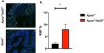
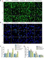
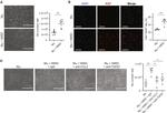

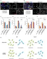


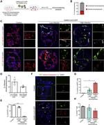
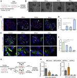


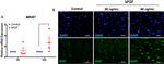
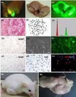

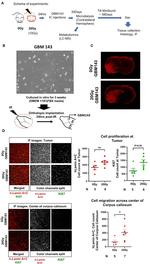


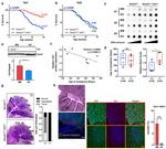


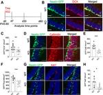


 in IHC (P)")
 in IHC (P)")
 in ICC/IF")
 in ICC/IF")
 in ICC/IF")
 in IHC (P)")
 in IHC (P)")
 in IHC (P)")
 in IHC")
 in IHC")
 in ICC/IF")
 in ICC/IF")
 in ICC/IF")
 in ICC/IF")
 in ICC/IF")
 in ICC/IF")
 in ICC/IF")
 in ICC/IF")
 in ICC/IF")
 in ICC/IF, Flow")
 in ICC/IF, Flow")
 in ICC/IF, Flow")
 in ICC/IF")
 in ICC/IF")
 in ICC/IF, IHC")
 in ICC/IF, IHC")
 in ICC/IF")
 in ICC/IF")
 in ICC/IF, IHC")
 in ICC/IF")
 in ICC/IF")
 in ICC/IF")
 in ICC/IF")
 in ICC/IF")
 in ICC/IF, IHC (P)")
 in ICC/IF, IHC")
 in ICC/IF, IHC")
 in ICC/IF")
 in ICC/IF")
 in ICC/IF")
 in ICC/IF")
 in ICC/IF")
 in ICC/IF")
 in ICC/IF")
 in ICC/IF")
 in ICC/IF")
 in ICC/IF, IHC")
 in ICC/IF, IHC")
 in ICC/IF")
 in ICC/IF")
 in ICC/IF")
 in ICC/IF")
 in ICC/IF, IHC")
 in ICC/IF, IHC")
 in ICC/IF")
 in ICC/IF")
 in ICC/IF")
 in ICC/IF")
 in ICC/IF")
 in ICC/IF")
 in WB, ICC/IF")
 in ICC/IF, IHC")
 in WB, ICC/IF")
 in ICC/IF")
 in ICC/IF")
 in WB, ICC/IF")
 in ICC/IF")
 in ICC/IF")
 in ICC/IF")
 in ICC/IF")
 in IHC, IHC (P)")
 in IHC (P)")
 in IHC, IHC (P)")
 in IHC (P)")
 in IHC (P), IHC (PFA)")
 in IHC (P)")
 in IHC (P)")
 in IHC (P)")
 in IHC, IHC (P)")
 in IHC, IHC (P)")
 in IHC (F)")
 in IHC (F)")
 in IHC, IHC (F)")
 in IHC (F)")
 in IHC")
 in IHC")
 in IHC")
 in IHC")
 in IHC")
 in IHC, IHC (PFA)")
 in IHC")
 in IHC")
 in IHC")
 in IHC")
 in IHC")
")
")

