Search


Citations & References (20)
Invitrogen™
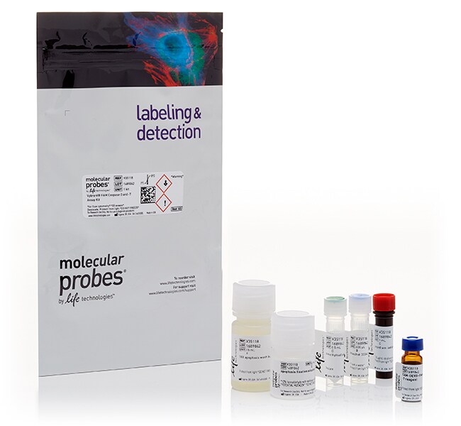
Vybrant™ FLICA Caspase Apoptosis Assay Kits for flow cytometry
Detect apoptosis via flow cytometry with Vybrant FLICA Caspase Assays that detect poly and caspase-3/7/8 activities.
Have Questions?
Change view

| Catalog Number | Enzyme |
|---|---|
| V35118 | Caspase 3/7 |
| V35117 | Poly Caspases |
| V35119 | Caspase 8 |
Catalog number V35118
Price (MXN)
-
Enzyme:
Caspase 3/7
Quickly detect caspase-mediated apoptosis by flow cytometry with Vybrant FAM caspase assay kits for poly-caspases and caspases 3 and 7. The Vybrant FAM caspase assay kits use fluorescent inhibitor of caspases (FLICA) methodology to detect and report caspase activity. Specifically, these caspase assay kits contain engineered FLICA substrates FAM-VAD-FMK (for poly-caspases), FAM-DEVD-FMK (for caspase-3/7), and FAM-LETD-FMK (for caspase-8). They also include Hoechst 33342 and propidium iodide stains, enabling simultaneous evaluation of membrane permeability and cell cycle by flow cytometry.
The Vybrant FAM caspase assay kits for flow cytometry employ a novel approach to detecting active caspases: the kits take advantage of FLICA methodology, which is an affinity label. This affinity label associates a fluoromethyl ketone (FMK) moiety, which can react covalently with a cysteine, with a caspase-specific amino acid sequence. Different amino acid recognition sequences are used to detect different caspases: valine-alanine-aspartic acid (VAD) for poly caspases (including caspase-1, -3, -4, -5, -6, -7, -8, and -9), aspartic acid-glutamic acid-valine-aspartic acid (DEVD) for caspase-3/7, and leucine-glutamic acid-threonine-aspartic acid (LETD) for caspase-8. A carboxyfluorescein (FAM) group is attached as a reporter.
The FLICA reagent is thought to interact with the enzymatic reactive center of an activated caspase via the recognition sequence, and then to attach covalently through the FMK moiety. The FLICA inhibitor is cell permeant and noncytotoxic. Unbound FLICA molecules diffuse out of the cell and are washed away; the remaining fluorescent signal is a direct measure of the amount of active caspase present at the time the inhibitor was added. The approximate excitation and emission peaks of the FLICA, propidium iodide, and Hoechst 33342 reagents are 488 nm/530 nm, 535 nm/6617 nm, and 350 nm/461 nm, respectively. The detection reagents included within the individual Vybrant FAM Poly Caspases Assay, Caspase-3 and -7 Assay, and Caspase-8 Assay apoptosis kits are 488/530 FAM-VAD-FMK reagent, 488/530 FAM-DEVD-FMK, and 488/530 FAM-LETD-FMK, respectively.
For Research Use Only. Not for use in diagnostic procedures.
Specifications
EnzymeCaspase 3/7
Excitation/EmissionFAM-DEVD-FMD: 388/530
PI: 535/617
Hoechst 33342: 350/461
PI: 535/617
Hoechst 33342: 350/461
Flow Cytometer Laser LinesUV, 488
For Use With (Equipment)Flow Cytometer
Label TypeOther Label(s) or Dye(s)
Label or DyeFAM, Hoechst 33342, Propidium Iodide
Product LineVybrant
Product TypeCaspase Assay Kit
Quantity25 Assays
Shipping ConditionRoom Temperature
Detection MethodFluorescence
FormatTube
Unit Size25 assays
Contents & Storage
1 vial FAM-DEVD-FMK caspase-3 and -7 reagent (FLICA reagent, lyophilized solid), 1 vial of Hoechst 33342 (400 μL, 1 mM in water), 1 vial of PI (1 mL, 250 μg/mL in water), 1 vial of DMSO (0.5 mL), 1 bottle of apoptosis fixative solution (10% formaldehyde with methanol in PBS, 6 mL), and 1 bottle of 10x apoptosis wash buffer. Store in refrigerator (2–8°C) and protect from light.
Frequently asked questions (FAQs)
What are the fluorescence excitation/emission maxima for the FAM-DEVD-FMK FLICA reagent, Hoechst 33342, and propidium iodide supplied in the Vybrant FAM Caspase-3 and -7 Assay Kit, for flow cytometry?
Citations & References (20)
Citations & References
Abstract
Monocyte Chemoattractant Protein-Induced Protein 1 (MCPIP1) Enhances Angiogenic and Cardiomyogenic Potential of Murine Bone Marrow-Derived Mesenchymal Stem Cells.
Journal:
PubMed ID:26214508
'The current evidence suggests that beneficial effects of mesenchymal stem cells (MSCs) toward myocardial repair are largely due to paracrine actions of several factors. Although Monocyte chemoattractant protein-induced protein 1 (MCPIP1) is involved in the regulation of inflammatory response, apoptosis and angiogenesis, whether MCPIP1 plays any role in stem cell-induced
Multiparametric evaluation of apoptosis: effects of standard cytotoxic agents and the cyanoguanidine CHS 828.
Journal:Mol Cancer Ther
PubMed ID:15141009
'A multiparametric high-content screening assay for measurement of apoptosis was developed. HeLa cells and lymphoma U-937 cells were exposed to cytotoxic drugs in flat-bottomed optical microtiter plates. After incubation, the DNA-binding dye Hoechst 33342, fluorescein-tagged probes that covalently bind active caspases and chloromethyl-X-rosamine to detect mitochondrial membrane potential (MMP) were
Flow cytometry-based apoptosis detection.
Journal:Methods Mol Biol
PubMed ID:19609746
'An apoptosing cell demonstrates multitude of characteristic morphological and biochemical features, which vary depending on the stimuli and the cell type. The gross majority of classical apoptotic hallmarks can be rapidly examined by flow and image cytometry. Cytometry thus became a technology of choice in diverse studies of cellular demise.
BH3-only protein Noxa regulates apoptosis in activated B cells and controls high-affinity antibody formation.
Journal:Blood
PubMed ID:22144184
The efficiency of humoral immune responses depends on the selective outgrowth of B cells and plasma cells that produce high affinity antibodies. The factors responsible for affinity maturation of B cell clones in the germinal center (GC) have been well established but selection mechanisms that allow clones to enter the
In vitro primary human lymphocyte flow cytometry based micronucleus assay: simultaneous assessment of cell proliferation, apoptosis and MN frequency.
Journal:Mutagenesis
PubMed ID:21791709
In order to minimise the number of positive in vitro cytogenetic results which are not confirmed in rodent carcinogenicity tests, biological systems that are p53 and DNA repair proficient should be recommended. Moreover, an appropriate cytotoxicity parameter for top dose selection should be considered. Recent International Conference on Harmonisation draft