Search
How Fluorescence Microscopy Works
 | Microscopy is a basic research toolMicroscopes are powerful research and discovery tools and have contributed to countless ground-breaking discoveries, over several centuries. Find basic definitions for common microscopy terms, information to help you understand differences between magnification and resolution, and how fluorescence can be used to improve contrast and increase resolution. |
What is a microscope?
A microscope magnifies objects; more lenses translate to higher magnification. In its most basic form, a microscope is simply a device that allows you to see things that are not visible to the naked eye by the use of lenses to magnify your sample. That means a magnifying glass is technically also a microscope. If you use light along with a lens, you’ve got something called an optic or light microscope. Figure 1. A magnifying glass is the simplest form of lens used to view objects. |
|

Brightfield vs. fluorescence microscopy
If you use additional lenses to increase your ability to magnify a sample, you’ve got a compound microscope. Most modern microscopes are compound microscopes, because the additional magnification gives a more enlarged image. If only white light is used for illumination, then it’s bright-field microscopy.
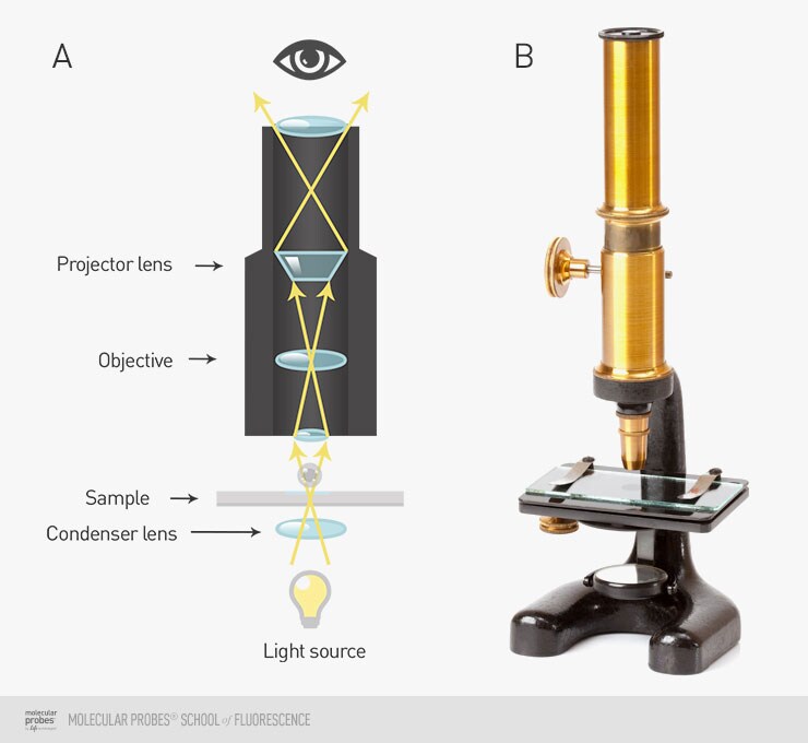
Figure 2.The light path through lenses and sample in basic brightfield microscopy (A). Antique 19th century drum-style compound microscope (B).
The difference between magnification and resolution
It’s pretty important to understand the difference between magnification and resolution when it comes to getting a good result when you’re doing fluorescence imaging. When we talk about magnification, we are referring to how much bigger an object appears when we look at it under the microscope.
In contrast, when we talk about resolution in a practical sense, we are referring to how much detail we can distinguish in our image, which can be subjective. In a more technical sense, resolution is limited by the refractive properties of light.
Using fluorescence can increase resolution
Resolution and magnification are not the same. In microscopy, we refer to the details visible in a magnified image as resolution. Magnification, or making something appear larger, doesn’t do you much good if you cannot see the detail in the sample you’ve magnified.
Bright-field microscopy relies on the differences in absorption of light due to differences in densities between various parts of the sample, which for our purposes is a cell. Because of this, bright-field microscopy is not great when you want to see many details in your cell. How do you increase your resolution? Add in fluorophores to stain structures in your sample and filters to illuminate it, focus the light emitted by the sample, add a sensitive detector, and voila! Now you can do fluorescence microscopy. Fluorescence microscopy gives you the advantage of better resolution by making various structures in the cells contrast better with their neighbors, as well as allowing you to collect images in more than one color.
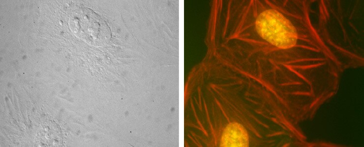
Figure 3. An image of the same field of BPAE cells captured using brightfield (left) and fluorescence (right) microscopy. Fluorescent labeling of the nucleus (yellow) and actin (red) makes it possible to see much more detailed cell structure.
Share
For Research Use Only. Not for use in diagnostic procedures.