Search

Materials Science
SEM in Geology
SEM EDS is a vital part of multi-modal, multi-scale analysis of rocks and minerals.
Join the conversation
bf5a- Apps/Techniques/Products/Resources/Contact Us
SEM mineral identification
Few disciplines rely as heavily on consistent, multi-scale observation and interpretation of features as the earth sciences. Everything from deep earth flow processes to more efficient methods of resource extraction are dependent on accurate sample characterization.
Thermo Fisher Scientific has built a family of imaging platforms and software solutions to facilitate data collection, visualization and analysis during complicated multi-modal, multi-scale characterization routines.
Petrology and mineralogy
Accurate textural analysis and the associated distribution of minerals within the rock texture are key to accurately describing the physical and chemical aspects of a rock system. Automated determination of mineralogy, based on scanning electron microscopy coupled with energy-dispersive X-ray spectroscopy (SEM-EDS), has established itself as a popular method for acquiring high-resolution images and chemical maps in the mining and mineral processing industries. Thermo Fisher Scientific has a 30-year history of delivering market-leading SEM-EDS technology. Here are just a few of the advantages afforded by our petrology and mineralogy solutions;
- Expand the number of samples that can be processed with routine petrologic and mineralogic analysis
- Build a strong statistical repository of mineral content based on automated analysis
- Automatically obtain accurate mineralogical identification
- Create a digital inventory of your thin sections and associated mineral assemblages
- Combine EDS elemental analysis, mineralogy and high-resolution imagery for advanced sample interpretation
- Analyze more samples for more robust statistics


Multi-scale analysis with geoscience software
While context is vital for textural analysis, it is especially important for geological or structural interpretation. Electron imaging captures a large number of modalities in a single acquisition, allowing for direct interpretation of composition and texture. However, simply observing a single frame, or even a series of images, out of context reduces the power of this analysis.
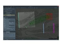
We provide a suite of automation software developed with the express purpose of preserving context. Thermo Scientific Maps Software is the cross-platform automation engine for our full line of electron microscopy imaging platforms. Maps Software takes the pain out of acquiring larger image mosaics within an easy to use and intuitive software environment.
- Preserve context with large, automatically acquired, high-resolution datasets
- Utilize system time efficiently
- Share observations with context for more collaborative data interpretation

Chemical zonation in a plagioclase feldspar sample. Albite is shown in dark blue, labradorite is shown in light blue.

Multi-scale analysis of a sandstone sample. The left image is a slice from a microCT dataset, the right 3 images are from subsequent SEM analysis that interrogated the clay mineralogy present in the pores.

Cross-section overview of the major rock types, revealing varied mineral and structural distribution.

Surface analysis of a rock cross-section. A single mineralogical dataset from SEM-EDS analysis is uniquely capable of telling a rich story on texture and composition.

Multi-scale analysis of a core plug with X-ray microtomography (microCT) and SEM. The initial sample, plus the extracted mini plug, were imaged with HeliScan microCT. A cross-section of the mini plug was then imaged with SEM. The SEM data has been correlated and segmented with Avizo Software.
Chemical zonation in a plagioclase feldspar sample. Albite is shown in dark blue, labradorite is shown in light blue.
Multi-scale analysis of a sandstone sample. The left image is a slice from a microCT dataset, the right 3 images are from subsequent SEM analysis that interrogated the clay mineralogy present in the pores.
Cross-section overview of the major rock types, revealing varied mineral and structural distribution.
Surface analysis of a rock cross-section. A single mineralogical dataset from SEM-EDS analysis is uniquely capable of telling a rich story on texture and composition.
Multi-scale analysis of a core plug with X-ray microtomography (microCT) and SEM. The initial sample, plus the extracted mini plug, were imaged with HeliScan microCT. A cross-section of the mini plug was then imaged with SEM. The SEM data has been correlated and segmented with Avizo Software.
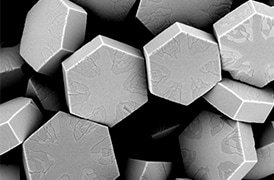
Fundamental Materials Research
Novel materials are investigated at increasingly smaller scales for maximum control of their physical and chemical properties. Electron microscopy provides researchers with key insight into a wide variety of material characteristics at the micro- to nano-scale.
Style Sheet for Komodo Tabs

3D Materials Characterization
Development of materials often requires multi-scale 3D characterization. DualBeam instruments enable serial sectioning of large volumes and subsequent SEM imaging at nanometer scale, which can be processed into high-quality 3D reconstructions of the sample.
_Technique_800x375_144DPI.jpg)
EDS Elemental Analysis
Thermo Scientific Phenom Elemental Mapping Software provides fast and reliable information on the distribution of chemical elements within a sample.
_Technique_800x375_144DPI.jpg)
3D EDS Tomography
Modern materials research is increasingly reliant on nanoscale analysis in three dimensions. 3D characterization, including compositional data for full chemical and structural context, is possible with 3D EM and energy dispersive X-ray spectroscopy.

Cross-sectioning
Cross sectioning provides extra insight by revealing sub-surface information. DualBeam instruments feature superior focused ion beam columns for high-quality cross sectioning. With automation, unattended high-throughput processing of samples is possible.

Multi-scale analysis
Novel materials must be analyzed at ever higher resolution while retaining the larger context of the sample. Multi-scale analysis allows for the correlation of various imaging tools and modalities such as X-ray microCT, DualBeam, Laser PFIB, SEM and TEM.
3D Materials Characterization
Development of materials often requires multi-scale 3D characterization. DualBeam instruments enable serial sectioning of large volumes and subsequent SEM imaging at nanometer scale, which can be processed into high-quality 3D reconstructions of the sample.
EDS Elemental Analysis
Thermo Scientific Phenom Elemental Mapping Software provides fast and reliable information on the distribution of chemical elements within a sample.
3D EDS Tomography
Modern materials research is increasingly reliant on nanoscale analysis in three dimensions. 3D characterization, including compositional data for full chemical and structural context, is possible with 3D EM and energy dispersive X-ray spectroscopy.
Cross-sectioning
Cross sectioning provides extra insight by revealing sub-surface information. DualBeam instruments feature superior focused ion beam columns for high-quality cross sectioning. With automation, unattended high-throughput processing of samples is possible.
Multi-scale analysis
Novel materials must be analyzed at ever higher resolution while retaining the larger context of the sample. Multi-scale analysis allows for the correlation of various imaging tools and modalities such as X-ray microCT, DualBeam, Laser PFIB, SEM and TEM.
Style Sheet for Instrument Cards Original



Style Sheet to change H2 style to p with em-h2-header class