Search


Citations & References (36)
Invitrogen™
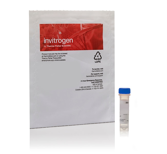
Cholera Toxin Subunit B (Recombinant) Conjugate
Recombinant cholera toxin B subunit (CT-B) conjugates bind with high affinity to GM1 ganglioside and support neuronal tracing, GM1 labeling, membrane trafficking, and membrane organization studies. Choose from Alexa Fluor™ 488, 555, 594, and 647 fluorescent conjugates, Biotin-XX, or horseradish peroxidase (HRP) to match your detection workflow.
Have Questions?
Change view

| Catalog Number | Conjugate | Quantity |
|---|---|---|
| C34776 | Alexa Fluor 555 | 100 μg |
| C22841 | Alexa Fluor 488 | 500 μg |
| C22843 | Alexa Fluor 555 | 500 μg |
| C34777 | Alexa Fluor 594 | 100 μg |
| C22842 | Alexa Fluor 594 | 500 μg |
| C34778 | Alexa Fluor 647 | 100 μg |
| C34779 | Biotin | 100 μg |
| C34780 also known as C-34780 | HRP (Horseradish Peroxidase) | 100 μg |
Catalog number C34776
Price (USD)
341.65
Online exclusive
360.00Save 18.35
Each
Conjugate:
Alexa Fluor 555
Quantity:
100 μg
Price (USD)
341.65
Online exclusive
360.00Save 18.35
Each
Recombinant cholera toxin B conjugates for GM1 labeling and neuronal tracing
Cholera toxin B subunit (CT-B) binds with high affinity to GM1 ganglioside on the cell surface and is widely used for neuronal tracing, GM1 labeling, endocytosis, and membrane trafficking studies. At neutral pH, CT-B forms a pentamer with multiple GM1-binding sites, enabling multivalent interactions with GM1-containing membranes.
These conjugates are made using recombinant cholera toxin B subunit without the enzymatically active A subunit of native cholera toxin.
Why use recombinant cholera toxin B conjugates?
- Label GM1 ganglioside: Use the high-affinity interaction between CT-B and GM1 to visualize and detect GM1-associated membrane structures.
- Trace neuronal projections: Use CT-B for well-established retrograde labeling of neuronal pathways.
- Study membrane trafficking: Follow CT-B/GM1 internalization and retrograde trafficking through endosomal pathways.
- Investigate membrane organization: Label GM1 and examine GM1-associated membrane domains in lipid raft-related studies.
- Use recombinant B subunit: Study GM1 binding and trafficking with CT-B lacking the enzymatically active A subunit of native cholera toxin.
- Choose multiple detection formats: Select fluorescent Alexa Fluor™ conjugates, Biotin-XX, or HRP to match fluorescence, affinity-based, or enzyme-based detection workflows.
Choose a CT-B conjugate for your detection workflow
Select from Alexa Fluor™ 488, 555, 594, and 647 CT-B conjugates for fluorescence imaging, Biotin-XX for avidin- or streptavidin-based detection, or HRP for enzyme-based detection.
Why choose from multiple conjugates?
- Match your imaging system: Select an Alexa Fluor™ conjugate compatible with available excitation sources, emission filters, and detectors.
- Support multicolor imaging: Choose spectrally distinct Alexa Fluor™ dyes for use with other fluorescent probes, antibodies, and cellular markers.
- Enable affinity-based detection: Use Biotin-XX conjugates with avidin- or streptavidin-based detection workflows.
- Enable enzyme-based detection: Use HRP conjugates for chromogenic or chemiluminescent detection workflows.
- Use one GM1-binding probe across workflows: Apply recombinant CT-B with different labels while maintaining the same GM1-binding biological probe.
Applications of cholera toxin B conjugates
Cholera toxin B conjugates are versatile probes for neuronal tracing, GM1 ganglioside labeling, endocytosis, and membrane trafficking studies.
Use CT-B conjugates to:
- Perform neuronal tracing, including widely used retrograde labeling of neuronal projections
- Label GM1 ganglioside on cell membranes
- Visualize GM1-associated membrane domains in lipid raft-related studies
- Study CT-B/GM1 internalization and retrograde trafficking through endosomal pathways
- Perform fluorescence imaging with Alexa Fluor™ 488, 555, 594, or 647 conjugates
- Perform avidin- or streptavidin-based detection with Biotin-XX conjugates
- Perform enzyme-based detection with HRP conjugates
For Research Use Only. Not for use in diagnostic procedures.
For Research Use Only. Not for use in diagnostic procedures.
Specifications
Label TypeAlexa Fluor Dyes
Product LineAlexa Fluor
Protein FormRecombinant
Protein SubtypeCholera Toxin
Quantity100 μg
Shipping ConditionRoom Temperature
ConjugateAlexa Fluor 555
FormLyophilized
RecombinantRecombinant
Unit SizeEach
Contents & Storage
Store in freezer (-5 to -30°C) and protect from light.
Frequently asked questions (FAQs)
I injected a fluorescent tracer, but cannot detect it after tissue is fixed and sectioned. What am I doing wrong?
Do you have a tracer that will only transport retrograde?
How do I know which tracer to choose for my experiment?
What products do you have for neuronal tracing?
Citations & References (36)
Citations & References
Abstract
Autoantigen Golgin-97, an effector of Arl1 GTPase, participates in traffic from the endosome to the trans-golgi network.
Journal:Mol Biol Cell
PubMed ID:15269279
'The precise cellular function of Arl1 and its effectors, the GRIP domain Golgins, is not resolved, despite our recent understanding that Arl1 regulates the membrane recruitment of these Golgins. In this report, we describe our functional study of Golgin-97. Using a Shiga toxin B fragment (STxB)-based in vitro transport assay,
Caveolin regulates endocytosis of the muscle repair protein, dysferlin.
Journal:J Biol Chem
PubMed ID:18096699
'Dysferlin and Caveolin-3 are plasma membrane proteins associated with muscular dystrophy. Patients with mutations in the CAV3 gene show dysferlin mislocalization in muscle cells. By utilizing caveolin-null cells, expression of caveolin mutants, and different mutants of dysferlin, we have dissected the site of action of caveolin with respect to dysferlin
Specialized cortical subnetworks differentially connect frontal cortex to parahippocampal areas.
Journal:J Neurosci
PubMed ID:22302828
'How information is manipulated and segregated within local circuits in the frontal cortex remains mysterious, in part because of inadequate knowledge regarding the connectivity of diverse pyramidal cell subtypes. The frontal cortex participates in the formation and retrieval of declarative memories through projections to the perirhinal cortex, and in procedural
Integrin-mediated adhesion regulates membrane order.
Journal:J Cell Biol
PubMed ID:16943184
'The properties of cholesterol-dependent domains (lipid rafts) in cell membranes have been controversial. Because integrin-mediated cell adhesion and caveolin both regulate trafficking of raft components, we investigated the effects of adhesion and caveolin on membrane order. The fluorescent probe Laurdan and two-photon microscopy revealed that focal adhesions are highly ordered;
Metastatic potential of mouse Lewis lung cancer cells is regulated via ganglioside GM1 by modulating the matrix metalloprotease-9 localization in lipid rafts.
Journal:J Biol Chem
PubMed ID:16636068
'To analyze mechanisms for cancer metastasis, we established high metastatic sublines from mouse Lewis lung cancer (P29) by repeated injection. Sublines established from the two subclones H7 and C4 commonly exhibited increased proliferation and invasion activity and reduced expression of ganglioside GM1, although they showed different preferences in their target