Search


Citations & References (16)
Invitrogen™
HCS CellMask™ Stains
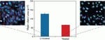
Easily visualize entire cells or individual cell structures during high-content screening (HCS) assays with HCS CellMask stains, which are available in a variety of colors for multiplexing flexibility and can be used immediately after fixation and permeabilization, or after antibody labeling.
Have Questions?
Change view

| Catalog Number | Description |
|---|---|
| H32720 | HCS CellMask Blue Stain |
| H32714 | HCS CellMask Green Stain |
| H32722 | HCS CellMask Near-IR Stain |
| H32713 | HCS CellMask Orange Stain |
| H32712 | HCS CellMask™ Red Stain |
| H32721 | HCS CellMask Deep Red Stain |
Catalog number H32720
Price (USD)
479.65
Online exclusive
500.00Save 20.35
Each
Description:
HCS CellMask Blue Stain
Price (USD)
479.65
Online exclusive
500.00Save 20.35
Each
Identify entire cells (cytoplasm and nucleus) during high-content screening (HCS) assays with the versatile HCS CellMask Red, CellMask Orange, CellMask Green, CellMask Blue, CellMask Deep Red, and CellMask Near-IR stains, which are fluorophores that can be used immediately after fixation and permeabilization, or after antibody labeling. They are available in a variety of colors for multiplexing protocols.
The versatile HCS CellMask Red, CellMask Orange, CellMask Green, CellMask Blue, CellMask Deep Red, and CellMask Near-IR stains are cell delineation tools for high content screening (HCS) platforms, label the entire cell (cytoplasm and nucleus) and provide an accurate backdrop against which the features of interest can be assessed. The HCS CellMask Red and Blue stains replace the HCS CellMask Red cytoplasmic/nuclear (Cat. No. H32711) and HCS CellMask Blue cytoplasmic/nuclear stains (Cat. No. H34558), respectively.
HCS CellMask stains can be applied to cells immediately after fixation or in the last step of multiplexing protocols, and they are compatible with detergent-based cell permeabilization protocols. If only nuclear staining is desired, HCS NuclearMask stains can be used for measuring DNA content in addition to enabling robust cell demarcation. HCS NuclearMask stains are also available in a choice of colors for multiplexing flexibility.
HCS CellMask stains can be applied to cells immediately after fixation or in the last step of multiplexing protocols, and they are compatible with detergent-based cell permeabilization protocols. If only nuclear staining is desired, HCS NuclearMask stains can be used for measuring DNA content in addition to enabling robust cell demarcation. HCS NuclearMask stains are also available in a choice of colors for multiplexing flexibility.
For Research Use Only. Not for human or animal therapeutic or diagnostic use.
Specifications
ColorBlue
DescriptionHCS CellMask Blue Stain
Detection MethodFluorescence
EmissionVisible
Excitation Wavelength Range346/442
For Use With (Equipment)High Content Instrument
Product LineCellMask
Quantity1 Set
Shipping ConditionRoom Temperature
Label TypeFluorescent Dye
Product TypeStain
Sub Cellular LocalizationCytoplasm, Cytosol, Nucleus
Unit SizeEach
Contents & Storage
Includes 1 set of vials, providing sufficient material to stain a total of ten 96-well microplates.
Store at ≤-20°C, desiccate, and protect from light.
Store at ≤-20°C, desiccate, and protect from light.
Frequently asked questions (FAQs)
I need a total cell stain, similar to HCS CellMask Blue stain, to label cytoplasm and nuclei in live cells. What do you recommend?
What dyes are used to make the CellMask stains?
Citations & References (16)
Citations & References
Abstract
Plk5, a polo box domain-only protein with specific roles in neuron differentiation and glioblastoma suppression.
Journal:Mol Cell Biol
PubMed ID:21245385
'Polo-like kinases (Plks) are characterized by the presence of a specific domain, known as the polo box (PBD), involved in protein-protein interactions. Plk1 to Plk4 are involved in centrosome biology as well as the regulation of mitosis, cytokinesis, and cell cycle checkpoints in response to genotoxic stress. We have analyzed
Transfer of polyglutamine aggregates in neuronal cells occurs in tunneling nanotubes.
Journal:
PubMed ID:23781027
Huntington's disease (HD) is a dominantly inherited neurodegenerative disease caused by CAG expansion in the huntingtin gene, which adds a homopolymeric tract of polyglutamine (polyQ) to the encoded protein leading to the formation of toxic aggregates. Despite rapidly accumulating evidences supporting a role for intercellular transmission of protein aggregates, little
Olivocochlear suppression of outer hair cells in vivo: evidence for combined action of BK and SK2 channels throughout the cochlea.
Journal:J Neurophysiol
PubMed ID:23282326
Cholinergic inhibition of cochlear hair cells via olivocochlear (OC)-efferent feedback is mediated by Ca(2+) entry through a9-/a10-nicotinic receptors, but the nature of the K(+) channels activated by this Ca(2+) entry has been debated (Yoshida N, Hequembourg SJ, Atencio CA, Rosowski JJ, Liberman MC. J Neurophysiol 85: 84-88, 2001). A recent
Acetylation modulates cellular distribution and DNA sensing ability of interferon-inducible protein IFI16.
Journal:Proc Natl Acad Sci U S A
PubMed ID:22691496
Detection of pathogenic nucleic acids is essential for mammalian innate immunity. IFN-inducible protein IFI16 has emerged as a critical sensor for detecting pathogenic DNA, stimulating both type I IFN and proinflammatory responses. Despite being predominantly nuclear, IFI16 can unexpectedly sense pathogenic DNA in both the cytoplasm and the nucleus. However,
Functional analysis of centrosomal kinase substrates in Drosophila melanogaster reveals a new function of the nuclear envelope component otefin in cell cycle progression.
Journal:Mol Cell Biol
PubMed ID:22751930
Phosphorylation is one of the key mechanisms that regulate centrosome biogenesis, spindle assembly, and cell cycle progression. However, little is known about centrosome-specific phosphorylation sites and their functional relevance. Here, we identified phosphoproteins of intact Drosophila melanogaster centrosomes and found previously unknown phosphorylation sites in known and unexpected centrosomal components.