Search
Invitrogen
Ki-67 Monoclonal Antibody (20Raj1), Brilliant Ultra Violet™ 737, eBioscience™
{{$productOrderCtrl.translations['antibody.pdp.commerceCard.promotion.promotions']}}
{{$productOrderCtrl.translations['antibody.pdp.commerceCard.promotion.promocode']}}: {{promo.promoCode}} {{promo.promoTitle}} {{promo.promoDescription}}. {{$productOrderCtrl.translations['antibody.pdp.commerceCard.promotion.learnmore']}}
{{$ctrl.translations['antibody.pdp.skuNotes.additionalInformation']}}:

FIGURE: 1 / 1
Ki-67 Antibody (367-5699-42) in Flow
Normal human peripheral blood cells were stimulated for 48 hours with immobilized CD3 Monoclonal Antibody, Functional Grade (Product # 16-0037-85). Cells were then surface-stained with CD19 Monoclonal Antibody, PE (Product # 12-0199-42) and stained intracellularly, using the Foxp3/Transcription Factor Staining Buffer Set (Product # 00-5523-00) and protocol, with Mouse IgG1 kappa Isotype Control, Brilliant Ultra Violet 737 (BUV737) (Product # 367-4714-81) (left) or Ki-67 Monoclonal Antibody, Brilliant Ultra Violet 737 (BUV737) (right). Viab... View More

Please note: We are reviewing Western blot images included in the antibody testing data in our catalog, including those provided by third parties. Unless expressly labeled or annotated as “raw-unedited”, Western blot images included in the antibody testing data in our catalog may have been edited, optimized or otherwise adjusted for presentation.

Product Details
367-5699-42
Applications
Tested Dilution
Publications
Product Specifications
Species Reactivity
Dog,
Human
Published species
Mouse
Host/Isotype
Mouse
/ IgG1, kappa
Recommended Isotype Control
Class
Monoclonal
Type
Antibody
Clone
20Raj1
Conjugate
Brilliant Ultra Violet™ 737
Brilliant Ultra Violet™ 737
Brilliant Ultra Violet™ 737
Excitation/Emission Max
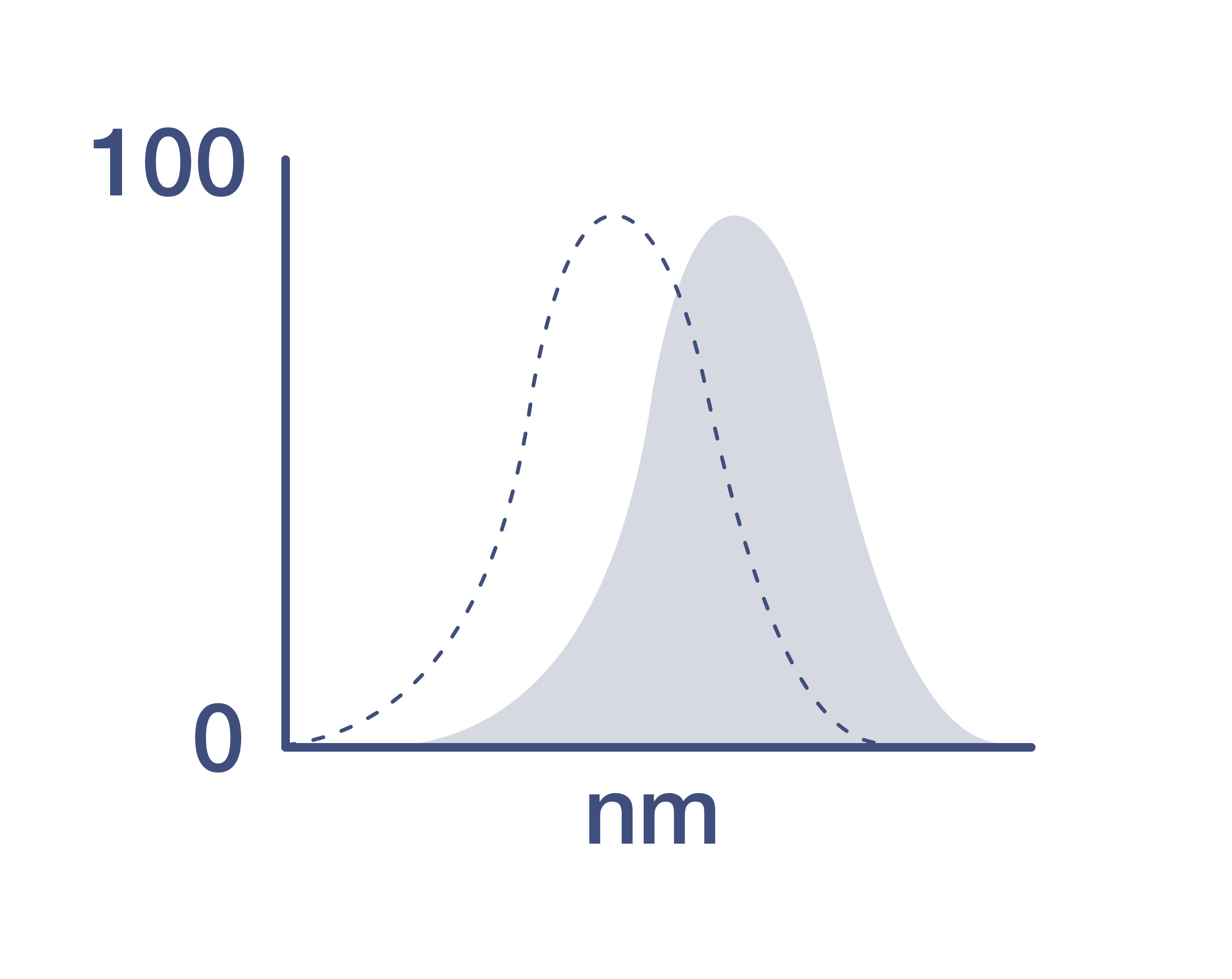
350/740 nm
View spectra
Form
Liquid
Concentration
5 µL/Test
Purification
Affinity chromatography
Storage buffer
PBS, pH 7.2, with BSA
Contains
0.09% sodium azide
Storage conditions
4°C, store in dark, DO NOT FREEZE!
Shipping conditions
Ambient (domestic); Wet ice (international)
RRID
Product Specific Information
Description
The monoclonal antibody 20Raj1 recognizes the human Ki-67 protein. Two isoforms of Ki-67 exist, a 345 and 395 kDa form that are expressed in dividing cells. Ki-67 is expressed in all cell types and is detectable during active phases of the cell cycle (G1, S, G2, and mitosis) but is absent from resting cells (G0). During interphase, Ki-67 expression is localized to the nucleus but redistributes to the chromosomes during mitosis and has specifically been found to associate with heterochromatin-bound proteins such as chromobox protein homolog 3 (CBX3). In studies of tumor cells, Ki-67 expression has been used as a marker for determining the fraction of proliferating cells within a given population of tumor cells. This monoclonal antibody 20Raj1 recognizes canine Ki-67.
Applications Tested
This 20Raj1 antibody has been pre-diluted and tested by intracellular staining followed by flow cytometric analysis of stimulated normal human peripheral blood cells using the Foxp3/Transcription Factor Staining Buffer Set (Product # 00-5523-00) and protocol. Please refer to "Staining Intracellular Antigens for Flow Cytometry, Protocol B: One step protocol for intracellular (nuclear) proteins" located at Flow Protocols . This may be used at 5 µL (0.125 µg) per test. A test is defined as the amount (µg) of antibody that will stain a cell sample in a final volume of 100 µL. Cell number should be determined empirically but can range from 10^5 to 10^8 cells/test.
Blocking Buffers
When using two or more Super Bright, Brilliant Violet™, Brilliant Ultra Violet™, or other polymer dye-conjugated antibodies in a staining panel, it is recommended to use Super Bright Complete Staining Buffer (Product # SB-4401) or Brilliant Stain Buffer (Product # 00-4409-75) to minimize any non-specific polymer interactions. Please refer to the datasheet for Super Bright Staining Buffer or Brilliant Stain Buffer for more information.
Light sensitivity
This tandem dye is sensitive to photo-induced oxidation. Please protect this vial and stained samples from light.
Fixation
• Samples can be stored in IC Fixation Buffer (Product # 00-8222) (100 µL of cell sample + 100 µL of IC Fixation Buffer) or 1-step Fix/Lyse Solution (Product # 00-5333) for up to 3 days in the dark at 4°C with minimal impact on brightness and FRET efficiency/compensation.
• Some generalizations regarding fluorophore performance after fixation can be made, but clone specific performance should be determined empirically.
• Our internal testing suggests that Brilliant Ultra Violet™ 737 (BUV737) is compatible with short-term methanol-based fixation, but should not be stored in buffers containing methanol for longer than one hour.
Excitation: 355 nm; Emission: 732 nm; Laser: Ultraviolet Laser.
BRILLIANT ULTRA VIOLET™ is a trademark or registered trademark of Becton, Dickinson and Company or its affiliates, and is used under license. Powered by Sirigen™.
Target Information
Ki-67 is a nuclear protein that is expressed during various stages in the cell cycle, particularly during late G1, S, G2, and M phases. The protein has a forkhead associated domain (FHA) through which it associates with euchromatin at the perichromosomal layer, the centromeric heterochromatin, and the nucleolus. Ki-67 is shown to have a cell cycle dependent topographical distribution with perinucleolar expression at G1, expression in the nuclear matrix at G2, and expression on the chromosomes during M phase. Ki-67 is commonly used as a proliferation marker because it is not detected in G0 cells, but increases steadily from G1 through mitosis. Ki-67 antibodies are useful in establishing the cell growing fraction in neoplasms. In neoplastic tissues, the prognostic value is comparable to the tritiated thymidine-labelling index. The correlation between low Ki-67 index and histologically low-grade tumors is strong. Ki-67 is routinely used as a neuronal marker of cell cycling and proliferation.
For Research Use Only. Not for use in diagnostic procedures. Not for resale without express authorization.
How to use the Panel Builder
Watch the video to learn how to use the Invitrogen Flow Cytometry Panel Builder to build your next flow cytometry panel in 5 easy steps.
Bioinformatics
Protein Aliases: antigen identified by monoclonal antibody Ki-67; antigen Ki67; Molecular Immunology Borstel antibody 1; proliferation-related Ki-67 antigen; protein phosphatase 1, regulatory subunit 105; RP11-380J17.2; unnamed protein product
Gene Aliases: KIA; MIB-; MIB-1; PPP1R105
UniProt ID: (Human) P46013
Entrez Gene ID: (Dog) 100686578, (Human) 4288
Disclaimer

Clicking the images or links will redirect you to a website hosted by BenchSci that provides third-party scientific content. Neither the content nor the BenchSci technology and processes for selection have been evaluated by us; we are providing them as-is and without warranty of any kind, including for use or application of the Thermo Fisher Scientific products presented.

Performance Guarantee
If an Invitrogen™ antibody doesn't perform as described on our website or datasheet,we'll replace the product at no cost to you, or provide you with a credit for a future purchase.*
Learn more
We're here to help
Get expert recommendations for common problems or connect directly with an on staff expert for technical assistance related to applications, equipment and general product use.
Contact tech support