Search Thermo Fisher Scientific
Disclaimer

Clicking the images or links will redirect you to a website hosted by BenchSci that provides third-party scientific content. Neither the content nor the BenchSci technology and processes for selection have been evaluated by us; we are providing them as-is and without warranty of any kind, including for use or application of the Thermo Fisher Scientific products presented.
Invitrogen
GL7 Monoclonal Antibody (GL-7 (GL7)), PerCP-eFluor™ 710, eBioscience™
")
FIGURE: 1 / 11
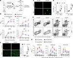
GL7 Antibody (46-5902-82) in Flow
C57Bl/6 splenocytes were unstimulated (left) or stimulated (right) with F (ab')2 Anti-Mouse IgM, u chain specific Functional Grade Purified (Product # 16-5890) and Anti-Mouse CD40 Functional Grade Purified (Product # 16-0401-82) for 3 days and then stained with Anti-Mouse CD19 FITC (Product # 11-0193-82) and 0.25 µg of Anti-Human/Mouse GL7 (T and B Cell Activation Marker) PerCP-eFluor® 710. Total viable cells, as determined by Fixable Viability Dye eFluor® 506, were used for analysis.


 in Flow")
 in Flow")
 in Flow")
 in Flow")
 in Flow")
 in Flow")
 in Flow")
 in Flow")
 in Flow")
 in Flow")
 in Flow")
Product Details
46-5902-82
Applications
Tested Dilution
Publications
Product Specifications
Species Reactivity
Human,
Mouse
Published species
Human,
Mouse
Host/Isotype
Rat
/ IgM
Recommended Isotype Control
Class
Monoclonal
Type
Antibody
Clone
GL-7 (GL7)
Conjugate
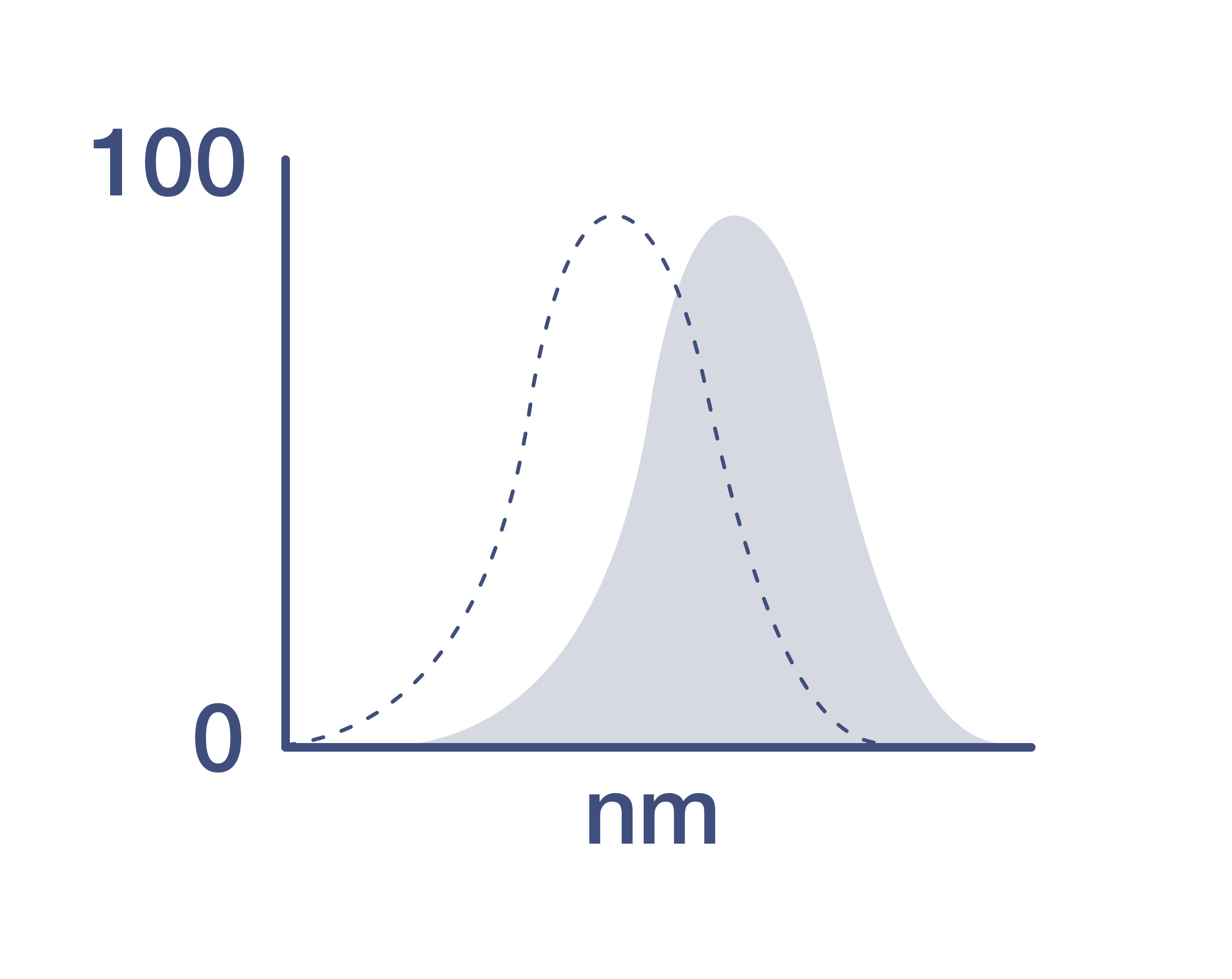
Excitation/Emission Max
482/708 nm
View spectra
Form
Liquid
Concentration
0.2 mg/mL
Purification
Affinity chromatography
Storage buffer
PBS, pH 7.2
Contains
0.09% sodium azide
Storage conditions
4° C, store in dark, DO NOT FREEZE!
Shipping conditions
Ambient (domestic); Wet ice (international)
RRID
AB_2573807
Product Specific Information
Description: This GL7 monoclonal antibody reacts with a cell-surface protein found on T and B lymphocytes activated in vitro, on bone marrow pre-B-II cells, germinal center B cells, and also human B cell lines Ramos and Daudi. There is strain variability with respect to antigen distribution on thymocytes and Con A-activated spleen cells, with expression in BALB/c greater than that in C57BL/6. GL7 is commonly used as a marker for mouse germinal center B cells. The epitope of GL7 has been identified as a sialic acid glycan moiety called Neu5Ac. This moiety is recognized by CD22.
Applications Reported: This GL-7 (GL7) antibody has been reported for use in flow cytometric analysis.
Applications Tested: This GL-7 (GL7) antibody has been tested by flow cytometric analysis of stimulated mouse splenocytes. This can be used at less than or equal to 0.5 µg per test. A test is defined as the amount (µg) of antibody that will stain a cell sample in a final volume of 100 µL. Cell number should be determined empirically but can range from 10^5 to 10^8 cells/test. It is recommended that the antibody be carefully titrated for optimal performance in the assay of interest.
PerCP-eFluor® 710 emits at 710 nm and is excited with the blue laser (488 nm); it can be used in place of PerCP-Cyanine5.5. We recommend using a 710/50 bandpass filter, however, the 695/40 bandpass filter is an acceptable alternative. Please make sure that your instrument is capable of detecting this fluorochrome.
Light sensitivity: This tandem dye is sensitive to photo-induced oxidation. Please protect this vial and stained samples from light.
Fixation: Samples can be stored in IC Fixation Buffer (Product # 00-8222) (100 µL of cell sample + 100 µL of IC Fixation Buffer) or 1-step Fix/Lyse Solution (Product # 00-5333) for up to 3 days in the dark at 4°C with minimal impact on brightness and FRET efficiency/compensation. Some generalizations regarding fluorophore performance after fixation can be made, but clone specific performance should be determined empirically.
Excitation: 488 nm; Emission: 710 nm; Laser: Blue Laser.
Filtration: 0.2 µm post-manufacturing filtered.
Target Information
GL7 is a cell-surface protein found on T and B lymphocytes activated in vitro, on bone marrow pre-B-II cells, germinal center B cells, and also human B cell lines Ramos and Daudi. There is strain variability with respect to antigen distribution on thymocytes and Con A-activated spleen cells, with expression in BALB/c greater than that in C57BL/6. GL7 is commonly used as a marker for mouse germinal center B cells. The epitope of GL7 has been identified as a sialic acid glycan moiety called Neu5Ac. This moiety is recognized by CD22.
For Research Use Only. Not for use in diagnostic procedures. Not for resale without express authorization.
Bioinformatics
Protein Aliases: GL7 antigen; Ly-77; Ly77

Performance Guarantee
If an Invitrogen™ antibody doesn't perform as described on our website or datasheet,we'll replace the product at no cost to you, or provide you with a credit for a future purchase.*
Learn more
We're here to help
Get expert recommendations for common problems or connect directly with an on staff expert for technical assistance related to applications, equipment and general product use.
Contact tech support