Search

Efficient, flexible, transformative flow cytometer with up to 14 colors
The Attune NxT Flow Cytometer is an advanced cell analyzer in which acoustic focusing fluidics is the key to both high sensitivity and high throughput.
Features
The Attune NxT Flow Cytometer shares common features with the Attune CytPix model.
Acoustic focusing for sensitivity
Fast, accurate acquisition
Rare event detection
Designed for flexibility
Novel optical design
Clog-resistant technology
Powerful, intuitive software
Biosafety hood compatible
For our imaging-enhanced flow cytometer, check out the Attune CytPix model.
Flow cytometry applications
Almost any standard flow cytometry application can benefit from the high sensitivity and throughput of the Attune NxT Flow Cytometer. See our Sample Data page for examples spanning infectious disease research, fluorescent proteins, CRISPR gene editing, immuno-oncology research, microbiology, plant ploidy, platelets, stem cells, and T cells.
One classic flow cytometry application is immunophenotyping. Flow cytometry is the method of choice for identifying cells within complex heterogeneous populations, as it allows for multiparameter analysis of thousands to millions of cells in a short time. Strong signal separation in the Attune NxT Flow Cytometer shows excellent separation of cell populations into subsets for immunophenotyping. A wide range of reagent choices—as well as the system’s automated compensation module, four spatially separated lasers, and 14 color choices—help to simplify multicolor panel design.

Gating strategy for 13-color immunophenotyping analysis of stained human whole blood using a stain/lyse protocol. Human whole blood cells were stained as described in the application note and acquired and analyzed on the Attune NxT Flow Cytometer. (A) Dead cells were excluded from the analysis by gating on live (propidium iodide–) cells in a dot plot. (B) CD45+ gating was used to select the leukocyte population from the lysed whole blood. (C) Lymphocytes and monocytes were identified based on forward and side scatter profiles. (D) Monocytes are found above lymphocytes on the scatter plot and express both CD14 and CD33. (F) Within the lymphocyte gate, immune cells can be subdivided based on their expression of CD3 (T cells), CD19 (B cells), or neither (NK cells). (E) B cells can be further characterized by HLA-DR and CD45RA expression. (G) T cells can be further subdivided into CD4+ (T helper cells) and CD8+ (cytotoxic T cells) subpopulations, while (J) regulatory T cells express CD4 and CD25. (H, K) CD62L identifies naive (TN) CD4 and CD8 T cells, while HLA-DR is expressed by activated T cells (TA). (I) Finally, NK cells lack B cell and T cell markers (CD19–CD3–) and express CD56.
Videos and demos


Laser and detector configurations
| Lasers | Laser configuration | Lasers included, No. of detection channels | Total detection channels* | Category Number | ||||
|---|---|---|---|---|---|---|---|---|
| Violet, 405nm | Blue, 488nm | Yellow, 561nm | Green, 532nm | Red, 637nm | ||||
| 1 | Blue
|
✝ | 4 | ✝ | ✝ | ✝ | 6 | A24864 |
| 2 | Blue/green
|
✝ | 3 | 4 | ✝ | 9 | A28995 | |
| Blue/yellow
|
✝ | 3 | 4 | ✝ | 9 | A24861 | ||
| Blue/red
|
✝ | 4 | ✝ | ✝ | 3 | 9 | A24863 | |
| Blue/violet
|
4 | 4 | ✝ | ✝ | ✝ | 10 | A24862 | |
| Blue/violet 6
|
6 | 3 | ✝ | ✝ | 11 | A29002 | ||
| 3 | Blue/green/red
|
✝ | 3 | 4 | 3 | 12 | A28997 | |
| Blue/red/yellow
|
✝ | 3 | 4 | 3 | 12 | A28993 | ||
| Blue/green/violet
|
4 | 3 | 4 | ✝ | 13 | A28999 | ||
| Blue/violet/yellow
|
4 | 3 | 4 | ✝ | 13 | A24859 | ||
| Blue/red/violet
|
4 | 4 | ✝ | ✝ | 3 | 13 | A24860 | |
| Blue/red/violet 6
|
6 | 3 | ✝ | 3 | 14 | A29003 | ||
| 4 | Blue/red/violet/green
|
4 | 3 | 4 | 3 | 16 | A29001 | |
| Blue/red/yellow/violet
|
4 | 3 | 4 | 3 | 16 | A24858 | ||
| Blue/red/yellow/violet 6
|
6 | 2 | 3 | 3 | 16 | A29004 | ||
|
*Number of detection channels includes all fluorescence channels as well as a forward scatter and a side scatter channel. ✝Laser upgrade available |
||||||||
Specifications
Optics: Fluorescence detection |
Laser power
|
Laser |
Wavelength (nm) |
Beam-shaping optics (BSO)* (mW) |
Diode power** (mW) |
Violet |
405 |
50 |
100 |
||
Blue |
488 |
50 |
100 |
||
Green |
532 |
100 |
140 |
||
Yellow |
561 |
50 |
100 |
||
Red |
637 |
100 |
140 |
||
* Amount of measured usable laser power after light has gone through the beam optics and shaping filters. ** Vendor-specified theoretical maximum. |
|||||
Laser excitation |
Optimized excitation for minimized stray laser-line noise and losses to reflection |
||||
Laser profile |
10 x 50 μm flat-top laser providing robust alignment |
||||
Emission filters |
Up to 14 color channels with wavelength-tuned photomultiplier tubes (PMTs); user-changeable, keyed filters |
||||
Laser separation |
150 μm |
||||
Optical alignment |
Fixed alignment with prealigned welded fiber; no user maintenance required |
||||
Onboard thermoelectric cooler |
No warm-up delay; fiber isn’t affected by “on/off” |
||||
Simmer mode |
Instant “on/off” reduces usage and/or aging by 10x; only turns on when acquiring samples; reports hours of usage |
||||
Flat-top laser specified at the flow cell |
Coefficient of variation (CV) <3% over the width of the flat-top laser |
||||
Upgradable |
Convenient field changes |
||||
Fluidics |
Flow cell |
Quartz cuvette gel coupled to 1.2 numerical aperture (NA) collection lens, 200 x 200 μm |
|||
Sample analysis volume |
20 μL–4 mL |
||||
Custom sample flow rates |
12.5–1,000 μL/min |
||||
Sample delivery |
Positive-displacement syringe pump for volumetric analysis |
||||
Sample tubes |
Accommodates tubes from 17 x 100 mm to 8.5 x 45 mm |
||||
Fluid-level sensing |
Active |
||||
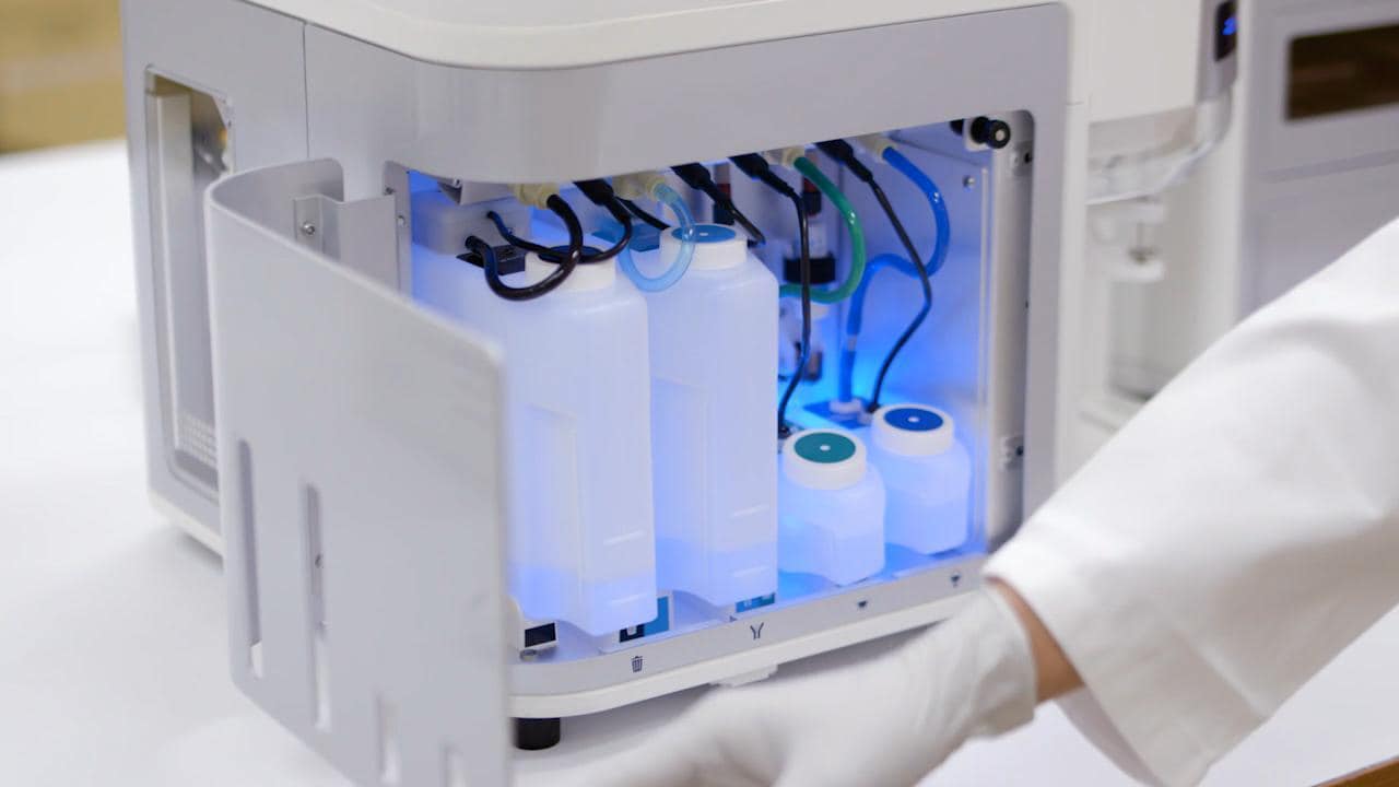
Standard fluid reservoirs |
1.8 L focusing fluid tank, 1.8 L waste tank, 175 mL shutdown solution tank, and 175 mL wash solution tank |
||||
Fluid storage |
All fluids stored within instrument |
||||
Extended fluidics option |
Configuration for 10 L fluid |
||||
Nominal fluid consumption |
1.8 L/day |
||||
Automated maintenance cycles |
≤15 min start-up and shutdown—deep clean, sanitize, and debubble modes |
||||
Performance: Fluorescence detection |
Fluorescence sensitivity |
≤80 molecules of equivalent soluble fluorochrome (MESF) for FITC, ≤30 MESF for PE, ≤70 MESF for APC |
|||
Fluorescence resolution |
CV <3% for the singlet peak of propidium iodide–stained chicken erythrocyte nuclei (CEN) |
||||
Data acquisition rate |
Up to 35,000 events/sec, 34 parameters, based on a 10% coincidence rate per Poisson statistics |
||||
Maximum electronic speed |
65,000 events/sec with all parameters |
||||
Carryover |
Single-tube format: <1% |
||||
Forward and side scatter sensitivity |
Able to discriminate platelets from noise |
||||
Forward and side scatter resolution |
Optimized to resolve lymphocytes, monocytes, and granulocytes in lysed whole blood |
||||
Forward scatter |
Photodiode detector with 488/10 nm bandpass filter |
||||
Side scatter |
PMT with default 488/10 nm bandpass filter; optional 405/10 nm bandpass filter |
||||
Fluorescence detectors |
14 individual detectors |
||||
Electronic pulse |
Measured area, height and width pulse for all detectors |
||||
Violet side scatter resolution |
Can be configured for violet side scatter to better resolve particles from noise |
||||
Minimum particle size |
0.2 μm on side scatter using submicron bead calibration kit from Bangs Laboratories—0.1 μm on side scatter under following conditions: Using an Attune NxT Flow Cytometer with standard 0.5 mm blocking configuration, an Invitrogen Attune NxT 488/10 Filter (Cat. No. 100083194), and Attune Focusing Fluid (Cat. No. 4488621, 4449791, or A24904) that has been passed through a 0.025 µm filter |
||||
Software |
Compensation |
Full matrix—automated and manual modes, on-plot compensation tools for fine adjustment; use of tubes and wells |
|||
Flow rate |
Precise flow rate control via software; no hardware adjustments |
||||
Live streaming |
Live update of statistics during acquisition of events up to 35,000 events/sec |
||||
Overlays |
Comparative analysis between samples; 3D view |
||||
Sample recovery |
System able to return unused samples |
||||
Concentration |
Direct concentration measurement without use of counting beads |
||||
Software layout |
Fully customizable for each user account |
||||
Bubble detection technology |
Stops automated run to preserve sample integrity |
||||
Maximum single-event file |
20 million with option to append |
||||
Heat map |
Set up for definition of plate layout; screening view for analysis for tubes and plates |
||||
Threshold |
Up to 4 individual thresholds with user option to apply Boolean logic |
||||
Gating |
Hierarchical gating with the ability to derive gates |
||||
Voltage |
User adjustable |
||||
Window extensions |
User adjustable |
||||
Area scaling factor (ASF) |
User adjustable |
||||
Acquisition settings |
Documented in FCS files and maintained upon import |
||||
Templates |
Create from existing experiments—instrument settings, workspaces, run protocols, heat map settings, and compensation settings optimized and defined previously |
||||
Tube-to-plate conversion |
One-click transition from tubes to plates and vice versa; no disassembly, no additional QC, no reboot required for conversion between plates and tubes |
||||
Graphics resolution |
Publication-quality images; support for TIF, PNG, BMP, JPG, GIF, and EMF; quickly copy and paste plots to any external application (e.g., Microsoft™ PowerPoint™ software) |
||||
User account administration |
Administrative creation of individual user accounts with designated roles, advanced setting permissions, management of individual accounts, user time tracking, and sample count |
||||
Quality and regulatory |
Instrument tracking |
Automated daily baseline and performance test with Levey-Jennings plots |
|||
Warranty |
1 year |
||||
Production verification testing |
Each instrument is tested and verified for assembly integrity and performance to specifications |
||||
Quality management system |
Manufacturing standards comply with the requirements of ISO 13485:2003 |
||||
Robust installation specifications |
Units installed by engineer; preplanning checklist, delivery, and installation; and performance validation compliance with standardized procedure |
||||
Regulatory status |
For Research Use Only |
||||
Computer |
Software requirements |
Invitrogen Attune Cytometric Software |
|||
Monitor |
23 in. flat panel (1,920 x 1,080 resolution); dual-monitor capability |
||||
Computer |
Minitower desktop |
||||
Operating system |
Microsoft™ Windows™ 10 64-bit |
||||
FCS format |
FCS 3.1, 3.0 |
||||
Processor |
Intel™ Core™ i7 processor |
||||
RAM |
32 GB |
||||
Hard drives |
2 x 2 TB SATA 3.0 Gb/s, 8 MB data burst cache; controller RAID1, integrated |
||||
Installation requirements |
Electrical requirements |
100–240 VAC, 50/60 Hz, <150 W |
|||
Heat dissipation |
<150 W |
||||
Temperature operating ranges |
15–30°C (59–86°F) |
||||
Operating humidity |
10–90%, noncondensing |
||||
Audible noise |
<65 dBA at 1.0 m |
||||
Instrument size (H x W x D) |
~40 x 58 x 43 cm (16 x 23 x 17 in.), including fluid bottles |
||||
Weight |
~29 kg (64 lb) |
||||
Related pages


For Research Use Only. Not for use in diagnostic procedures.