Search Thermo Fisher Scientific
Akt Signaling Pathway: The Master Regulator in Human Disease
Dissect the Akt Pathway Using ABfinity Antibodies, Luminex Multiplex Assays, and More
The serine–threonine kinase Akt (also known as protein kinase B) is a central convergence node in a broadly influential signaling network. Akt activation serves as a master switch of these cellular signaling pathways, generating a multitude of intracellular responses through a plethora of downstream targets and interacting partners (Figure 1). Because of its pivotal role in cell signaling, and the consequences of that signaling in diseases ranging from cancer and diabetes to neurodegeneration, Akt is one of the most actively studied kinases in both basic research and drug development. Here we highlight our newest products for analyzing the Akt signaling pathway and its downstream targets.
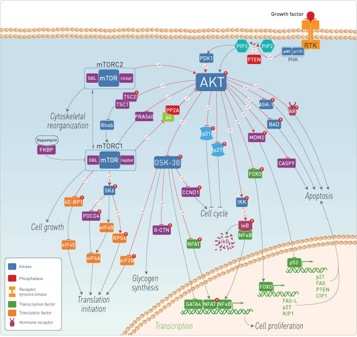 Figure 1. The Akt signaling pathway.
Akt Phosphorylation Leads to Activation
Akt is expressed as three isoforms—AKT1/PKBα, AKT2/PKBβ, and AKT3/PKBγ [1]. An amino‑terminal pleckstrin homology (PH) domain, a central serine–threonine catalytic domain, and a small carboxy‑terminal regulatory domain are present in all three isoforms. The PH domain binds phosphatidylinositol-3,4-bisphosphate (PIP2) and phosphatidylinositol-3,4,5-trisphosphate (PIP3), products of phosphatidylinositol 3-kinase (PI3K). This binding causes Akt to translocate to the plasma membrane, where it becomes phosphorylated by phosphoinositide-dependent kinase 1 (PDK1) on Thr308 in the activation loop of the catalytic domain.
Thr308 phosphorylation leads to activation; however, full activation requires phosphorylation at a second site (Ser473). Current evidence suggests the mTOR–rictor complex (mTORC2) is the primary kinase for the second phosphorylation event (Figure 1), although other kinases like integrin-linked kinase (Ilk) [2], PDK1 [3], DNA-dependent protein kinase (DNA-PK) [4], and ataxia telangiectasia mutated (ATM) have also been identified [5].
Activated Akt targets downstream signaling substrates to regulate cell processes that are associated with tumor development, including regulators of apoptosis, gene transcription, cell cycle progression, and cellular metabolism (Figure 1). Dysregulation in the Akt signaling pathway, as well as in downstream pathways, is therefore one of the hallmarks of cancer, making Akt a critical target for the development of anticancer drugs and therapies [6].
Validated Products for Multiple Assay Platforms
We have developed a wide selection of Novex primary antibodies, including phosphorylation site–specific (phosphospecific) antibodies that allow you to analyze differential patterns of protein phosphorylation in the Akt pathway. These products include our new high-quality ABfinity recombinant monoclonal antibodies, which are validated for multiple applications including western blot analysis (Figure 2), immunocytochemistry studies (Figure 3), indirect ELISA (Figure 4), and flow cytometry assays (Figure 5). Produced by cloning and expressing functionally screened, immunogen-specific antibody genes in mammalian cells, the ABfinity™ recombinant monoclonal antibodies provide superior specificity, sensitivity, and consistency in immunoassays; see page 18 for a more complete description of the innovative ABfinity recombinant antibody technology.
In addition to ABfinity antibodies, antibody pairs, and ELISA kits, we also offer Novex multiplex assay panels for measuring the levels of total and phosphorylated proteins in the Akt pathway using the Luminex platform. The new Akt Pathway Total Magnetic 7-Plex and Phospho Magnetic 7-Plex Panels provide accurate quantitation of seven different proteins simultaneously in cell lysates and tissue homogenates (Figure 6). These magnetic bead–based Luminex assays simplify the wash steps while increasing both throughput and precision. Our Luminex multiplex assays are suitable for use with the Luminex 100/200, FLEXMAP 3D, and MAGPIX systems.
![Western blot of 3T3 cell lysates labeled with anti–AKT [pS473] ABfi nity™ recombinant rabbit monoclonal antibody](/content/dam/LifeTech/migration/images/technical-reference-library/newsletters-journals/bioprobes-journal/bioprobes-issues-2012/bioprobes-67.par.31441.image.275.308.1.dat-bp67-s007415-akt-fig2-gif.gif) | Figure 2. Western blot of NIH/3T3 cell lysates labeled with anti–AKT [pS473] ABfinity recombinant rabbit monoclonal antibody. Anti–AKT [pS473] ABfinity monoclonal antibody was used at 0.1 µg/mL to label AKT [pS473] in lysates of untreated 3T3 (lane 1) or PDGF-treated (lane 2) NIH/3T3 cells. |
![Immunocytochemistry of mouse fibroblasts labeled with anti–AKT [pS473] ABfi nity™ recombinant rabbit monoclonal antibody](/content/dam/LifeTech/migration/images/technical-reference-library/newsletters-journals/bioprobes-journal/bioprobes-issues-2012/bioprobes-67.par.82019.image.275.278.1.dat-bp67-s007416-akt-fig3-jpg.gif) | Figure 3. Immunocytochemistry of mouse fibroblasts labeled with anti–AKT [pS473] ABfinity recombinant rabbit monoclonal antibody. Mouse fibroblast cells were labeled with anti–AKT [pS473] ABfinity monoclonal antibody at 5 µg/mL, followed by green-fluorescent Alexa Fluor 488 goat anti-rabbit secondary antibody. Nuclei were stained with blue-fluorescent Hoechst 33342 nuclear counterstain. |
![Indirect ELISA of anti–AKT [pS473] ABfinity™ recombinant rabbit monoclonal antibody](/content/dam/LifeTech/migration/images/technical-reference-library/newsletters-journals/bioprobes-journal/bioprobes-issues-2012/bioprobes-67.par.88551.image.275.262.1.dat-bp67-s008166-akt-fig4-gif.gif) | Figure 4. Indirect ELISA of anti–AKT [pT308] ABfinity recombinant rabbit monoclonal antibody. Indirect ELISA was performed using various dilutions of anti–AKT [pT308] ABfinity monoclonal antibody to detect recombinant AKT [pT308] protein coated onto the plate. |
![Flow cytometry of Jurkat cells labeled with anti–AKT [pS473] ABfinity™ recombinant rabbit monoclonal antibody](/content/dam/LifeTech/migration/images/technical-reference-library/newsletters-journals/bioprobes-journal/bioprobes-issues-2012/bioprobes-67.par.77897.image.275.252.1.dat-bp67-s007418-akt-fig5-gif.gif) | Figure 5. Flow cytometry of Jurkat cells labeled with anti–AKT [pS473] ABfinity recombinant rabbit monoclonal antibody. Jurkat cells were incubated with (red trace) or without (green trace) 50 µM LY294002 for 1 hr prior to being fixed and permeabilized using FIX & PERM reagents (An Der Grub Bio Research GmbH). Cells were then stained with 1 µg anti–AKT [pS473] ABfinity monoclonal antibody, followed by Alexa Fluor 488 goat anti-rabbit secondary antibody. The blue trace represents secondary antibody alone. |
 | Figure 6. Multiplex quantitation of Akt pathway signaling proteins using the Luminex platform. MCF-7 cells were treated with insulin to induce protein phosphorylation, and then cell lysates were prepared. Protein concentrations in the MCF-7 cell lysates were measured for seven phosphorylated proteins—AKT [pS473], GSK-3β [pS9], IGF-1R [pYpY1135/1136], IR [pYpY1162/1163], IRS-1 [pS312], p70S6K [pTpS421/424], PRAS40 [pT246]—using the Akt Pathway Phospho Magnetic 7-Plex Panel and the Luminex 200 system. |
Find Many More Products Online for Akt Pathway Research
We have highlighted just a few of the many products that we offer for Akt pathway research. Our page on Akt pathway products has much more information on our complete selection of antibodies, assays, and proteins associated with the Akt signaling pathway, all arranged in a comprehensive and easily navigable format. There you’ll find:
- ABfinity recombinant monoclonal and oligoclonal antibodies
- Bead-based Luminex multiplex assay kits
- Novex antibodies and antibody pairs
- Novex ELISA kits
- Gibco growth factors and cytokines
References
- Vivanco I, Sawyers CL (2002) Nature Rev Cancer 2:489–501.
- Persad S, Attwell S, Gray V et al. (2000) Proc Natl Acad Sci U S A 97:3207–3212.
- Balendran A, Casamayor A, Deak M et al. (1999) Curr Biol 9:393–404.
- Feng J, Park J, Cron P et al. (2004) J Biol Chem 279:41189–41196.
- Dong LQ, Liu F (2005) Am J Physiol Endocrinol Metab 289:E187–E196.
- Liu P, Cheng H, Roberts TM et al. (2009) Nat Rev Drug Discov 8:627–644.
Resources
Article Download
Get a copy of this article as it appears in the print version of BioProbes 67.
Download Now (4.7 mb)
Learn More About
For Research Use Only. Not for use in diagnostic procedures.