Search
Ready-to-Use Reagents Shift the Focus to Results
ReadyProbes™ Reagents Are the Ones We Use in Our Labs
The use of live-cell fluorescence imaging is becoming increasingly popular due to its unmatched spatiotemporal resolution of dynamic cellular events that cannot be captured with fixed-cell analysis. In addition to advances in optics, sensor technology, computing power, and software tools, this shift has been enabled by more stable, brighter, and highly specific fluorophores, including fluorescent proteins, organic dyes, and quantum dots. The result has been widespread adoption of fluorescence imaging in cell-based research, where scientists can apply these new tools to areas from developmental and stem cell biology to medical research, drug discovery, and environmental studies. Fixed-cell fluorescence imaging remains a fundamental method of analysis in applications in which live-cell protocols are either incompatible, such as immunocytochemical studies, or impractical, such as high-content screening.
Despite these advances, fluorescence imaging is often considered difficult or inaccessible, with complex instrumentation and a myriad of often bewilderingly similar reagents that must be stored frozen and then diluted several hundred-fold before use. Moreover, troubleshooting fluorescent labeling protocols can be time-consuming, particularly for live-cell imaging where exposure to nonphysiological conditions can adversely impact the system under study and obscure results.
Focus on the Biology, Not the Protocol
We have applied our 30 years of fluorescence experience to the development of ready-to-use reagents that facilitate several of the procedures required for today’s cell biology research. The first set of these ReadyProbes™ reagents and kits addresses core fluorescence imaging needs (Table 1). ReadyProbes™ reagents are formulated for maximum convenience, without sacrificing any of their optical or physicochemical properties. These reagents are provided with concise protocols and user-friendly packaging that includes recyclable, recycled, and sustainable materials where possible.
Table 1. ReadyProbes™ ready-to-use reagents for fluorescence imaging.
| ReadyProbes™ Product* | Quantity | Cat. No. | |
|---|---|---|---|
| Live-cell imaging reagents | |||
Live Cell Imaging Solution | 500 mL | A14291DJ | |
BackDrop™ Background Suppressor | 6 x 2.5 mL | R37603 | |
LIVE/DEAD® Cell Imaging Kit | 1 kit | R37601 | |
NucBlue™ Live Cell Stain | 6 x 2.5 mL | R37605 | |
| Fixed-cell imaging reagents | |||
NucBlue™ Fixed Cell Stain | 6 x 2.5 mL | R37606 | |
Image-iT® Fixation/Permeabilization Kit | 1 kit | R37602 | |
| *Find more information on our complete selection of ReadyProbes™ reagents and kits. | |||
Live Cell Imaging Solution for Improved Optical Clarity
When imaging live cells, it is imperative that environmental factors do not induce stress, which can alter the cell state or perturb the process or system under study. Many cells—including stem cells, primary cells, and neurons—are highly sensitive to even the relatively modest pH changes that may occur within minutes when cell cultures growing in carbonate-buffered media are removed from the incubator (Figure 1). Live Cell Imaging Solution (LCIS) is a physiological medium based on the classic Ringer’s formulation and contains a live cell–compatible HEPES buffer for extended buffering capacity. This medium has universal utility in cell-based research, providing a solution for live-cell imaging, dye loading, and wash steps.
Compared with standard media, Live Cell Imaging Solution has better optical clarity, which leads to improved signal-to-noise ratios and reduced background levels in fluorescence imaging experiments (Figure 2). In the absence of environmental chambers, we recommend minimizing exposure of the cell cultures to temperature and osmolarity changes by reducing the time cells are outside the incubator and maximizing the volume of liquid in which they reside.
 | Figure 1. Change in pH after cells are removed from the incubator. pH changes over time are shown for three cell types (CHO-M1, U2OS, and BPAE) kept on a lab bench in Live Cell Imaging Solution (LCIS) and three different media (Ham’s F-12 medium, McCoy’s medium, and DMEM). Note that LCIS containing metabolically active cells maintains a stable physiological pH at room temperature for at least 4 hours. |
 Figure 2. Improved optical clarity with Live Cell Imaging Solution. CHO-M1 cells stained with (A)NucBlue™ Live Cell Stain (a Hoechst 33342 formulation) or (B)calcein AM and (C) MMM cells stained with LysoTracker® Red DND-99 were imaged in (top row) Live Cell Imaging Solution (LCIS) or (bottom row) Ham’s F-12 medium with 10% FBS (CHO-M1 cells) or MEM with 10% FBS (MMM cells). Figure 2. Improved optical clarity with Live Cell Imaging Solution. CHO-M1 cells stained with (A)NucBlue™ Live Cell Stain (a Hoechst 33342 formulation) or (B)calcein AM and (C) MMM cells stained with LysoTracker® Red DND-99 were imaged in (top row) Live Cell Imaging Solution (LCIS) or (bottom row) Ham’s F-12 medium with 10% FBS (CHO-M1 cells) or MEM with 10% FBS (MMM cells). |
NucBlue™ Nuclear Stains for Live- or Fixed-Cell Imaging
The NucBlue™ Live and NucBlue™ Fixed Cell Stains are special formulations of the Hoechst 33342 and DAPI nucleic acid stains, respectively, which are among the most commonly used counterstains in fluorescence imaging. Unlike the Hoechst 33342 and DAPI dyes, which often require frozen storage and dilution before use, the NucBlue™ Live and NucBlue™ Fixed Cell Stains are provided ready to use in ultraconvenient dropper bottles that can be stored right next to your microscope or workstation. Just add two drops per milliliter to each of your cell samples and they are ready for imaging (Figure 3)—no pipetting, calculations, or dilutions required.
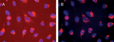 Figure 3. Suppression of background signal with BackDrop™ Background Suppressor. U2OS cells were grown in complete medium (DMEM) with 20% FBS and transduced with CellLight® Mitochondria-RFP. Following overnight incubation, cells were counterstained with 2 drops of NucBlue™ Live Cell Stain per mL of cell sample, incubated for 20 min, and imaged (A) without and (B) with the addition of 2 drops of BackDrop™ Background Suppressor per mL of cell sample. Identical exposure and gain settings were used for both images. Figure 3. Suppression of background signal with BackDrop™ Background Suppressor. U2OS cells were grown in complete medium (DMEM) with 20% FBS and transduced with CellLight® Mitochondria-RFP. Following overnight incubation, cells were counterstained with 2 drops of NucBlue™ Live Cell Stain per mL of cell sample, incubated for 20 min, and imaged (A) without and (B) with the addition of 2 drops of BackDrop™ Background Suppressor per mL of cell sample. Identical exposure and gain settings were used for both images. |
BackDrop™ Background Suppressor for Live-Cell Imaging
Background signals from culture vessels, media components, and nonspecific labeling increase signal-to-noise and reduce the sensitivity of fluorescence experiments. BackDrop™ Background Suppressor effectively suppresses blue-, green-, and red-fluorescent background signals in live-cell imaging samples, typically eliminating the need to wash off excess dye or remove media from cells before imaging intracellular fluorescent labels (Figure 3). Provided in a convenient dropper bottle, BackDrop™ Background Suppressor can sit right on your bench where you need it; simply add two drops per milliliter of sample and image. The degree of background suppression provided by the BackDrop™ reagent varies with fluorophores, concentration, labeling efficiency, cell type, and other conditions, but can often be significant.
LIVE/DEAD® Cell Imaging Kit
Assays for cell viability, cytotoxicity, and cell death are commonplace in all areas of cell biology research, from the care and maintenance of cell lines to the dissection of cellular pathways and the discovery of drug mechanisms of action. The LIVE/DEAD® Cell Imaging Kit is a sensitive two-color fluorescence cell viability assay optimized for the commonly used FITC and Texas Red® filter sets. Packaged for workflow convenience, this kit allows you to quickly and effortlessly determine the fraction of live and dead cells in a population, producing results after a single 15-minute incubation. For the simultaneous discrimination of live and dead cells, this assay relies on two fluorophores—a membrane-permeant dye for live-cell labeling and a membrane-impermeant dye that stains dead and dying cells in which the plasma membrane integrity has been compromised—which have been carefully selected for their brightness, compatible spectral properties, and ease of use in imaging workflows.
Image-IT® Fixation/Permeabilization Kit for Fixed-Cell Imaging
The new Image-iT® Fixation//Permeabilization Kit provides ready-to-use, room temperature–stable solutions that allow you to fix, permeabilize, block, and wash cells in preparation for antibody staining and imaging. The high-quality components and optimized formulations help preserve cell morphology and reduce background staining, thus improving signal-to-noise ratios and image quality. Cells are treated sequentially with the 1X fixative, permeabilization solution, and blocking solution according to a simplified, easy-to-follow protocol. Care was taken to optimize the reagents for both preservation of 3D cellular structure and adequate antibody access to intracellular antigenic sites. In extensive quality-control testing with both nuclear and cytoplasmic targets (Figure 4)—including fluorescent proteins, organic dyes, antibodies, and Click-iT® EdU reagents—the reagents in this kit showed equal or superior performance when compared with standard fixation and permeabilization reagents.
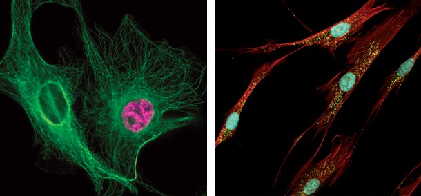
Figure 4. Fixed-cell imaging with the Image-iT® Fixation/Permeabilization Kit. (A) BPAE cells were pulsed with EdU, fixed and permeabilized using the Image-iT® Fixation/Permeabilization Kit, labeled with a mouse monoclonal anti–β-tubulin antibody followed by an Alexa Fluor® 488 goat anti-mouse secondary antibody, and then stained with Alexa Fluor® 488 azide provided in the Click-iT® EdU Alexa Fluor® 647 Imaging Kit. The image was acquired on a Nikon upright microscope (60x). (B)Neonatal human dermal fibroblasts (HDFn) were fixed and permeabilized using the Image-iT® Fixation/Permeabilization Kit. Peroxisomes were labeled with a rabbit anti-PMP70 antibody followed by an Alexa Fluor® 488 goat anti-rabbit secondary antibody; actin was stained using Alexa Fluor® 594 Phalloidin; nuclei were stained with NucBlue™ Fixed Cell Stain. The image was acquired on a Nikon upright microscope (20x).
Check Back for More ReadyProbes™ Reagents
We are continuing to develop user-friendly reagents and approaches to facilitate fluorescence imaging for beginners and experts alike. As always, we welcome your comments, thoughts, and suggestions. Learn more about our current selection of ReadyProbes™ reagents and kits.
Resources | |
| Article Download Get a copy of this article as it appears in the print version of BioProbes 67. | Learn More About: |
FOR RESEARCH USE ONLY. NOT INTENDED FOR ANY ANIMAL OR HUMAN THERAPEUTIC OR DIAGNOSTIC USE.
For Research Use Only. Not for use in diagnostic procedures.