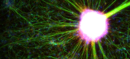
Preserve the fluorescent signal from your sample
Loss of fluorescence through irreversible photobleaching processes can lead to a significant reduction in sensitivity, particularly when target molecules are of low abundance or when excitation light is of high intensity or long duration. The mechanisms associated with photobleaching are also implicated in phototoxicity, which may adversely impact cell viability and data quality.
To minimize photobleaching of experimental samples, we have developed a series of antifade reagents that increase fluorophore photostability in both live-cell and fixed-cell samples. We offer choices for immediate imaging and for long-term storage, plus formulations that include a nuclear stain to combine mounting and counterstaining in a single step. Reduced photobleaching means longer tracking for time-course experiments, higher sensitivity, and better quantitation from fluorescent signals.
Select the antifade that matches your fluorescence imaging needs
ProLong Glass | ProLong Diamond | ProLong Gold | |
---|---|---|---|
Learn more | |||
General specifications | |||
Form | Ready-to-use hard-setting mountant | ||
Refractive index | 1.52 (after curing) | 1.47 (after curing) | 1.47 (after curing) |
What type of imaging am I planning? | |||
Specimen type | Animal cells/tissue (fixed) | ||
Live-cell imaging | |||
Fixed-cell, immediate imaging | |||
Fixed-cell, long-term imaging (up to 10 µm sample thickness) |
![]() |
![]() |
![]() |
Fixed-cell, long-term imaging (up to 150 µm sample thickness) |
![]() |
||
What type of microscope objective can produce best results?
|
|||
Oil immersion objective |
![]() |
||
Glycerol-corrected objective |
![]() |
![]() |
|
Water- or air-corrected objective | |||
What fluorophores does my sample include? | |||
Alexa Fluor dyes |
![]() |
![]() |
![]() |
Traditional dyes |
![]() |
![]() |
|
Fluorescent proteins |
![]() |
![]() |
|
Do I want to add counterstain in the mountant? | |||
Counterstain with DAPI |
![]() |
![]() |
![]() |
Counterstain with SYTOX Deep Red |
![]() |
![]() |
![]() |
No counterstain |
![]() |
![]() |
![]() |
SlowFade Glass | SlowFade Diamond | SlowFade Gold | |
---|---|---|---|
Learn more | |||
General specifications | |||
Form | Ready-to-use soft-setting mountant | ||
Refractive index | 1.52 | 1.42 | 1.42 |
What type of imaging am I planning? | |||
Specimen type | Animal cells/tissue (fixed) | ||
Live-cell imaging | |||
Fixed-cell, immediate imaging |
![]() |
![]() |
![]() |
Fixed-cell, long-term imaging (up to 10 µm sample thickness) | |||
Fixed-cell, long-term imaging (up to 500 µm sample thickness) |
![]() |
||
What type of microscope objective can produce best results? (All objective types are compatible with these reagents) | |||
Oil immersion objective | |||
Glycerol-corrected objective |
![]() |
![]() |
![]() |
Water- or air-corrected objective | |||
What fluorophores does my sample include? | |||
Alexa Fluor dyes |
![]() |
![]() |
![]() |
Traditional dyes |
![]() |
![]() |
|
Fluorescent proteins |
![]() |
![]() |
|
Do I want to add counterstain in the mountant? | |||
Counterstain with DAPI |
![]() |
![]() |
![]() |
Counterstain with SYTOX Deep Red |
|
||
No counterstain |
![]() |
![]() |
![]() |
ProLong Live | |
---|---|
Learn more | |
General specifications | |
Form | 100x concentrated liquid |
Refractive index | 1.3 |
What type of imaging am I planning? | |
Specimen type | Animal cells/tissue (live) |
Live-cell imaging |
![]() |
Fixed-cell, immediate imaging | |
Fixed-cell, long-term imaging (up to 10 µm sample thickness) | |
Fixed-cell, long-term imaging (up to 150 µm sample thickness) | |
What type of microscope objective can produce best results? (All objective types are compatible with these reagents) | |
Oil immersion objective | |
Glycerol-corrected objective |
![]() |
Water- or air-corrected objective | ![]() |
What fluorophores does my sample include? | |
Alexa Fluor dyes |
![]() |
Traditional dyes |
![]() |
Fluorescent proteins |
![]() |
Do I want to add counterstain in the mountant? | |
Counterstain with DAPI |
|
Counterstain with SYTOX Deep Red | |
No counterstain |
![]() |
ProLong Glass (curing) |
SlowFade Glass (non-curing) |
CytoVista Reagents (non-curing) |
|
---|---|---|---|
Learn more | Learn more | Learn more | |
General Specifications | |||
Form | Ready-to-use, curing (hard-setting) mountant For immediate imaging of samples up to 150 mm thick |
Ready-to-use non-curing (soft-set) mountant For immediate imaging of samples up to 500 mm thick |
Ready-to-use, non-curing (soft-setting) family of reagents 3D Cell Culture clearing and staining kit for immediate imaging of 3D cell culture (organoids and spheroids up to 1 mm thick) Tissue clearing and staining kit for immediate imaging of thick tissue (up to 10 mm) |
Refractive index | 1.52 (after curing) | 1.52 | 1.48 (organoids/spheroids up to 1 mm thick) 1.53 (tissue samples up to 10 mm thick) |
What type of imaging am I planning? | |||
Specimen type | Animal cells/tissue (fixed) | Animal cells/tissue (fixed) | Animal cells/tissue (fixed) |
Fixed-cell imaging, immediate imaging |
![]() |
![]() |
|
Fixed-cell, long-term imaging |
![]() |
||
What is the thickness of my sample? | |||
Up to 10 μm |
![]() |
||
Up to 150 μm |
![]() |
||
Up to 500 μm |
![]() |
||
Up to 1 mm |
![]() |
||
Up to 10 mm |
![]() |
||
What type of microscope objective can produce best results? (All objective types are compatible with these reagents) | |||
Oil immersion objective |
![]() |
![]() |
![]() |
Glycerol-corrected objective |
![]() |
||
What fluorophores does my sample include? | |||
Alexa Fluor dyes |
![]() |
![]() |
![]() |
Traditional dyes |
![]() |
![]() |
![]() |
Fluorescent proteins |
![]() |
![]() |
![]() |
Do I want to add counterstain in the mountant? | |||
Counterstain with DAPI |
![]() |
![]() |
|
Counterstain with SYTOX Deep Red |
![]() |
||
No counterstain |
![]() |
![]() |
![]() |
Image-iT Plant Clearing and Mounting Reagents for Imaging | |
---|---|
Learn more | |
General Specifications | |
Form | Ready-to-use reagents for clearing and mounting plant tissue for imaging |
Refractive index | 1.45 (clearing and soft-set mounting option) 1.415 (hard-set mounting option) |
What type of imaging am I planning? | |
Specimen type | Plant cells/tissue section/whole tissue (fixed) |
Live-cell imaging | |
Fixed-cell imaging |
![]() |
Fixed-cell imaging, long-term imaging (up to 10 μM sample thickness) |
![]() |
Fixed-cell imaging, long-term imaging (up to 150 μM sample thickness) |
![]() |
What type of microscope objective can produce best results? (All objective types are compatible with these reagents) | |
Oil immersion objective | |
Glycerol-corrected objective |
![]() |
Water- or air-corrected objective | |
What fluorophores does my sample include? | |
Most common histological stains |
![]() |
Fluorescent proteins | |
Do I want to add counterstain in the mountant? | |
Counterstain with DAPI | |
Counterstain with SYTOX Deep Red | |
No counterstain |
![]() |
Greener products from Thermo Fisher Scientific
For added convenience and a reduction in the environmental impact associated with cold storage requirements, our antifade reagents are provided in dropper bottles with storage right at your bench. In addition, these reagents are formulated with reduced sodium azide to help minimize environmental and health hazard exposure risks.
Learn more about these greener product alternatives
Resources
Publications
Poster
Research tools
Cell Analysis Learning Center
Access cell analysis application resources for more success as you plan and execute your experiments.
Support
Cell Analysis Support Center
Find tips, troubleshooting help, and resources for your cell analysis workflow.
5 Steps resources
For Research Use Only. Not for use in diagnostic procedures.