Search
New APEX® Kits
IgG primary antibodies are usually available only in small quantities, can be expensive, and are packaged with stabilizing proteins such as bovine serum albumin (BSA). These stabilizing proteins can interfere with the amine-reactive reagents used to covalently attach a fluorescent label. Unfortunately, procedures to remove these stabilizing proteins may cause significant loss of antibody.
The APEX® Antibody Labeling Kits (Table 1) provide a convenient means of directly labeling a small amount of IgG antibody (10–20 µg) with a fluorescent label or biotin-XX moiety, while allowing you to easily remove contaminants (such as BSA) without losing antibody. These kits include all the reagents required to perform five separate IgG antibody labeling reactions using one of the superior Molecular Probes® fluorescent labels or biotin-XX. Additional APEX® kit configurations are now available, allowing you to select from Alexa Fluor®, Oregon Green® 488, or Pacific Blue™ fluorescent labels, or biotin-XX, a unique Molecular Probes® product that provides a 14-atom spacer between the antibody and the biotin moiety.
Table 1. Spectral Characteristics and Applications of the Labels Available in the APEX® Antibody Labeling Kits.
* Fluorescence excitation (Ex) and emission (Em) maxima, in nm.
† Newest additions to the APEX® Antibody Labeling Kit product line.
| Fluorophore or Biotin Label | Ex/Em * | Application |
|---|---|---|
| Alexa Fluor® 488 | 496/519 | Imaging or flow cytometry |
| Alexa Fluor® 555 | 555/565 | Imaging |
| Alexa Fluor® 568 † | 578/603 | Imaging |
| Alexa Fluor® 594 | 590/617 | Imaging |
| Alexa Fluor® 647 | 650/665 | Imaging or flow cytometry |
| Oregon Green® 488 | 496/524 | Imaging or flow cytometry |
| Pacific Blue™ | 416/451 | Imaging or flow cytometry |
| Biotin-XX † | NA | Imaging or flow cytometry |
† Newest additions to the APEX® Antibody Labeling Kit product line.
Easy Labeling Procedure
The APEX® labeling procedure is ingeniously easy. The kits contain pipette tips preloaded with a solid-phase resin that captures the antibody (Figure 1); simply apply the amine-reactive fluorescent label or biotin-XX moiety and your IgG antibody of choice to the resin. A fluorescent or biotin-XX–conjugated IgG antibody is formed, and the conjugate is then eluted from the resin using elution buffer. The IgG antibody conjugate is ready for use in an imaging or flow cytometry assay in as little as 2.5 hours, with minimal hands-on time. The typical yield of conjugated IgG antibody from the APEX® kits ranges from 40 to 80%.
 | Figure 1. Diagram of an APEX® antibody labeling tip. |
Time and Effort Without Sacrificing Performance
Using directly labeled antibodies eliminates the background fluorescence commonly observed when secondary antibodies bind nonspecifically to the sample. In addition, directly labeled antibodies allow you to use more than one same-species antibody in a single staining experiment. Effective conjugation of IgG antibodies using APEX® antibody labeling technology is demonstrated in Figure 2.
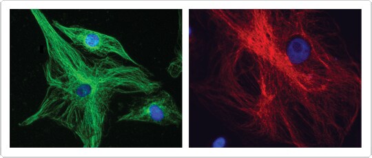
Figure 2. Effective conjugation of IgG antibody with or without BSA. Bovine pulmonary artery endothelial (BPAE) cells were fixed and permeabilized, then (left panel) treated with mouse anti–α-tubulin, detected with secondary antibody (goat anti-mouse) labeled using the APEX® Biotin-XX Kit, and counterstained with Alexa Fluor® 488 Streptavidin (green fluorescence); or (right panel) treated with mouse anti–α-tubulin, and detected with secondary antibody (goat anti-mouse) labeled using the APEX® Alexa Fluor® 568 kit (red fluorescence) in the presence of 1% BSA. Nuclei were stained with blue-fluorescent DAPI.
Antibody and Protein Labeling Kits for Diverse Applications
In addition to the APEX® antibody labeling kits, we offer several antibody and protein labeling kits that use our superior Alexa Fluor® fluorescent dyes or Qdot® nanocrystals. Each type of kit has been optimized for a specific application (Table 2). Monoclonal antibody labeling kits, Qdot® antibody conjugate kits, SAIVI™ antibody labeling kits, and Zenon® IgG labeling kits are also available. Learn more about our diverse selection of labeling technologies at APEX® Antibody Labeling Kits.
Table 2. Molecular Probes® Antibody and Protein Labeling Kits.
| Amount of IgG | Product | Key Features |
|---|---|---|
| <1–20 µg | Zenon® IgG Labeling Kits |
|
| 10–20 µg | APEX® Antibody Labeling Kits |
|
| 20–100 µg | Microscale Protein Labeling Kits |
|
| 100 µg | Monoclonal Antibody Labeling Kits |
|
| 300 µg | Qdot® Antibody Conjugation Kits |
|
| 1 mg | Protein Labeling Kits |
|
| 0.3–5 mg | SAIVI™ Antibody Labeling Kits |
|
For Research Use Only. Not intended for any animal or human therapeutic or diagnostic use.
For Research Use Only. Not for use in diagnostic procedures.
Quick Product View
See a complete listing of the products discussed in this article.
View products
Learn More
Find more information on our diverse selection of labeling technologies.
- Learn More about Antibody labeling
Download This Article
Get a copy of this article as it appears in the print version of BioProbes 63.
