Search
Fluorescent proteins provide dynamic and spatial information at the level of cells, tissues, and organisms, and have enabled tremendous insights in biological research over the past several decades. Traditional methods for using fluorescent proteins are constrained by low expression efficiency and cytotoxicity associated with lipid-based transfection, and the accessibility of these tools has been limited to researchers skilled in molecular biology. The baculovirus-based CellLight® reagents are rapidly transforming fluorescent protein–based live-cell imaging from the domain of a handful of experts into a simple and powerful technique available to everyone.
Powered by the BacMam 2.0 platform, CellLight® reagents offer a drastically simplified workflow, high efficiency of transduction in a broad range of cell types, and exquisite intracellular targeting to organelles and other subcellular structures. These reagents are compatible with traditional fluorescent reagents—for multiplex labeling using a wide array of fluorescent probes—ultimately enabling a deeper understanding of biological processes within the context of living, functioning cells (Figure 1) or fixed and permeabilized cells.
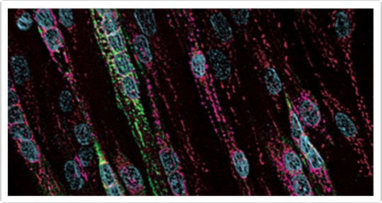
Figure 1. Imaging primary cells with CellLight® reagents. Primary human skeletal muscle cells (GIBCO® HSkM-S cells) were cultured in poly-D-lysine–coated glass-bottomed petri dishes and transduced with CellLight® ER-GFP and CellLight® Golgi-RFP in complete medium to label the endoplasmic reticulum (green) and the Golgi apparatus (magenta), respectively. The following day, the cells were loaded with 1 µg/mL Hoechst 33342 (to label nuclei, blue) and 25 nM MitoTracker® Deep Red (to label mitochondria, red) for 5 min at 37°C in DPBS. Cells were washed three times and imaged using standard DAPI/FITC/TRITC/Cy®5 filters.
How CellLight® Reagents Work
Molecular Probes® CellLight® reagents are baculovirus-packaged DNA constructs for the expression of fluorescent proteins fused to signal peptides, for specific targeting of key cellular structures (Table 1). CellLight® reagents are powered by BacMam technology, which combines highly efficient baculovirus gene delivery with a powerful mammalian promoter for expression of targeted fluorescent proteins [1,2]. Since mammalian cells do not recognize baculoviral promoters and genes, and baculoviruses cannot replicate in mammalian cells, CellLight® reagents are safe for both your cells and your laboratory personnel (i.e., biosafety level 1). In addition, they provide a simple, footprint-free method for transient expression of targeted fluorescent proteins and prolonged visualization of subcellular structures. CellLight® reagents are enabling tools for live-cell fluorescence imaging, and are also compatible with formaldehyde-based fixed-cell analyses.
| CellLight® Reagent |
CFP
(488/510)* |
GFP
(488/510)* |
RFP
(555/584)* |
|---|---|---|---|
| Actin | |||
| Cytoplasm | |||
| Endoplasmic Reticulum | |||
| Early Endosomes | |||
| Golgi Apparatus | |||
| Histones (Histone 2B) | |||
| Lysosomes | |||
| MAP4 | |||
| Mitochondria | |||
| Nucleus | |||
| Peroxisomes | |||
| Plasma Membrane | |||
| Synaptophysin | |||
| Talin | |||
| Tubulin | |||
| BacMam 2.0 Transduction Control | |||
| Null virus (control) |
* Excitation and emission maxima, in nm. † BacMam 1.0.
Amazing Images, Amazingly Simple Workflow
CellLight® reagents are supplied ready for immediate use—simply add these baculovirus suspensions to your cells, incubate overnight to allow protein expression to occur, then visualize the results (Figure 2). There is no need to purify plasmid, perform time-consuming subcloning, or perform quality control for vector integrity. Furthermore, the baculovirus delivery system offers reproducible expression using a simple and robust workflow. Compared to lipid-mediated transfection, which requires DNA quantitation and optimization of lipid–DNA complex formation, the workflow is uncomplicated, and the reagents are noncytotoxic. Unlike expression vectors, CellLight® reagents are delivered without dye-loading chemicals, and unlike conventional stains, they help provide labeling independently of organelle function (e.g., mitochondrial membrane potential). Unlike antibodies, they do not require fixation and permeabilization of samples. Furthermore, CellLight® reagents can be titrated to provide fine tuning of expression levels. CellLight® reagents offer high cotransduction efficiency; therefore, multiple BacMam reagents can be used simultaneously to visualize several organelles or structures in each cell (Figure 3).
 | Figure 2. Simple one-step gene delivery with BacMam 2.0 reagents. |
 | Figure 3. Imaging stem cell–derived cardiomyocytes with CellLight® reagents. Cardiomyocytes were incubated overnight with CellLight® Actin-GFP and CellLight® Mitochondria-RFP in complete medium. Cells were stained the following day with Hoechst 33342, then imaged using standard DAPI/FITC/TRITC filters. The striations of actin filaments characteristic of muscle cells are clearly evident. |
Better Transduction of More Cell Types
The new BacMam 2.0 system employs an updated capsid protein that improves transduction efficiency, and enhanced genetic elements that drive stronger expression of downstream sequences. These improvements extend the utility of this popular gene delivery platform to cell types such as primary neurons that could not be transduced with the original BacMam 1.0 system. For other cell types, including some stem cells and CHO cells, BacMam 2.0 provides more efficient transduction [3–5]. And for some intractable cell types such as fully differentiated neurons (Figure 4) and cardiomyocytes (Figure 3), BacMam 2.0 is the most effective method for gene delivery. While the new BacMam 2.0 platform precludes the need for the BacMam Enhancer Reagent in many cell types, the reagent improves delivery and expression in some cells, including immortalized T cells (e.g., Jurkat cells).
 | Figure 4. Live-cell imaging of neurons using CellLight® Tubulin-RFP. Hippocampal tissue from postnatal day 3 rats was harvested and dissociated in neural culture medium, then plated onto glial feeder cultures. Cells were resuspended at 50,000 cells/mL in complete neural culture medium plus mitotic inhibitors. The medium was removed from the feeder cultures and replaced with 2 mL of the neural cell suspension. CellLight® Tubulin-RFP (50 µL) was added to the plates, and cells were incubated overnight. Fluorescence was observed in neurons after 48 hr in culture. |
Control Reagents for Optimizing Fluorescent Protein Expression
The BacMam GFP transduction control encodes nontargeted emerald GFP (emGFP) for extremely bright and photostable labeling of the entire cell. In addition to its utility for trial and optimization of BacMam 2.0 gene delivery in your cell model, the BacMam GFP transduction control has been successfully used to visualize neuronal growth cone dynamics in live rat hippocampal neurons with striking results view the movie on Real-Time Visualization of Neuronal Growth Cone Dynamics Using BacMam 2.0). We also offer the CellLight® Null reagent, which transduces cells but lacks mammalian expression elements. This negative control is designed to help identify potential baculovirus-mediated effects on cells, and to determine background fluorescence relative to cells that have been transduced.
Lights. Cells. Action.
CellLight® reagents are specifically designed for visualizing dynamic events in live cells with high spatial and temporal resolution. Reagents for most targets are available with either emGFP or TagRFP for convenient multiplexing and colocalization studies, and are compatible with other fluorescent proteins, classic fluorescent dyes, and Qdot® nanocrystals for even more multiplexing options. To date, over 90 cell types have been shown to be effectively transduced using BacMam delivery technology. Learn more about CellLight® Ready-to-Use Fluorescent Protein Based Reagents.
- Kost TA, Condreay JP, Jarvis DL (2005) Nat Biotechnol 23:567–575.
- Fornwald JA, Lu Q, Wang D et al. (2007) Methods Mol Biol 388:95–114.
- Kaneko H, Suzuki H, Abe T et al. (2006) Biochem Biophys Res Commun 349:1220–1227.
- Hirasawa T, Yoshikawa K, Nakakura Y et al. (2007) J Biotechnol 131:34–44.
- Zeng J, Du J, Lin J et al. (2009) Mol Ther 17:1585–1593.
For Research Use Only. Not intended for any animal or human therapeutic or diagnostic use.
For Research Use Only. Not for use in diagnostic procedures.
Quick Product View
See a complete listing of the products discussed in this article.
View products
Learn More
Find more information on ready-to-use fluorescent protein–based reagents.
- Learn More about CellLight® Reagents
Download This Article

 |
| Live-cell imaging of neuronal growth cone dynamics in cultured rat hippocampal neurons using BacMam 2.0 GFP transduction control and CellLight™ MAP4-RFP. |