Search

Materials Science
SEM in Geology
SEM EDS is a vital part of multi-modal, multi-scale analysis of rocks and minerals.
Join the conversation
SEM mineral identification
Few disciplines rely as heavily on consistent, multi-scale observation and interpretation of features as the earth sciences. Everything from deep earth flow processes to more efficient methods of resource extraction are dependent on accurate sample characterization.
Thermo Fisher Scientific has built a family of imaging platforms and software solutions to facilitate data collection, visualization and analysis during complicated multi-modal, multi-scale characterization routines.
Petrology and mineralogy
Accurate textural analysis and the associated distribution of minerals within the rock texture are key to accurately describing the physical and chemical aspects of a rock system. Automated determination of mineralogy, based on scanning electron microscopy coupled with energy-dispersive X-ray spectroscopy (SEM-EDS), has established itself as a popular method for acquiring high-resolution images and chemical maps in the mining and mineral processing industries. Thermo Fisher Scientific has a 30-year history of delivering market-leading SEM-EDS technology. Here are just a few of the advantages afforded by our petrology and mineralogy solutions;
- Expand the number of samples that can be processed with routine petrologic and mineralogic analysis
- Build a strong statistical repository of mineral content based on automated analysis
- Automatically obtain accurate mineralogical identification
- Create a digital inventory of your thin sections and associated mineral assemblages
- Combine EDS elemental analysis, mineralogy and high-resolution imagery for advanced sample interpretation
- Analyze more samples for more robust statistics


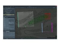
Multi-scale analysis with geoscience software
While context is vital for textural analysis, it is especially important for geological or structural interpretation. Electron imaging captures a large number of modalities in a single acquisition, allowing for direct interpretation of composition and texture. However, simply observing a single frame, or even a series of images, out of context reduces the power of this analysis.
We provide a suite of automation software developed with the express purpose of preserving context. Thermo Scientific Maps Software is the cross-platform automation engine for our full line of electron microscopy imaging platforms. Maps Software takes the pain out of acquiring larger image mosaics within an easy to use and intuitive software environment.
- Preserve context with large, automatically acquired, high-resolution datasets
- Utilize system time efficiently
- Share observations with context for more collaborative data interpretation

Chemical zonation in a plagioclase feldspar sample. Albite is shown in dark blue, labradorite is shown in light blue.

Multi-scale analysis of a sandstone sample. The left image is a slice from a microCT dataset, the right 3 images are from subsequent SEM analysis that interrogated the clay mineralogy present in the pores.

Cross-section overview of the major rock types, revealing varied mineral and structural distribution.

Surface analysis of a rock cross-section. A single mineralogical dataset from SEM-EDS analysis is uniquely capable of telling a rich story on texture and composition.

Multi-scale analysis of a core plug with X-ray microtomography (microCT) and SEM. The initial sample, plus the extracted mini plug, were imaged with HeliScan microCT. A cross-section of the mini plug was then imaged with SEM. The SEM data has been correlated and segmented with Avizo Software.
Chemical zonation in a plagioclase feldspar sample. Albite is shown in dark blue, labradorite is shown in light blue.
Multi-scale analysis of a sandstone sample. The left image is a slice from a microCT dataset, the right 3 images are from subsequent SEM analysis that interrogated the clay mineralogy present in the pores.
Cross-section overview of the major rock types, revealing varied mineral and structural distribution.
Surface analysis of a rock cross-section. A single mineralogical dataset from SEM-EDS analysis is uniquely capable of telling a rich story on texture and composition.
Multi-scale analysis of a core plug with X-ray microtomography (microCT) and SEM. The initial sample, plus the extracted mini plug, were imaged with HeliScan microCT. A cross-section of the mini plug was then imaged with SEM. The SEM data has been correlated and segmented with Avizo Software.

Investigación sobre materiales fundamentales
Se investigan nuevos materiales a escalas cada vez más pequeñas para lograr el máximo control de sus propiedades físicas y químicas. La microscopía electrónica proporciona a los investigadores información clave sobre una amplia variedad de características materiales a escala nanométrica.
Style Sheet for Komodo Tabs

Caracterización de materiales en 3D
El desarrollo de materiales suele requerir caracterización en 3D en varias escalas. Los instrumentos DualBeam permiten el corte en secciones en serie de grandes volúmenes y la posterior adquisición de imágenes SEM a escala de nanómetro, las cuales se pueden procesar en reconstrucciones 3D de la muestra de alta calidad.
_Technique_800x375_144DPI.jpg)
Análisis elemental EDS
EDS proporciona información de composición vital sobre las observaciones de microscopio electrónico. En concreto, nuestros exclusivos sistemas de detectores Super-X y Dual-X añaden opciones para mejorar el rendimiento y/o la sensibilidad, permitiendo optimizar la adquisición de datos para cumplir con sus prioridades de investigación.
_Technique_800x375_144DPI.jpg)
Tomografía EDS en 3D
La investigación de materiales modernos depende cada vez más del análisis a nanoescala en tres dimensiones. La caracterización en 3D, incluidos los datos de composición para el contexto químico y estructural completo, es posible con EM en 3D y espectroscopia de rayos X dispersiva.

Corte transversal
El corte transversal proporciona una visión adicional, ya que descubre información de la subsuperficie. Los instrumentos DualBeam tienen columnas FIB para poder realizar el corte transversal con alta calidad. Con la automatización, se puede realizar el procesamiento de muestras de alto rendimiento sin supervisión.

Análisis de escala múltiple
Los novedosos materiales se deben analizar a una resolución cada vez mayor, manteniendo el contexto más amplio de la muestra. El análisis de escala múltiple permite la correlación de varias herramientas y modalidades de obtención de imágenes, tales como microTC de rayos X, DualBeam, PFIB láser, SEM y TEM.
Caracterización de materiales en 3D
El desarrollo de materiales suele requerir caracterización en 3D en varias escalas. Los instrumentos DualBeam permiten el corte en secciones en serie de grandes volúmenes y la posterior adquisición de imágenes SEM a escala de nanómetro, las cuales se pueden procesar en reconstrucciones 3D de la muestra de alta calidad.
Análisis elemental EDS
EDS proporciona información de composición vital sobre las observaciones de microscopio electrónico. En concreto, nuestros exclusivos sistemas de detectores Super-X y Dual-X añaden opciones para mejorar el rendimiento y/o la sensibilidad, permitiendo optimizar la adquisición de datos para cumplir con sus prioridades de investigación.
Tomografía EDS en 3D
La investigación de materiales modernos depende cada vez más del análisis a nanoescala en tres dimensiones. La caracterización en 3D, incluidos los datos de composición para el contexto químico y estructural completo, es posible con EM en 3D y espectroscopia de rayos X dispersiva.
Corte transversal
El corte transversal proporciona una visión adicional, ya que descubre información de la subsuperficie. Los instrumentos DualBeam tienen columnas FIB para poder realizar el corte transversal con alta calidad. Con la automatización, se puede realizar el procesamiento de muestras de alto rendimiento sin supervisión.
Análisis de escala múltiple
Los novedosos materiales se deben analizar a una resolución cada vez mayor, manteniendo el contexto más amplio de la muestra. El análisis de escala múltiple permite la correlación de varias herramientas y modalidades de obtención de imágenes, tales como microTC de rayos X, DualBeam, PFIB láser, SEM y TEM.
Hoja de estilo para tarjetas originales instrumentos


Style Sheet to change H2 style to p with em-h2-header class