Search


Diagrama del volumen y resolución que se puede analizar por varios instrumentos
en el flujo de trabajo de análisis de escala múltiple.
en el flujo de trabajo de análisis de escala múltiple.
A medida que los materiales continúan avanzando, cada vez es más importante examinarlos no solo a resoluciones cada vez más altas, sino también obtener estas observaciones dentro de su contexto macroscópico relevante. Esto requiere correlacionar diferentes modos de imagen con las mismas coordenadas para una visión contextual real. Las mediciones también deben obtenerse con la suficiente rapidez para su aplicación práctica en entornos reales de control de procesos y análisis de fallos. Thermo Fisher Scientific ofrece un flujo de trabajo completo para la observación de materiales, combinando imágenes correlacionadas a diferentes escalas con información adicional, como la composición química.
El análisis de escala múltiple comienza con la observación a micro escala con técnicas espectroscópicas no destructivas. La microtomografía computarizada por rayos X (microCT) realiza una representación completa en 3D de la muestra mediante barridos de rayos X en serie. Estos barridos, o tomogramas 2D, se combinan digitalmente para formar la estructura 3D. Con la microtomografía computarizada Heliscan de Thermo Scientific, la serie de barridos circulares se sustituye por un barrido único helicoidal continuo. Esto permite un barrido más rápido a una dosis más baja, aumentando la precisión y la cantidad de información obtenida. Las observaciones de la microtomografía computarizada pueden proporcionar una resolución de hasta 400 nm, lo que la convierte en una herramienta ideal para el estudio no destructivo de la muestra antes de la caracterización de mayor resolución.
Una vez identificada una región de interés, se utiliza la instrumentación DualBeam (microscopía electrónica de barrido y haz de iones enfocado, FIB-SEM) para un análisis de superficie más cercano y la extracción de muestras. (Tenga en cuenta que el haz de iones enfocado puede consistir en una fuente de iones de metal líquido, galio, o un FIB de plasma.) El SEM permite el análisis de superficies a nanoescala, mientras que el FIB/PFIB se utiliza para el corte en secciones en serie, o para extraer una fina laminilla de muestra para su observación posterior con la microscopía electrónica de transmisión (TEM). La adición de un láser de femtosegundo al PFIB-SEM permite una preparación de muestras aún más rápida, corte transversal o corte en secciones en serie. El análisis TEM posterior proporciona una caracterización de materiales a escala atómica para obtener una visión completa de la composición elemental y estructural de una muestra.
La microscopía real de escala múltiple genera imágenes fiables y de alta calidad en todos los instrumentos, al tiempo que los alinea con precisión en una representación completa de la muestra. Con el software de automatización y análisis de datos de Thermo Scientific, todo el flujo de trabajo a gran escala se convierte en un procedimiento guiado y rutinario que se puede integrar fácilmente en el entorno de control de calidad o proceso.

The multi-scale analysis workflow offered by Thermo Fisher Scientific integrating software and hardware.
*Maps is not directly integrated with mCT, it can load processed CT data.
**AutoScript is not available yet on HeliScan and TEM microscopes.
*Maps is not directly integrated with mCT, it can load processed CT data.
**AutoScript is not available yet on HeliScan and TEM microscopes.

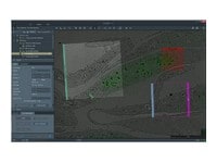
Multi-scale analysis workflow applied to the casing of an automotive oil filter (a glass-fiber-reinforced polymer composite).

HeliScan MicroCT analysis used in the correlative study of defects in an oil filter casing made of a glass-fiber-reinforced composite. Here a 40 μm³ voxel size was used to capture the entire filter (100×100×210 mm³) at 70 kV in 18 hours. A lateral cross-section is shown. For a full view of the 3D reconstruction, see the corresponding video.

HeliScan MicroCT analysis used in the correlative study of defects in an oil filter casing made of a glass-fiber-reinforced composite. Here a 5 μm³ voxel size was used to observe a portion of the casing in greater detail (13×13×50 mm³), obtained at 70 kV in 7 hours. A lateral cross-section is shown. For a full view of the 3D reconstruction, see the corresponding video.
.")
Following microCT analysis of an oil filter casing, a region of interest is identified for serial sectioning with an oxygen plasma FIB-SEM instrument. Using Auto Slice and View Software, serial 25-nm-thick slices were removed from the sample surface, which was imaged with SEM between each slice.
.")
Following microCT analysis of an oil filter casing, a region of interest is identified for serial sectioning with a DualBeam instrument. Here the results of the analysis are reconstructed in Avizo Software as a 3D representation of the region, clearly showing the glass fibers that reinforce the material.

SEM image of an oil filter casing, taken after serial sectioning of a region of interest with oxygen plasma FIB on the Helios Hydra DualBeam. High-resolution serial-sectioning data reveals a void within the nanometer-thin multilayer coating shell. This adds vital information to the microCT data, where the region of interest appears as dense particles.
Análisis de microtomografía computarizada HeliScan utilizado en el estudio correlativo de defectos en una carcasa de filtro de aceite fabricada por un compuesto reforzado con fibra de vidrio.
Caracterización del fallo del material de una pieza Inconel 718 aditivamente fabricada con aditivos con análisis de escala múltiple. Realizado en colaboración con la Universidad de Manchester.
Multi-scale analysis workflow applied to the casing of an automotive oil filter (a glass-fiber-reinforced polymer composite).
HeliScan MicroCT analysis used in the correlative study of defects in an oil filter casing made of a glass-fiber-reinforced composite. Here a 40 μm³ voxel size was used to capture the entire filter (100×100×210 mm³) at 70 kV in 18 hours. A lateral cross-section is shown. For a full view of the 3D reconstruction, see the corresponding video.
HeliScan MicroCT analysis used in the correlative study of defects in an oil filter casing made of a glass-fiber-reinforced composite. Here a 5 μm³ voxel size was used to observe a portion of the casing in greater detail (13×13×50 mm³), obtained at 70 kV in 7 hours. A lateral cross-section is shown. For a full view of the 3D reconstruction, see the corresponding video.
Following microCT analysis of an oil filter casing, a region of interest is identified for serial sectioning with an oxygen plasma FIB-SEM instrument. Using Auto Slice and View Software, serial 25-nm-thick slices were removed from the sample surface, which was imaged with SEM between each slice.
Following microCT analysis of an oil filter casing, a region of interest is identified for serial sectioning with a DualBeam instrument. Here the results of the analysis are reconstructed in Avizo Software as a 3D representation of the region, clearly showing the glass fibers that reinforce the material.
SEM image of an oil filter casing, taken after serial sectioning of a region of interest with oxygen plasma FIB on the Helios Hydra DualBeam. High-resolution serial-sectioning data reveals a void within the nanometer-thin multilayer coating shell. This adds vital information to the microCT data, where the region of interest appears as dense particles.
Análisis de microtomografía computarizada HeliScan utilizado en el estudio correlativo de defectos en una carcasa de filtro de aceite fabricada por un compuesto reforzado con fibra de vidrio.
Caracterización del fallo del material de una pieza Inconel 718 aditivamente fabricada con aditivos con análisis de escala múltiple. Realizado en colaboración con la Universidad de Manchester.

Control de calidad
El control y garantía de calidad son esenciales en la industria moderna. Ofrecemos una gama de herramientas de EM y espectroscopía para el análisis multiescala y multimodal de defectos, lo que le permite tomar decisiones fiables e informadas para el control y la mejora de procesos.

Investigación sobre materiales fundamentales
Se investigan nuevos materiales a escalas cada vez más pequeñas para lograr el máximo control de sus propiedades físicas y químicas. La microscopía electrónica proporciona a los investigadores información clave sobre una amplia variedad de características materiales a escala nanométrica.

Investigación de baterías
El desarrollo de baterías se realiza mediante análisis multiescala con microCT, SEM y TEM, espectroscopía Raman, XPS y visualización y análisis 3D digital. Aprenda cómo este enfoque proporciona la información estructural y química necesaria para crear mejores baterías.

Investigación sobre polímeros
La microestructura polimérica determina las características y el rendimiento del material a granel. La microscopía electrónica permite un análisis exhaustivo en microescala de la morfología y composición de los polímeros para aplicaciones de control de calidad e I+D.

Investigación sobre metales
La producción eficaz de metales requiere un control preciso de las inclusiones y precipitados. Nuestras herramientas automatizadas pueden realizar varias tareas cruciales para el análisis de metales, incluyendo el recuento de nanopartículas, el análisis químico EDS y la preparación de muestras de TEM.

Investigación sobre catálisis
Los catalizadores son cruciales para la mayoría de los procesos industriales modernos. Su eficacia depende de la composición microscópica y la morfología de las partículas catalíticas; EM con EDS es ideal para estudiar estas propiedades.

Gas y petróleo
A medida que la demanda de petróleo y gas continúa, existe la necesidad constante de una extracción eficiente y eficaz de hidrocarburos. Thermo Fisher Scientific ofrece una amplia gama de soluciones de microscopía y espectroscopía para una gran variedad de aplicaciones de la ciencia del petróleo.

Investigación geológica
Las ciencias geológicas están basadas en la observación uniforme y precisa de múltiples escalas de características dentro de las muestras de roca. SEM-EDS, combinado con software de automatización, permite el análisis directo a gran escala de la composición de la textura y los minerales para la investigación de la petrología y la mineralogía.

Fibras y filtros
El diámetro, la morfología y la densidad de las fibras sintéticas son parámetros clave que determinan la vida útil y la funcionalidad de un filtro. La microscopía electrónica de barrido (SEM) es la técnica ideal para investigar rápida y fácilmente estas características.

Pruebas de materiales para automóviles
Todos los componentes de un vehículo moderno están diseñados para garantizar la máxima seguridad, eficacia y rendimiento. La caracterización detallada de materiales de automoción con microscopía electrónica y espectroscopía informa sobre decisiones cruciales sobre procesos, mejoras de productos y nuevos materiales.
Hoja de estilo para tarjetas originales instrumentos


Style Sheet for Media Gallery Tabs